28-03-2026 07:55
 Marc Detollenaere
Hello everybody,Yesterday I found a number of whit
26-03-2026 15:31
 Åke Widgren
Hello,I found this one in October last year, on r
27-03-2026 15:23
Gernot FriebesHi,this Trichopezizella deviates from typical T. b
25-03-2026 10:35
Hulda Caroline HolteHello,I collected this species growing on a dead b
27-03-2026 15:08
Gernot FriebesHi,I'm looking for help with this coelomycete on C
27-03-2026 10:47
Åge OterhalsI have tentatively identified this Stictis to S. f
24-03-2026 21:37
Elisabeth StöckliBonsoir,Sur bois (tronc) très pourri de conifère


25-03-2026 22:23
Marc Detollenaere
Dear Forum,On a debarked stem of Tilia, we found s
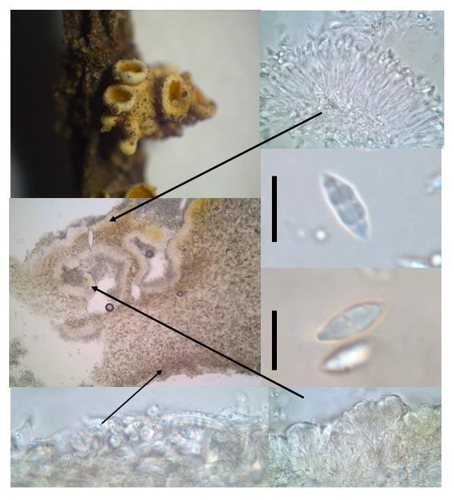
Stromatic cupulate coelomycete from Belize
Joanne Taylor,
07-09-2017 00:57
An interesting stromatic cupulate coelomycete was collected in Belize. it was growing on a seedling tree, on the stem and whether it was pathogenic or not, was unlcear. The description is below. It is very distinctive so surely must be described?
Creamy white stroma on host tissue (approximately 5mm), unclear whether it is bursting out of cuticle or superficial. Forming several cupulate conidiomata, often with stromatic short fat finger like projections.
In section hymenium can be convoluted and folded while appearing flat from above, orange coloured. The excipulum consists of textura intricata cells which are hyaline, thick walled with a narrow lumen, have crystalline encrustations and vary little from the stromatic tissue except in the upper margin where they are appear in a clear matrix.
The conidiogenous cells line the hymenium and are densely packed in some sort of matrix and will not separate even after treatment with 10% KOH. Conidiophores cylindrical/filiform, branched? or not, producing cylindrical filiform conidiogenous cells with small collarettes and are possibly percurrent with annelations.
Conidia are hyaline, fusiform with a truncate base and an apiculate apex, three septate with a germslit particularly visible in KOH (9-10 x 3 um).