19-05-2026 10:27
Patrice TANCHAUDBonjour, récolte récente sur terre retournée i
04-06-2026 18:39
Gernot FriebesHi,I collected this species in two different locat
22-05-2026 13:29
Gernot FriebesHi,I am curious to hear your opinion on this mater
04-06-2026 10:50
 François Freléchoux
Bonjour, J'ai trouvé hier un petit asco observé
04-06-2026 07:02
François Freléchoux
Bonjour, Voici la description d'une espèce qui p
04-06-2026 13:34
Gernot FriebesHi,I am interested to hear your opinion on this Le
04-06-2026 11:36
Gernot FriebesHi,found on Vaccinium myrtillus.Asci: IKI –, 8-s
18-10-2022 00:12
Valencia Lopez Francisco JavierHola amigos/asRecientemente encontré esta colecci
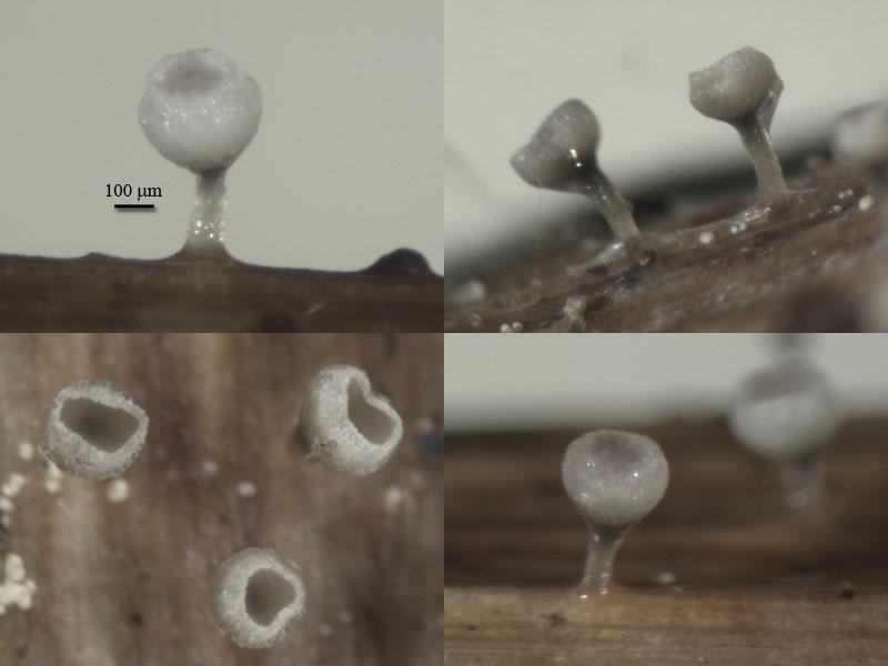
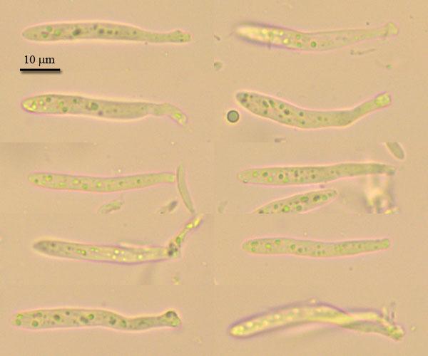
Apothecia goblet-shaped, receptacle deep-cupulate, to 0,5 mm in diam, stipe thin (100 mk), the same high as cup, all frb up to 1 mm high; stipe brownish, translucent, receptacle brownish at base, lighter to white at margin (when dry edge powdery from incrustation), edge rised under hymenium surface forming narrow collar.
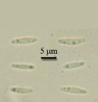
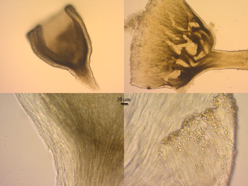
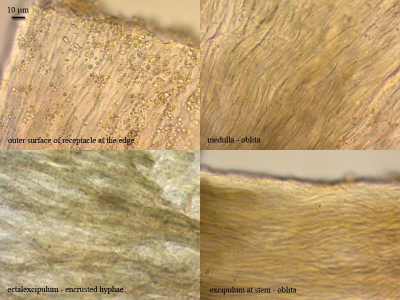
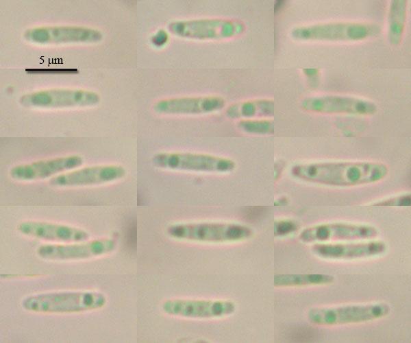
Excipulum from textura oblita, but outer layer of receptacle formed by porrecta, hyphae with rough walls (brown); margin from textura oblita, with abundant crystals; asci clavate, with crozier, with small euamyloid pore, 33,5-43 x 4,2-5,2; paraphyses lanceolate (not clear difference in two types), septate at base, slightly exceeding the asci, up to 3 mk broad in largest part; spores narrow-ellipsoid, with small guttules, 8 (7-9,4) x 1,7 (1,5-2,2) (N=18).
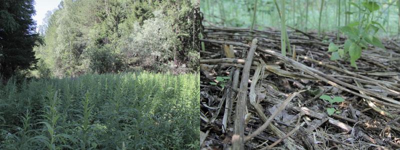
On dead stems of Glyceria triflora at forest edge, N61,090492 E69,480253, 26.06.2012.
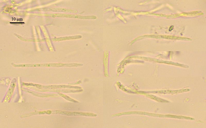

You do not have any micropics in vital state? Here I suspect multiguttulate paraphyses as typical of Cyathicula.
Useful should also bi a photo of the apical rings in IKI. If you compare their shape with those of Crocicreas gramineum, we could perhaps see a distinct difference.
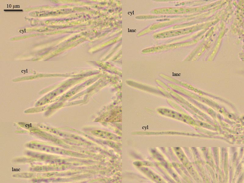
You say paraphyses lanceolate, but I see also cylindrical ones.
Zotto
i will send you vital photo,
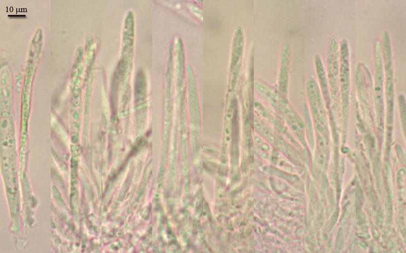
there are pictures of ring, it it differrent.
right, i was confused with paraphyses, they were badly seen in previous specimen. Since all hymenial parts smaller, differences not so clear. But now i checked again and think there are also two types, lanceolate and narrow (these rarely seen).
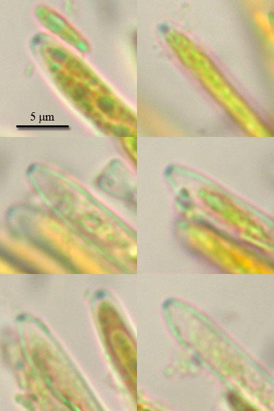
i am not sure about VBs since lack of experience seing them in vital, but what would you say?
On your spore photo I think that two spores are alive (lower left, central right). You say KOH, is this true for all spore photos?
I will compare with Cyathicula starbaeckii.