
04-06-2026 11:36
Gernot FriebesHi,found on Vaccinium myrtillus.Asci: IKI –, 8-s
05-06-2026 12:10
 François Freléchoux
Capitotricha sp. sur Lonicea caerulea Caractères
05-06-2026 11:02
Thomas Læssøehttps://svampe.databasen.org/observations/10596691

19-05-2026 10:27
Patrice TANCHAUDBonjour, récolte récente sur terre retournée i
04-06-2026 18:39
Gernot FriebesHi,I collected this species in two different locat
22-05-2026 13:29
Gernot FriebesHi,I am curious to hear your opinion on this mater
04-06-2026 10:50
François Freléchoux
Bonjour, J'ai trouvé hier un petit asco observé
Tiny black ascomycet 1
Gernot Friebes,
02-06-2009 20:22
can you help me with this ascomycet? Host was Picea. The spores are 14,5-16 x 5,5-6,5 µm and slightly brownish when mature, the asci are IKI- and 8-spored.
Best wishes,
Gernot
Gernot Friebes,
02-06-2009 20:22
Re:Tiny black ascomycet 1
spores
Gernot Friebes,
02-06-2009 20:23
Re:Tiny black ascomycet 1
ascus in lugol
Jacques Fournier,
03-06-2009 10:49

Re:Tiny black ascomycet 1
Hi Gernot,
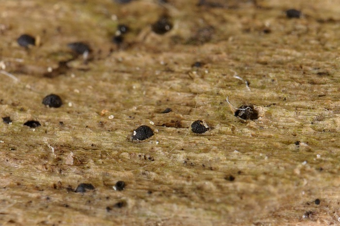
it's impossible to give you any clue without decent data on the morphology of the ascomata, peridium, asci and paraphyses. Please understand black dots with unicellular ascospores can fit hundreds of ascomycetes.
First what are the black dots on wood surface? very small superficial ascomata, necks of immersed ones? then you need a cross section to show the degree of immersion and thickness of the peridium.
Are there setae or any hyphal appendages? What the ostiole looks like?
Asci seem unitunicate on your photo but this should be assessed prior to any further microscopic study. IKI is not the best medium to show the presence of an apical apparatus, try others. What paraphyses look like, if any?
Then, and only then you come to ascospores.
If you follow this protocol not only it will be easier for us to give you an idea, but you also will progress. The same for your other finding.
Best wishes,
Jacques
Gernot Friebes,
03-06-2009 14:17
Re:Tiny black ascomycet 1
Hello Jaques,
thanks for your response. I am very sorry about the few informations, I did not know a lot about these special characters because in my literature they are not mentioned. In the future I will try to do better!
For these two findings I will try to give answers to your protocol these days. Unfortunately I have no time to do it immediately.
By the way, is there any general literature you can recommend me for tiny ascomycetes like this, to get a little overview? Or is it mainly about searching in many different books and publications?
Best wishes,
Gernot
Jacques Fournier,
03-06-2009 14:51
Re:Tiny black ascomycet 1
taxonomy of pyrenomycetes and loculoascomycetes is moving frequently and no comprehensive account is available, as far as I know. In best cases recent monographs exist for some families or genera, otherwise you have to find your way in a tangle of various publications. I suggest you to first focus on those that have been well monographed, it's already a great deal of work!
Best wishes,
Jacques
Best wishes,
Jacques
Gernot Friebes,
03-06-2009 20:27
Re:Tiny black ascomycet 1
Hi Jaques,
thanks for your literature-tips. So I will try it with easier genera but if I find something which is not very easy I cannot do any different than trying to determine it :).
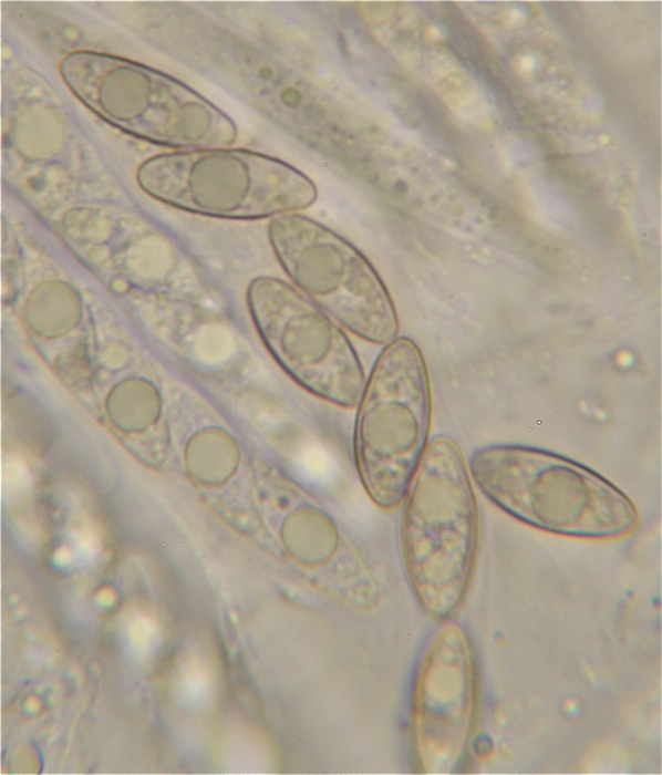
Anyway, I tried to make up for what you asked in your protocol and here are the results of ascomycet 1. The asci are bitunicate (I think) and there are very long and thin paraphyses. The ascomata are semi-immersed and the surface is cracked. I did not manage a proper section but maybe the new information are enough to something. If not, no problem, I will keep the finding until I get a binocular form better pictures.
Best wishes,
Gernot
ascomata:
thanks for your literature-tips. So I will try it with easier genera but if I find something which is not very easy I cannot do any different than trying to determine it :).
Anyway, I tried to make up for what you asked in your protocol and here are the results of ascomycet 1. The asci are bitunicate (I think) and there are very long and thin paraphyses. The ascomata are semi-immersed and the surface is cracked. I did not manage a proper section but maybe the new information are enough to something. If not, no problem, I will keep the finding until I get a binocular form better pictures.
Best wishes,
Gernot
ascomata:
Gernot Friebes,
03-06-2009 20:34
 8066.pdf
Jacques Fournier,
03-06-2009 20:47
Re:Tiny black ascomycet 1
for the moment I have no clue about your fungus. Judging from your photos it's difficult to assess whether asci are bitunicate or not. I bet they are unitunicate, please use other stains until you have a good image of apex mature and immature asci.
Also try to evaluate how they dehisce when a gentle pressure is applied to the cover slip.
Are the paraphyses simple or ramified, septate or not?
more soon,
Jacques
Also try to evaluate how they dehisce when a gentle pressure is applied to the cover slip.
Are the paraphyses simple or ramified, septate or not?
more soon,
Jacques
Gernot Friebes,
03-06-2009 20:55
Re:Tiny black ascomycet 1
ok, I will have a closer look about the asci. Luckily I have enough ascomata left. Which stains do you recommend me for looking at the asci?
The paraphyses are simple and septate.
Best wishes,
Gernot
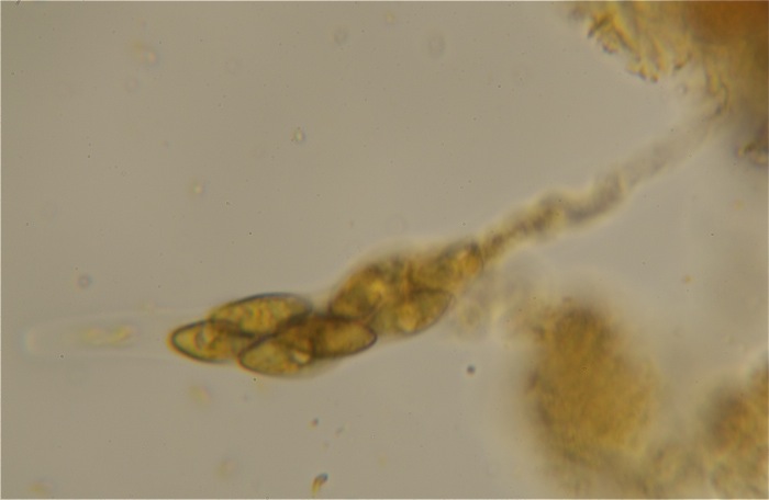
oh, what I nearly forgot: around some ascomata there are thick, brownish hyphae
The paraphyses are simple and septate.
Best wishes,
Gernot
oh, what I nearly forgot: around some ascomata there are thick, brownish hyphae
Jacques Fournier,
03-06-2009 21:10
Re:Tiny black ascomycet 1
dilute blue Waterman ink or Congo red are good stains to begin with. Melzer is much better than IKI to show the apical structure of bitunicate asci. Chlorazol black often proves very efficient, but is less easily available. Otherwise just try any mounting medium until something shows up!
Immersed ascomata with such brown hyphae around and unitunicate asci usually are long-beaked. Is there any chance the beaks have been broken? If yes you should see a small stump on top of the ascomata.
Immersed ascomata with such brown hyphae around and unitunicate asci usually are long-beaked. Is there any chance the beaks have been broken? If yes you should see a small stump on top of the ascomata.
Gernot Friebes,
03-06-2009 21:29
Re:Tiny black ascomycet 1
thanks again for the information about the stains.
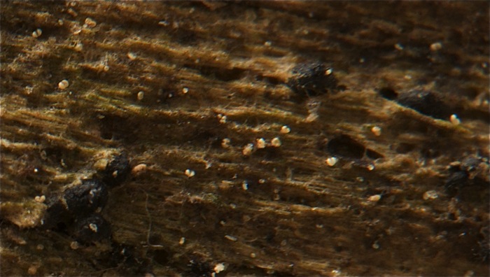
Actually, these little dots are very confusing. As I tried to find a ascomata of the "tiny black ascomycet 2", suddenly there where greenish, muriform ascospores. So there is another species growing in the midst of the ascomata I actually was looking for. Macroscopically I cannot see any differences.
The same happend with this finding - I tried to examine it under the microscope and suddenly there were two-celled, browinsh, verrucous spores and very long hair, so another different species. As I looked carefully I could see the differences between the two species: hairy ascomata with tiny necks (the new species) and round ascomata with no neck (the species I show here). It can be assumed that the ascomata I show here do not have a neck.
Jacques Fournier,
03-06-2009 21:44
Re:Tiny black ascomycet 1
sure you will turn crazy if you have two mixed collections, which makes at least four different ascomycetes appearing as similar black dots! I figure you begin to guess why it is so important to record a number of characters prior to a possible identification.
Gernot Friebes,
03-06-2009 21:55
Re:Tiny black ascomycet 1
I never doubted that these characters are important, I just were not aware of them ;). In the future I will try to show as many characters as possible so you, and maybe other users, do not have to ask so many questions.
With the two new ascomycetes I found between my tiny black ascomycetes I can start trying to figure out these characters properly. I hope you will not have so many circumstances then as you had here!
With the two new ascomycetes I found between my tiny black ascomycetes I can start trying to figure out these characters properly. I hope you will not have so many circumstances then as you had here!
Jacques Fournier,
03-06-2009 22:04
Re:Tiny black ascomycet 1
no problem, it was a good opportunity to show members of the forum that in most cases an ascomycete seen from the sky along with the spores do not make sufficient basis for an identification. Good luck with your mixed tiny immersed hairy beaked things!
Jacques
Jacques