
26-03-2026 15:31
 Åke Widgren
Hello,I found this one in October last year, on r
27-03-2026 15:23
Gernot FriebesHi,this Trichopezizella deviates from typical T. b
25-03-2026 10:35
Hulda Caroline HolteHello,I collected this species growing on a dead b
27-03-2026 15:08
Gernot FriebesHi,I'm looking for help with this coelomycete on C
27-03-2026 10:47
Åge OterhalsI have tentatively identified this Stictis to S. f
24-03-2026 21:37
Elisabeth StöckliBonsoir,Sur bois (tronc) très pourri de conifère


25-03-2026 22:23
 Marc Detollenaere
Dear Forum,On a debarked stem of Tilia, we found s
24-03-2026 15:44
Åge OterhalsI hope someone can confirm the name of this collec
Niesslia-like Fruit Bodies but Microscopy Varies
Peter Thompson,
27-07-2017 15:30
I have found fruit bodies which resemble the genus Niesslia macroscopically, but with differing microscopy.
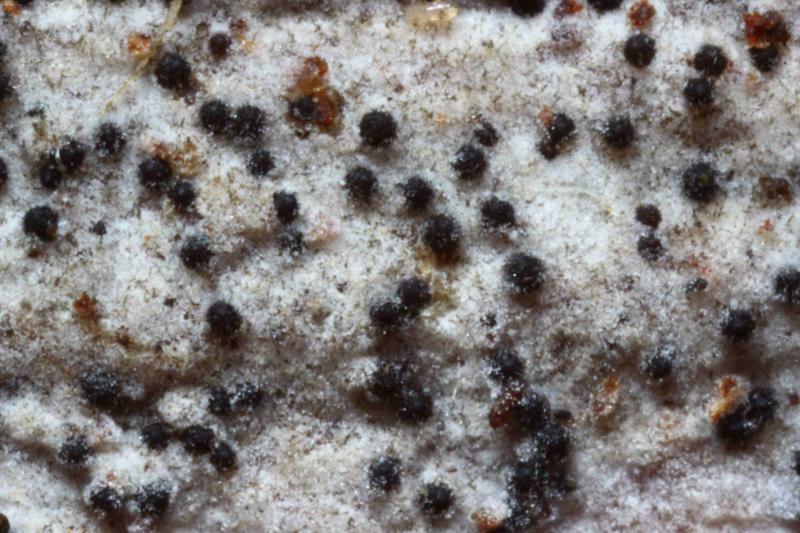
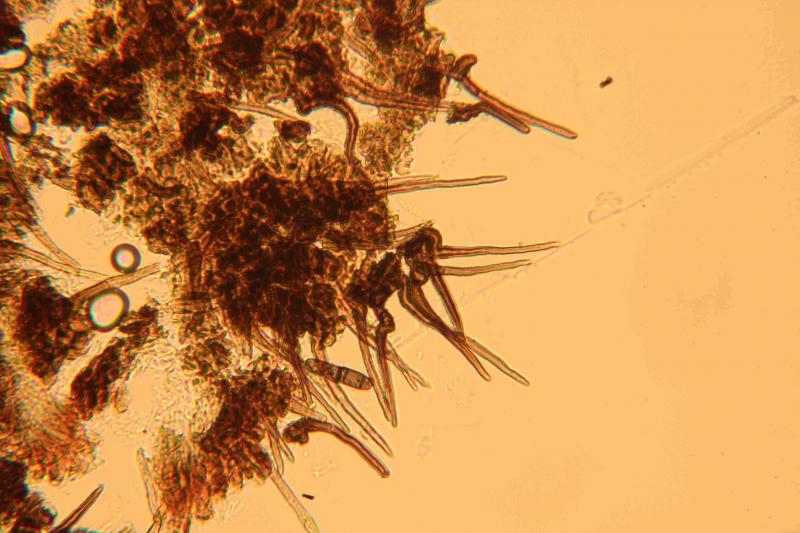
The tiny, spherical fruit bodies have short, thick walled, pointed brown hairs and rest on the surface of their host.
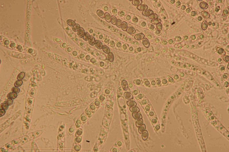
Microscopically, the asci are arranged in whorls, containing eight pale brownish green spores, typically with one large guttule in each. The spores are in a single row in the asci and measure 8.5 - 10 x 5 - 6 um. The asci do not react to Melzers, but the spores inside them 'lose' their guttules.
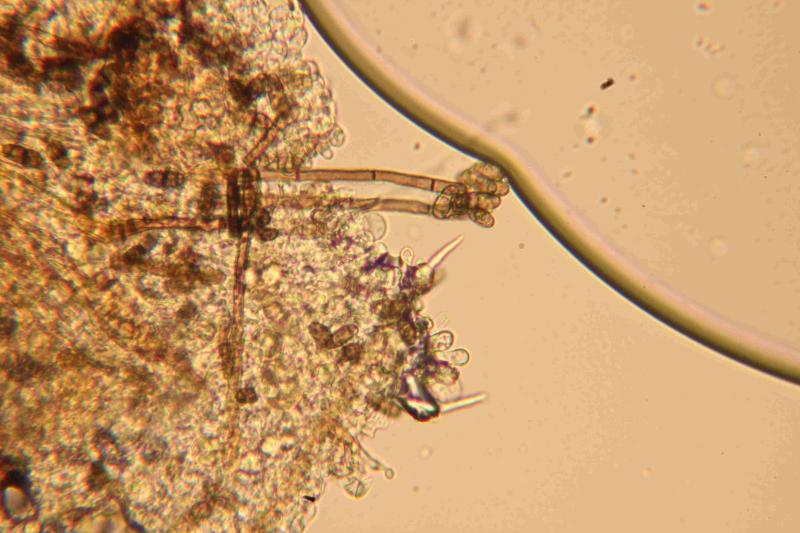
Although the host is well decayed wood of Salix, there is an effuse greyish white layer covering its surface - possibly a resupinate from the basidiomycota. The conidial state of Chaetosphaerella phaeostroma is also present, thinly distributed, but right across the sample.
I have attached photos of the fruit bodies, asci and spores, hairs and effuse greyish matter.
I wonder if anyone has any ideas about what this tiny, hairy, black, spherical ascomycete might be?
Thank You,
Peter.
Pascal RIBOLLET,
28-07-2017 14:30
Re : Niesslia-like Fruit Bodies but Microscopy Varies
Hello Peter,
Did you consider Helminthosphaeria ? The key by Miller et al. (2014) gives H. hyphodermiae, associated with resupinate basidio and with 1-celled spores...
Cheers
Pascal
Did you consider Helminthosphaeria ? The key by Miller et al. (2014) gives H. hyphodermiae, associated with resupinate basidio and with 1-celled spores...
Cheers
Pascal
Peter Thompson,
28-07-2017 16:01
Re : Niesslia-like Fruit Bodies but Microscopy Varies
Hello Pascal,
Thank you for pointing me towards Helminthosphaeria.
I will need to re-examine my sample to check for septa in some of the spores, because Miller et al, (2014) has led me to another document describing H. hyphodermiae and H. odontiae.
It seems very likely that the conidia and conidiophores, which I thought were those of the Diplosporium state of Chaetosphaerella phaeostroma, might instead be those of the Diplococcium state of Helminthosphaeria odontiae. The conidia and conidiophores of H. hyphodermiae seem different.
With Best Wishes,
Peter.
Peter Thompson,
31-07-2017 09:52
Re : Niesslia-like Fruit Bodies but Microscopy Varies
Hello Pascal,
I have looked at the sample again and see that some spores have a septum towards the base. This, the hairs with more pointed tips and the spore sizes clearly indicate that my sample keys out as Helminthosphaeria odontiae.
There seem to be no previous British records in either of the national databases.
With Best Wishes,
Peter.