
08-04-2026 20:33
 Vasileios Kaounas
Found 07-04-26, in Abies cephalonica. Diameter 1,
08-04-2026 10:39
FRANCIS FOUCHIERBonjour , je recherche en pdf cet article: KORF R

06-04-2026 21:36
 Viktorie Halasu
Hello, could anyone please send me the article wi
06-04-2026 19:40
 David Gibbs
Help with this one much appreciated, on rotting Fa
06-04-2026 11:07
Louis DENYBonjour forum, Trouvé sur bois de feuillu très d
06-04-2026 16:24
Juuso ÄikäsLast Tuesday I found some tiny white Helotiales gr
 Hello everyone,
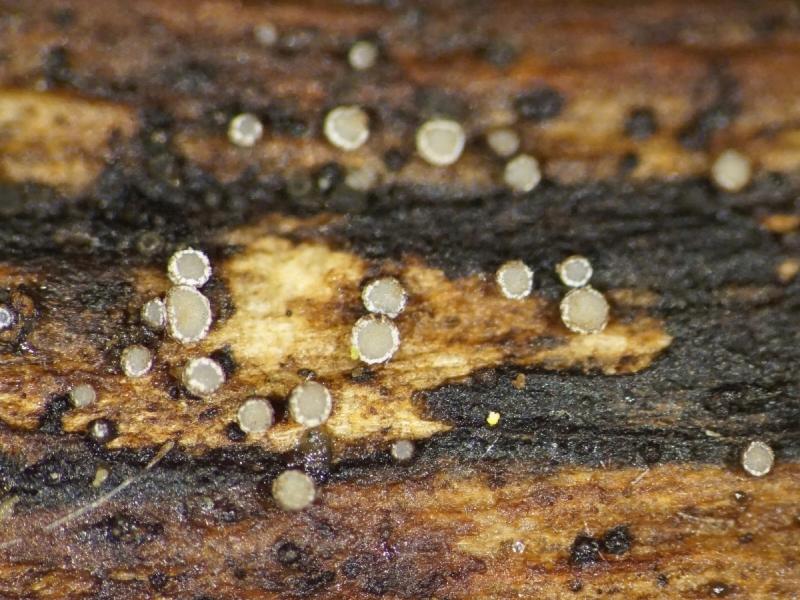
On dead wood of Cytisus scoparius I found a group of tiny apothecia. Does anyone recognizes this species or is familiar with the group in which this species should be found?
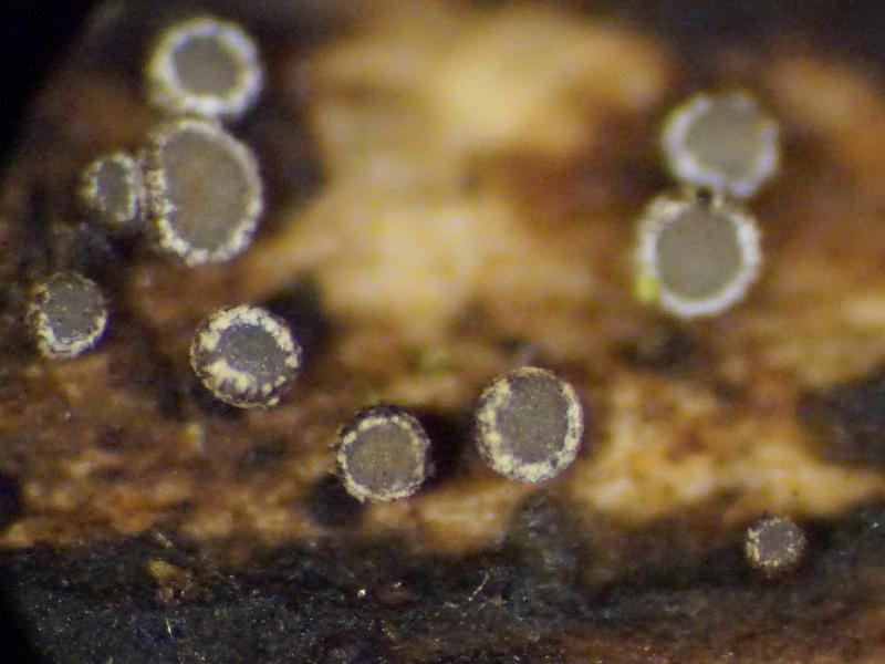
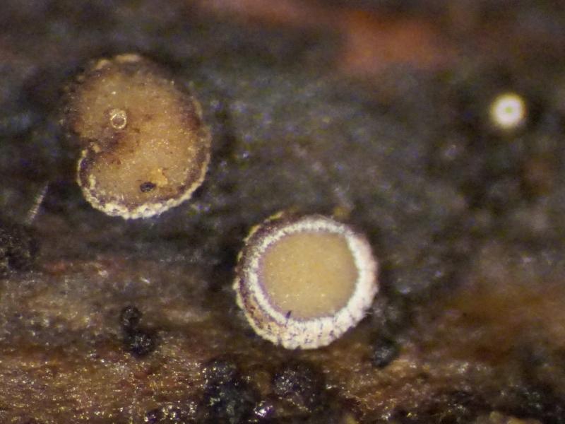
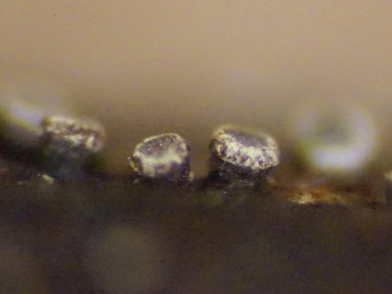
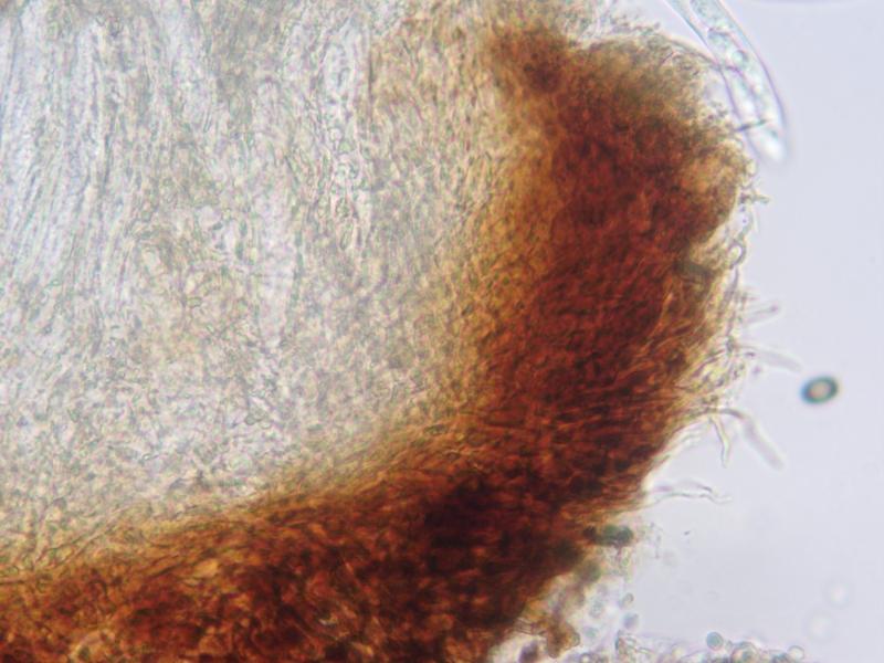
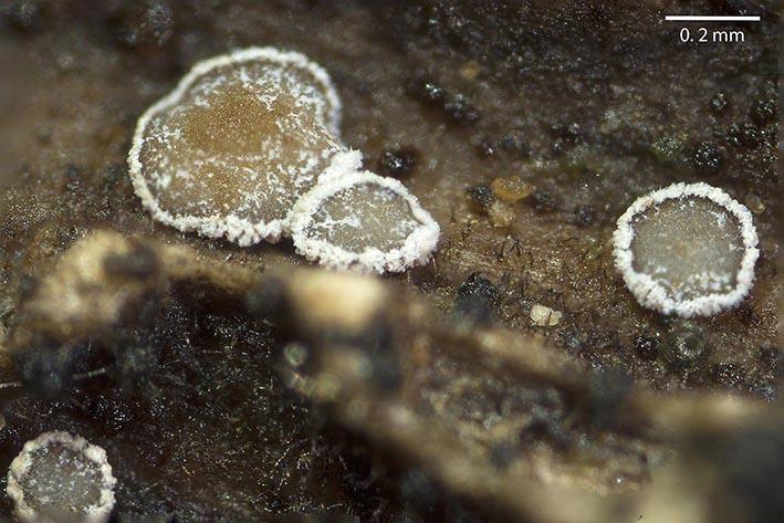
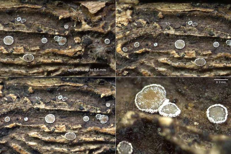
Apothecia young barrel-shaped, dark brown and with a white flaky edge around the opening, older cup-shaped, up to 0.5 mm diam., dark brown with still the white edge present, sessile, base broadly attached to the substrate, base blackish brown, hymenium grey when young, older yellowish grey.
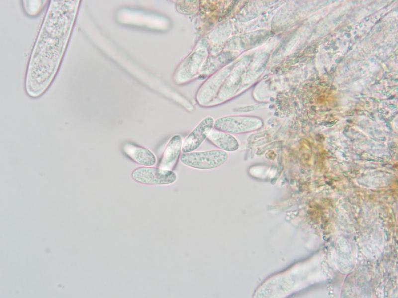
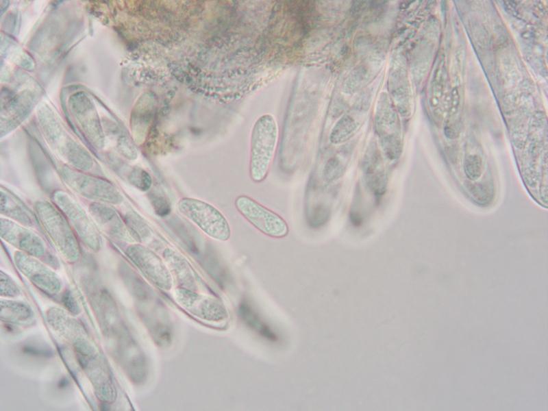
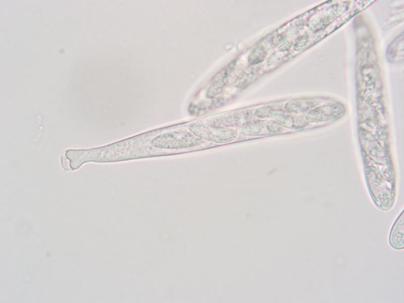
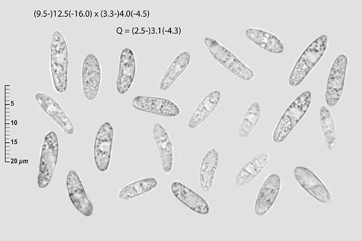
Spores 12,5-14,3 x 4,4-5,2 (10 spores meassured),
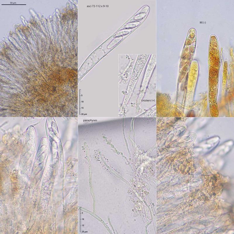
Asci 87-107x8,-9,5 ?m. J-, with clamps,
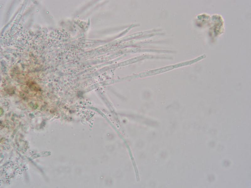
Paraphyses thread-like, septated, sometimes splitting, upto 2 ?m diam.
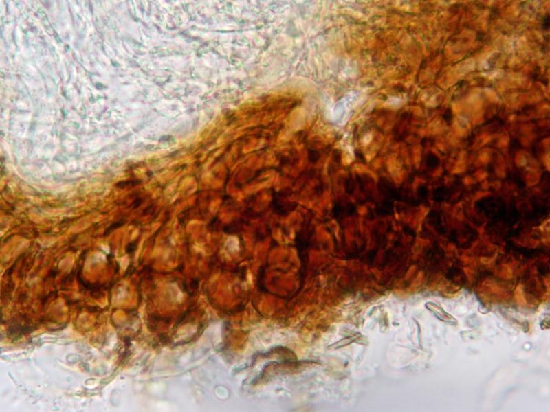
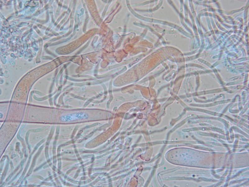
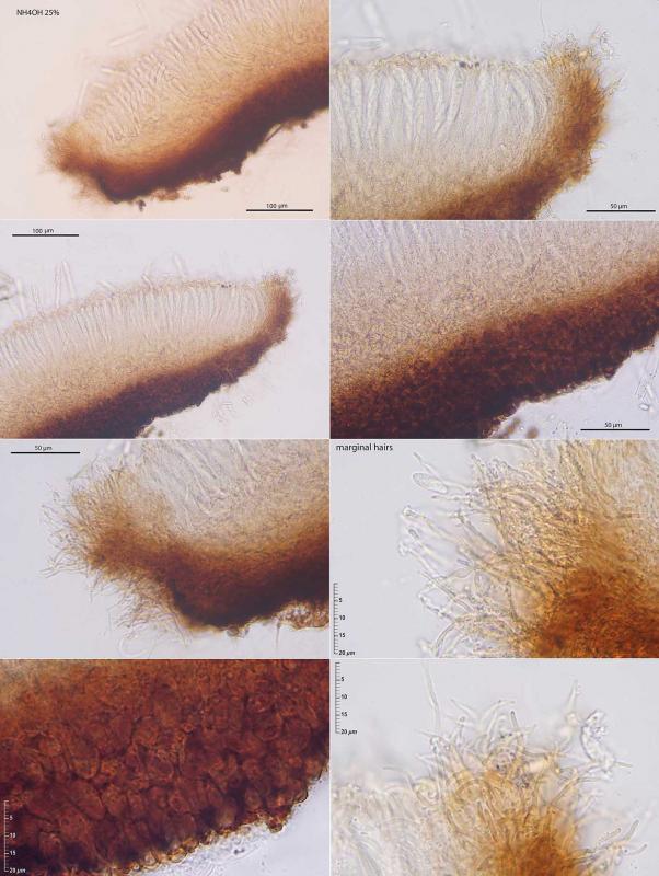
Excipulum composed of (only?) brown, globose thick-walled cells, towards de margin transitioning into hyphae, but I don't know whether this means the apothecia have marginal hairs,
The white flakes consist of microscopically small grains which release immediately in water.
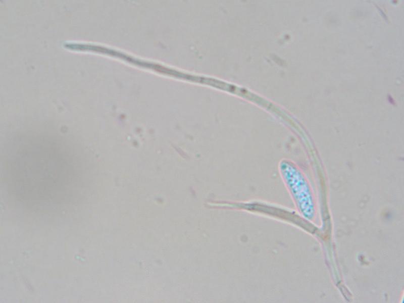
Pretty! I bet this is Cordieritidaceae and that it has glycogen bodies in the spores like Patinella sanguineoatra. And the granules in hymenium are also reminiscent of that species. Yours is of course something else but perhaps related. I would be interested to sequence this, if you can spare piece of the collection.
Thank you,
Adam


Enrique and Zotto, Thanks also for your reply. It's a known species (good to know), but apparently not officially described yet.
Zotto, indeed, when I checked whether the granules would dissolve in KOH, to my surprise, most of the tissue dissolved. I found this rather strange that I was reluctant to mention it (and also to avoid wasting apothecia). I thought I'd wait and see what suggestions would come.
My photos aren't ready yet. I'll send them to you tomorrow and I'll also share them here.
I've now soaked some apothecia and repeated the test. The excipulum discolored slightly (not as strongly as yesterday), and at the beginning only a very fleeting light brown discoloration was visible. Not spectacular or intense.
Does it make any difference to the reaction whether fresh or dried material is used?
Here my pics collected by my friend Javi Mateos on branches of Genista florida still attached to the shrub (may 2023, 1000 m of altitude).
In reality, the apos (up tp 0.6 mm) did not look very 'sanguine'.
The ionomidotic reaction exists, but it was very weak. If anyone needs the photos at higher resolution, please request them from me. I have an ITS sequence that I already sent you. The LSU was invalid and I couldn't use it.
Do you know were the protologue of this species is?