
08-04-2026 20:33
 Vasileios Kaounas
Found 07-04-26, in Abies cephalonica. Diameter 1,
08-04-2026 10:39
FRANCIS FOUCHIERBonjour , je recherche en pdf cet article: KORF R

06-04-2026 21:36
 Viktorie Halasu
Hello, could anyone please send me the article wi
06-04-2026 19:40
 David Gibbs
Help with this one much appreciated, on rotting Fa
06-04-2026 11:07
Louis DENYBonjour forum, Trouvé sur bois de feuillu très d
06-04-2026 16:24
Juuso ÄikäsLast Tuesday I found some tiny white Helotiales gr
Ascomycete on Polystichum proliferum
Heather Merrylees,
17-09-2025 10:50
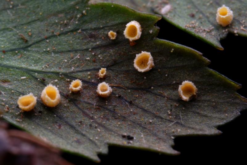
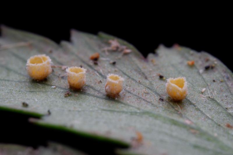
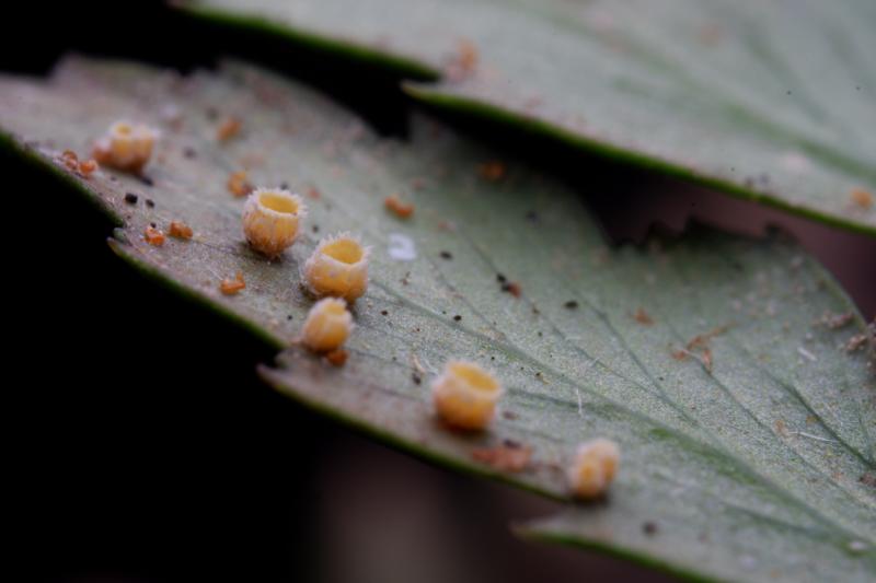
I am hoping for any advice on the identification of this ascomycete I found growing on the underside of Polystichum proliferum in Victoria, Australia. A specimen is lodged at the National Herbarium of Victoria (MEL). There was no reaction in Melzer's reagent.
Recorded on iNaturalist as: https://www.inaturalist.org/observations/299481745
Looking forward to learning more and thanks in advance for your assistance.
Best regards,
Heather Merrylees
Hans-Otto Baral,
17-09-2025 11:40

Re : Ascomycete on Polystichum proliferum
Hello, I am quite unsure about the asci and spores. The latter are filiform?
The structure of the conglutinate hairs is also not easy to see. Would be good to have some oil immersion photos. And if you have Lugol, please test the asci herewith for possible hemiamyloidity (or use Melzer to a KOH-pretrated mount).
Heather Merrylees,
17-09-2025 11:59
Re : Ascomycete on Polystichum proliferum
Thanks so much for your swift reply and recommendations Hans-Otto.
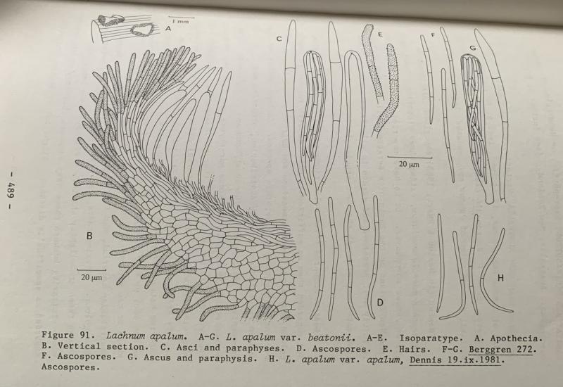
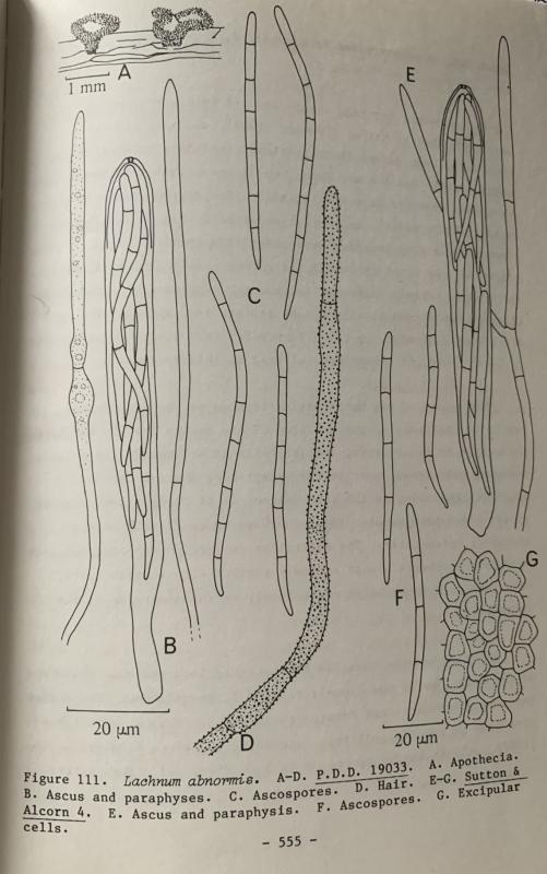
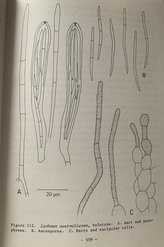
I agree the ascospores appear filiform - also septate (3?) and quite pointed at the ends. I've attached some of the illustrations from Spooner's Helotiales of Australasia for comparison that seemed the closest.
I'll get back to the microscope this Friday and will update accordingly. Many thanks!
I agree the ascospores appear filiform - also septate (3?) and quite pointed at the ends. I've attached some of the illustrations from Spooner's Helotiales of Australasia for comparison that seemed the closest.
I'll get back to the microscope this Friday and will update accordingly. Many thanks!
Heather Merrylees,
28-11-2025 09:45
Re : Ascomycete on Polystichum proliferum
Hi there!
I have had another try at the microscopy - (still very much a beginner). Please find attached a pdf of the results. I pre-treated/imaged in KOH, and then added MLZ to the same preparation after removing as much KOH as possible. Please let me know if you have any suggestions - I will have to find some Lugol. Many thanks in advance for your help and advice on this.
Best regards,
Heather Merrylees
 Ascomycete-KOH-MLZ-0001.pdf
Hans-Otto Baral,
28-11-2025 10:45
Re : Ascomycete on Polystichum proliferum
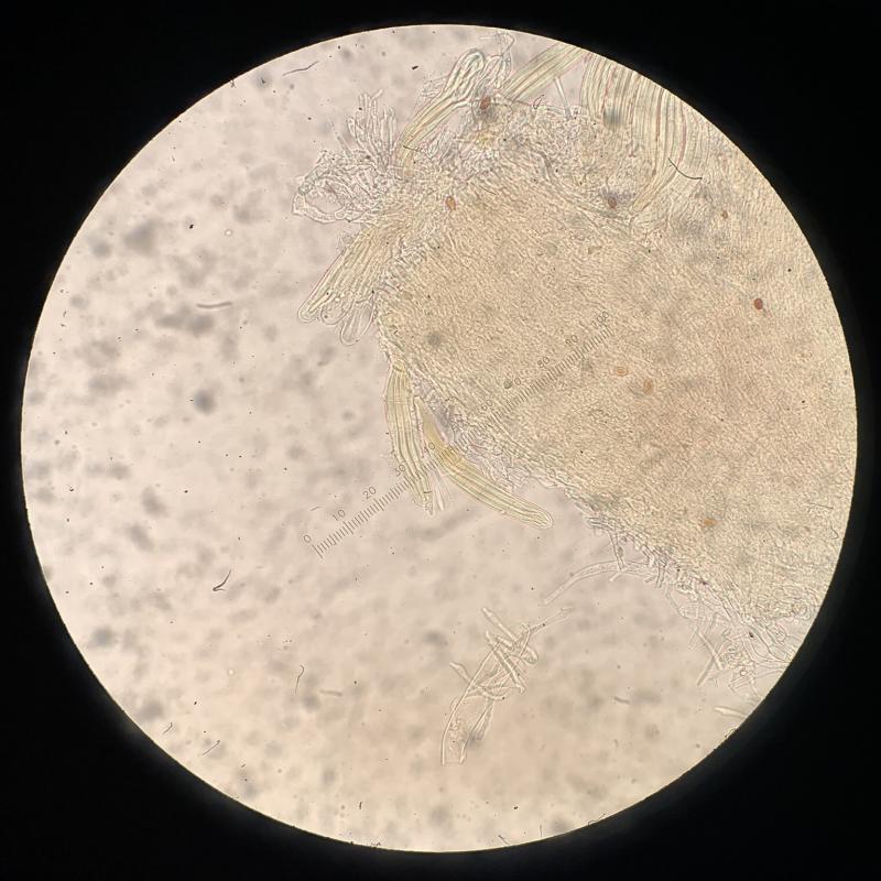
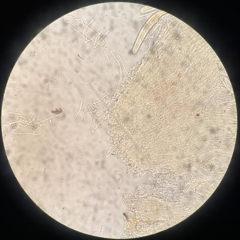
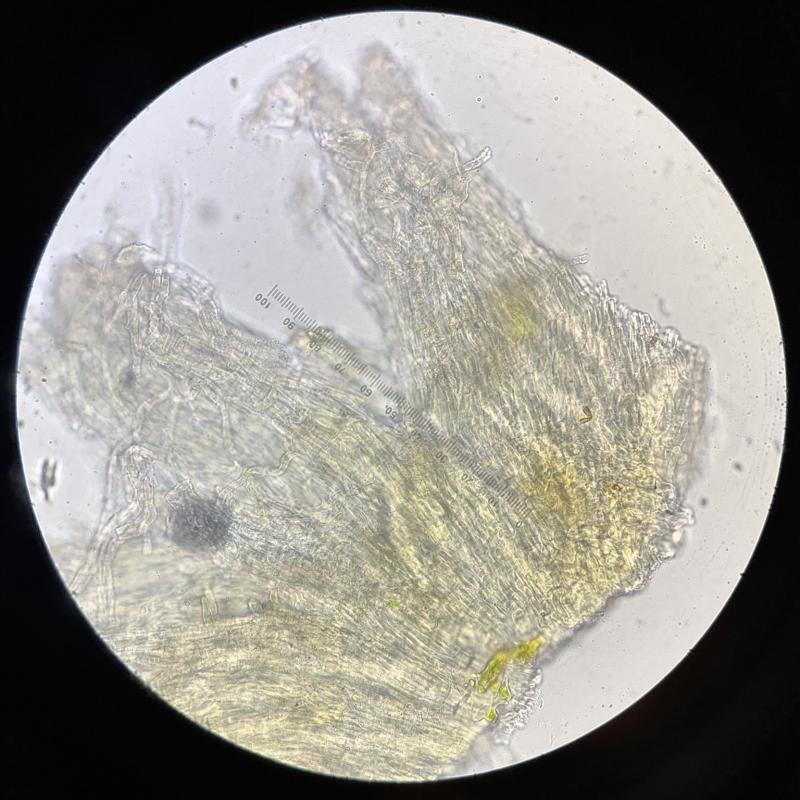
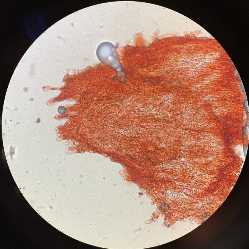
Hi, our pics are very instructive and of high resolution. The strongly raised margin reminds me of Solenopezia, but the hairs are almost smooth. The protruding lanceolate-spathulate brownish paraphyses are remarkable. Did you measure the spores? Do you have a scale for your pics? I suppose the spores are 5-7-septate. On the first photo with the spores there is an immature ascus on the left which looks like having an amyloid ring. But on the overnext photo the mature ascus apex has a pronounced apical chamber. I cannot see iodine in any of your pics. If you have no Lugol, try Melzer after mounting in KOH and washing with water.
With the spore size I can try my databse for fern discos with such a size.
Heather Merrylees,
28-11-2025 11:53
Re : Ascomycete on Polystichum proliferum
Hi Hans-Otto,
Please find attached the updated PDF with a few extra photos - all photos now have scale bars in the lower right corner. I'm afraid I did not find any clear spores outside of asci to measure - the rough estimate based on the scale bar appears to be approx. 100 µm x 5 µm? I understand it is best to measure spores outside of the asci. Also - I have indicated whether the image is KOH only or Melzers (after KOH pre-treatment). The KOH was drawn from under the cover slip with tissue, and replaced with Melzers which is perhaps not optimal technique - next time I will wash with water as well. Learning lots!
Many thanks,
Heather
Hans-Otto Baral,
29-11-2025 10:26
Re : Ascomycete on Polystichum proliferum
You did not measure the spores now? I arrive at 90-97 x 4.5-4.7 µm.
My database has only Gorgoniceps boltonii and Belonium equisetinum, both on Equisetum.