10-06-2026 13:41
 François Freléchoux
Bonjour à nouveau, Voici une trouvaille d'hier.
09-06-2026 18:32
Camille MertensSur morceau de roseau immergé 0,5 - 0,7 mm de dia
10-06-2026 11:53
Steve ClementsBonjour, This disco is abundant on dead stems of
10-06-2026 12:54
Steve ClementsBonjour encore, Pouvez-vous m'aider, s'il vous pl
10-06-2026 10:45
François Freléchoux
Bonjour à nouveau, Encore une détermination qui
08-06-2026 10:16
 Spooren Marco
I don`t have a clou about this fungus,it is not in
10-06-2026 09:24
François Freléchoux
Bonjour, J'imagine que cette détermination ne do
08-06-2026 17:00
François BartholomeeusenGood day everyone, On June 5 2026, I collected de
07-06-2026 15:10
 William Slosse
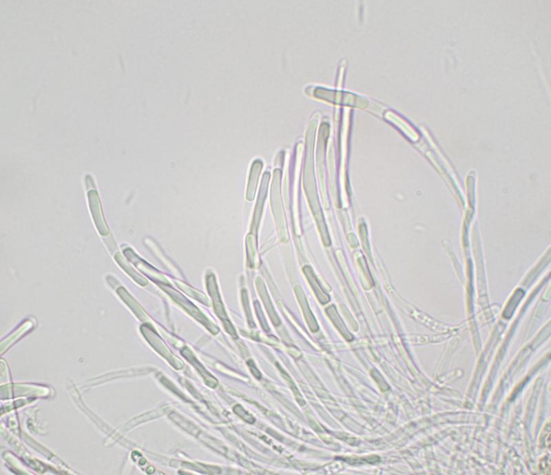
Hello everyone,On 05-06-26, I found following asco
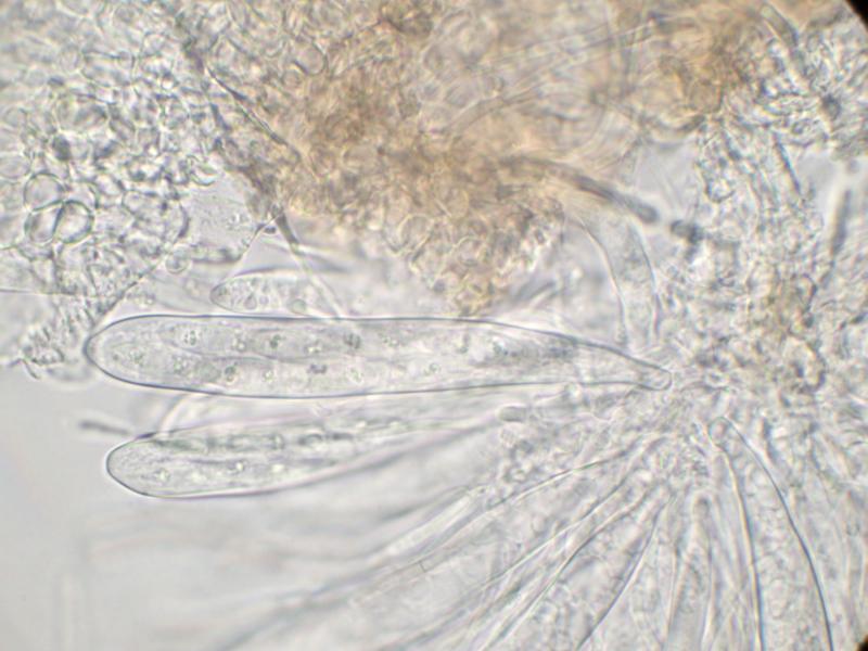
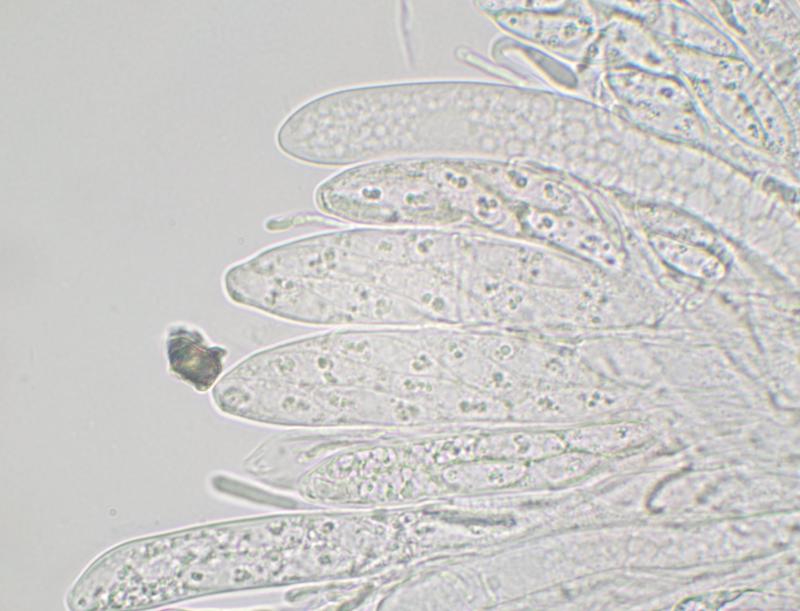
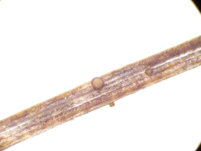
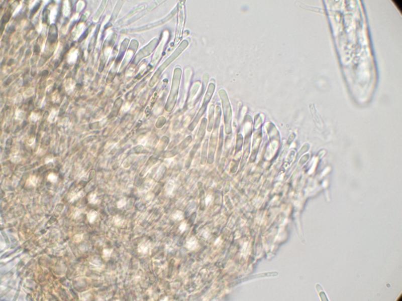
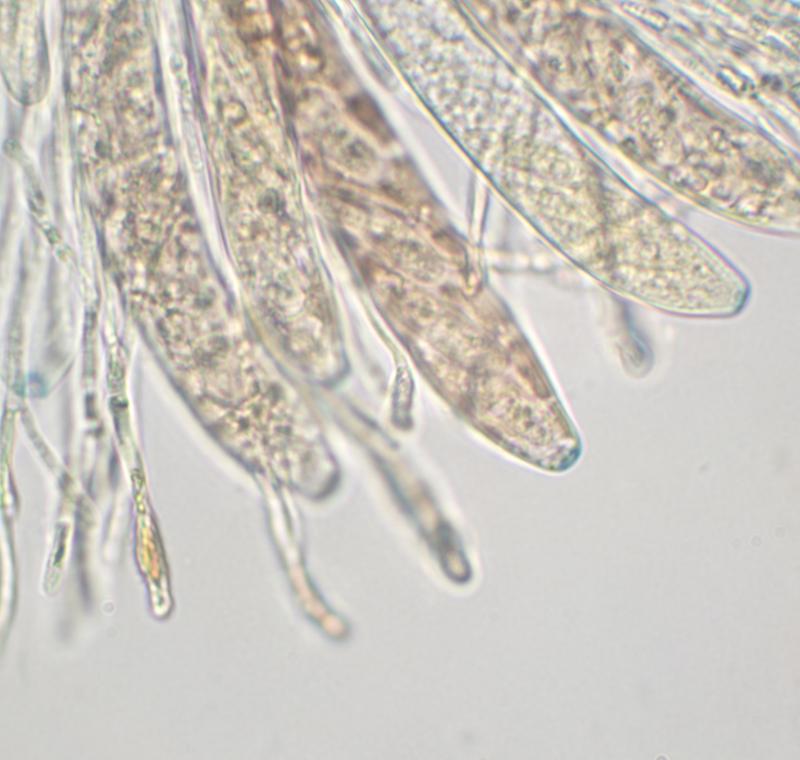
Apothecia sessile; cup-shaped; ca 300 µm diam.; hymenium pale grey; exterior brownish orange.
Excipulum brown textura globulosa.
Paraphyses narrowly cylindrical (2-2.5 µm wide), sometimes swollen at apex; sometimes branched; cylindrical refractive VB in upper part.
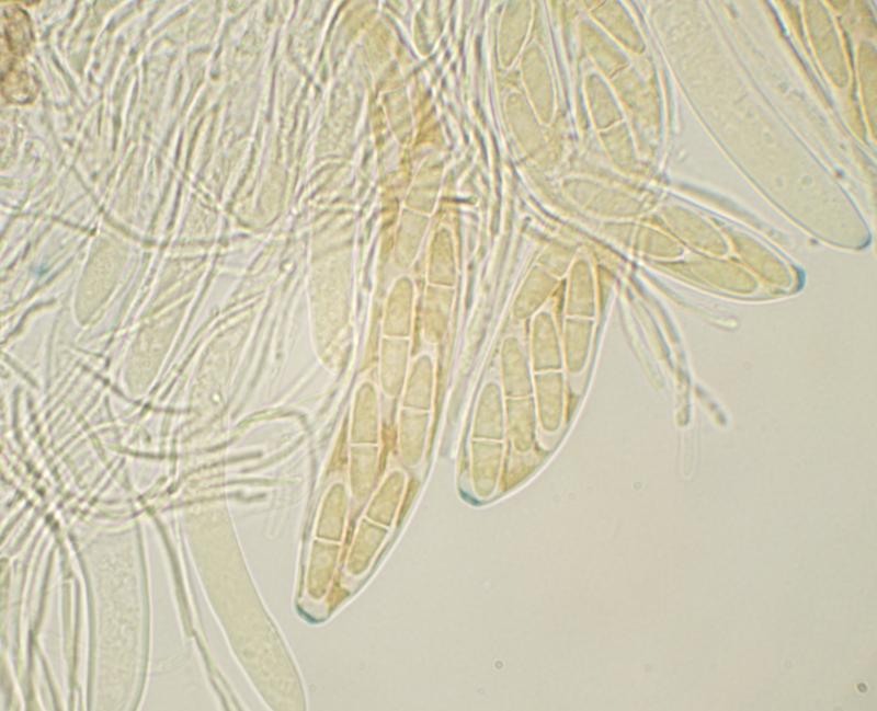
Asci clavate; ca 100-110 x 13-16 µm; 8-spored (biseriate); IKI+ blue; with shallow apical ring.
Ascospores fusiform; hyaline; free spores 21-23 x 6 µm; mostly 1-septate, but free spores sometimes 2-septate; scattered small OBs, mainly near ends of spore.
This seems to resemble Nimbomollisia (Niptera) eriophori. I didn't notice gelatinous sheaths on the spores when examining the specimen but the image of spores in the ascus in MLZ seems to show some sort of gelatinous structure at the ends of the spores. Some of the paraphyses have swollen apices but this feature isn't as well developed as I would have expected in Nimbomollisia.
I'd be grateful for a second opinion.
Thanks
Marcus

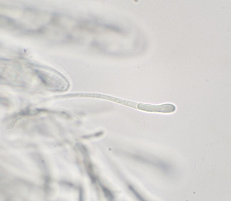
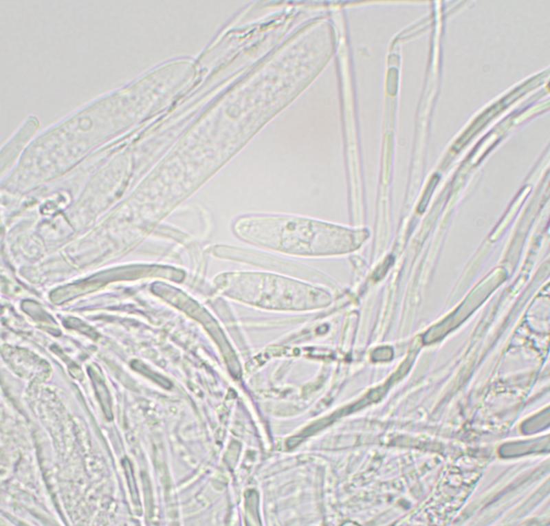
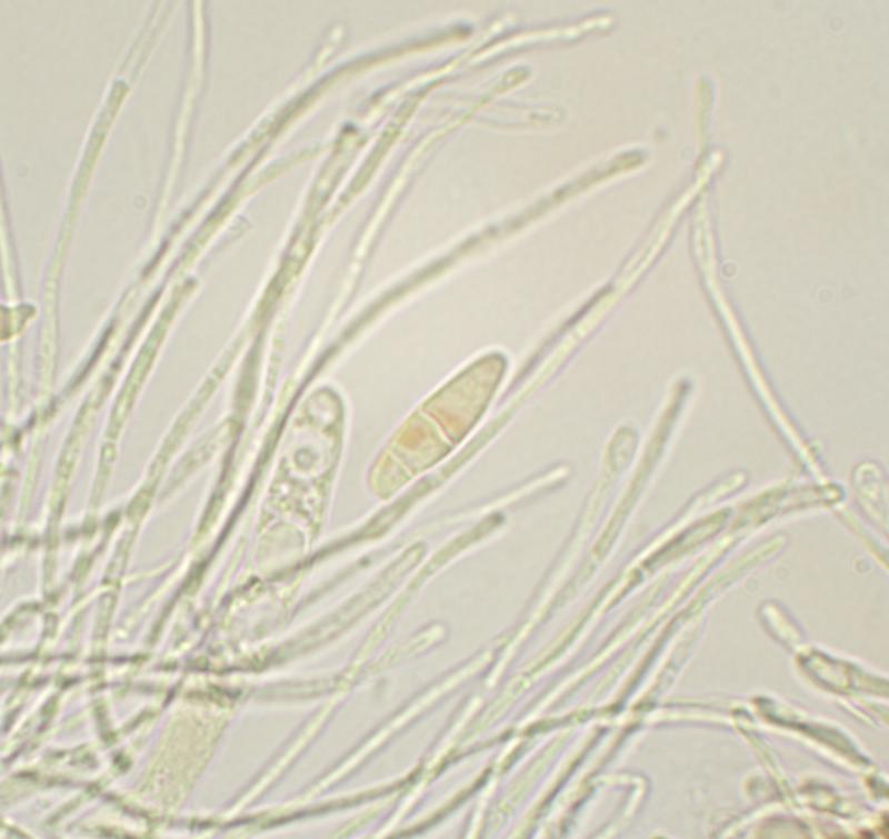

There were very few free spores. Spores in the asci were difficult to see clearly (see images) but I couldn't see any obvious caps or sheaths.