
19-04-2026 21:23
Steve ClementsBonjour, I found this anamorphic fungus on old pl
19-04-2026 20:46
Steve Clements1 mm diameter approx spherical conidiophores on pl
12-04-2026 17:56
 Hardware Tony
Found on dead stems in February earlier this year
17-04-2026 19:16
 Enrique Rubio
Hi to everybodyI would appreciate any assistance r
14-04-2026 05:32
Ethan CrensonHi all, A few weeks back a friend pointed out som
17-04-2026 15:14
 Bruno Coué
Bonjour.Récoltes du 16/04/2026, sur feuilles mort
12-04-2026 15:52
Gernot FriebesHi,I'm looking for help with this anamorph collect
14-04-2026 21:52
Gernot FriebesHi,found on dead leaves of Carex elata. Conidia: 4
16-04-2026 22:09
Buckwheat PeteHello, I'd like to ask about this older specimen:
15-04-2026 19:33
Fátima Durán ManzanequeHi!! I need help, I found this Ascomycete but I d

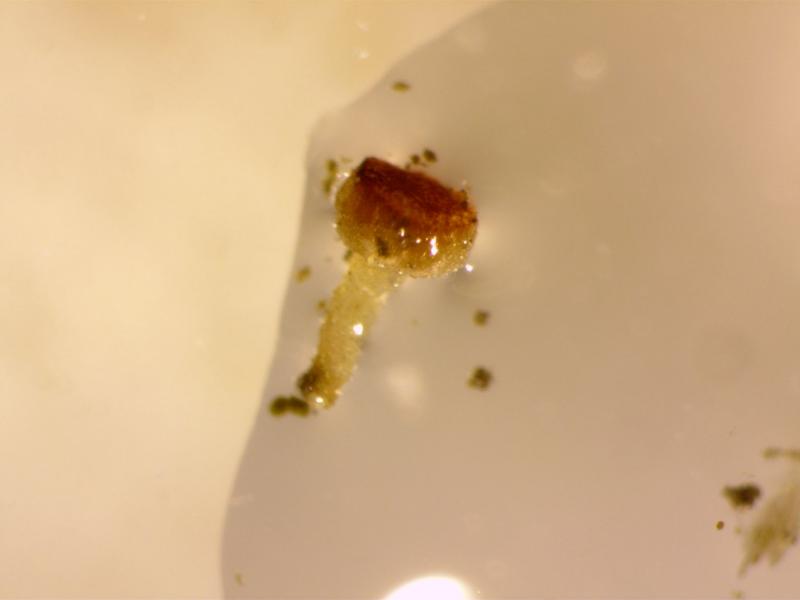
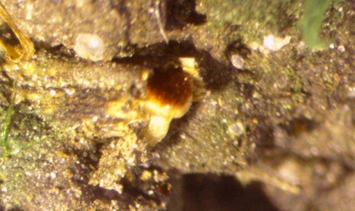
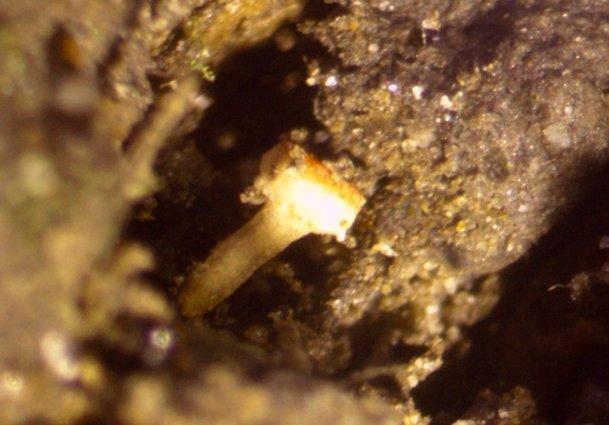
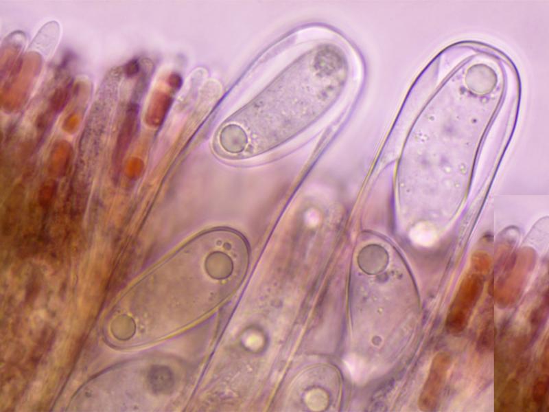
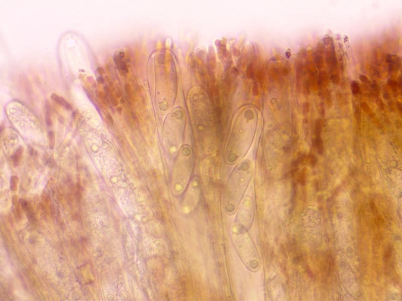
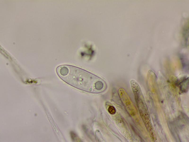
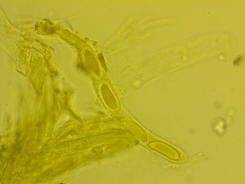
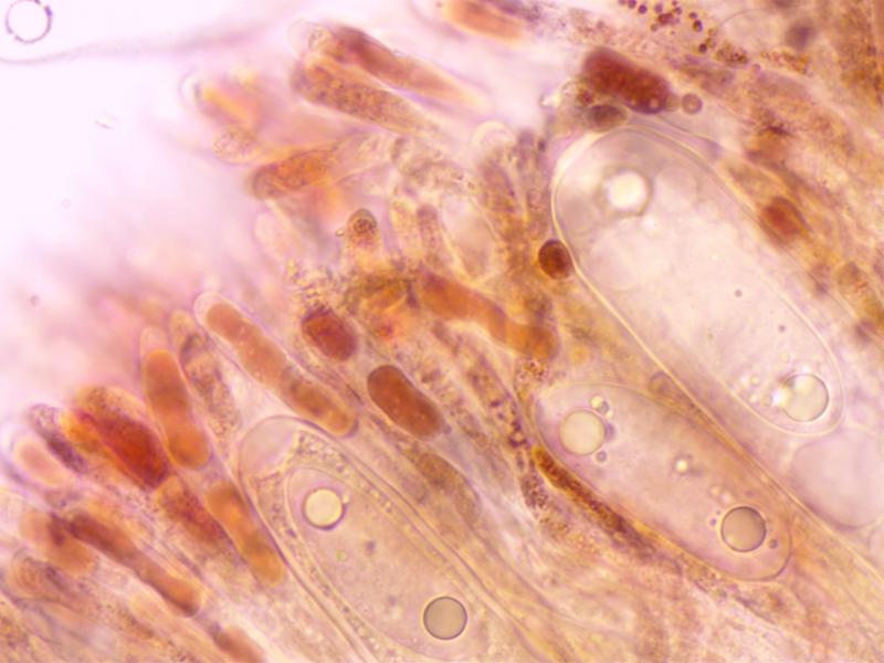
Hi All,
I have found a strange and distinctive Ascomycete growing on damp soil close to a brook. Only a single apothecium thus far.
Apothecium: c1x0.5mm. Hymenium convex, bright red, not clearly demarced from the excipulum.
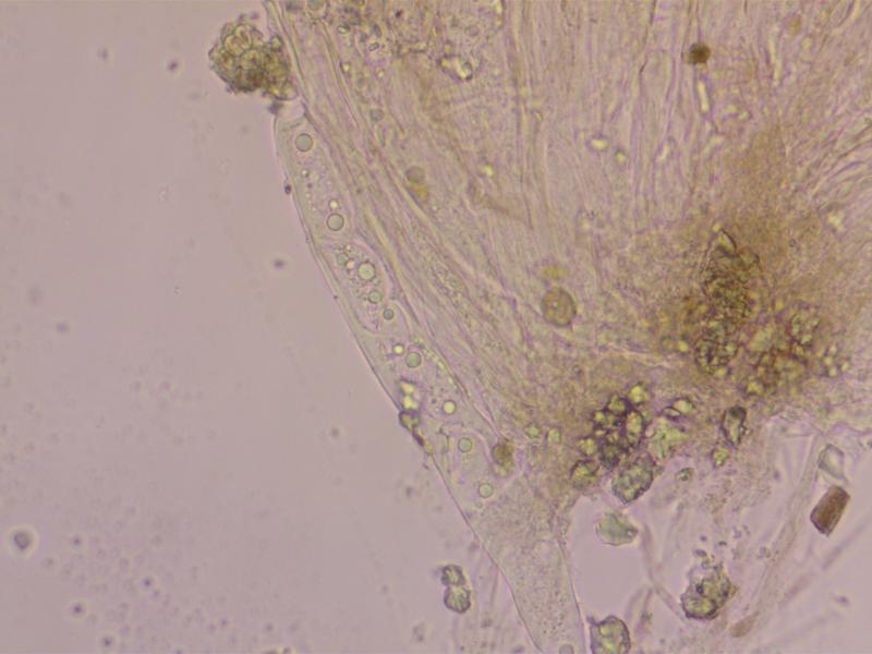
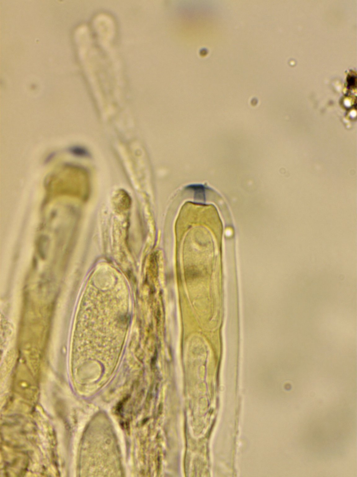
Asci: Cylindric, 250-325x18-25. 4-5 spored, uniseriate, with differetial maturation of the ascospores. Ascus pore blueing in Melzer' after treatment with KOH.
Ascospores:44-50x15-19. Ellipsoid-cylindric, some curved and some with strangely attenuated apices. With polar guttules. With a gelatinous coat.
Paraphyses with red pigment, swollen to 6-7.
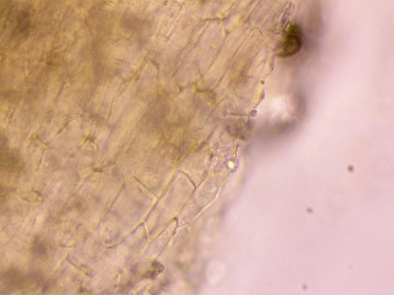
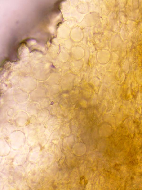
Excipulum: Textura Globosa.
I would be very interested in any thoughts on this. Thanks in advance.
Charles.
 rsz-red-stipitate-disco-asci-7-rhiwlas-pentraeth-7420-0001.jpg
Hi Zotto,
Yes, the t. prismatica is from the stipe. Have not tested with Lugol and very little material remains (on a couple of slides) Will definitely look out for more material-not far from where I live in Pentraeth, Anglesey.
Hi Zotto,
Its the ascus width that I had got wrong-it should be 250-325x18-25, so really quite large. Being Sclerotiniaceous I imagine the fungus would have been associated with plant material perhaps with a sclerotium but, unfortunately it quickly became detached from the substrate. There was some Mnium hornum fairly close but I don't think the fungus was associated with it. The site was in broad leaved woodland with ash etc.