
08-04-2026 10:39
FRANCIS FOUCHIERBonjour , je recherche en pdf cet article: KORF R

06-04-2026 21:36
 Viktorie Halasu
Hello, could anyone please send me the article wi
06-04-2026 19:40
 David Gibbs
Help with this one much appreciated, on rotting Fa
06-04-2026 11:07
Louis DENYBonjour forum, Trouvé sur bois de feuillu très d
06-04-2026 16:24
Juuso ÄikäsLast Tuesday I found some tiny white Helotiales gr
05-04-2026 20:40
 Robin Isaksson
Hi!Found i Japan on bark of Abies sp. Spores 35-4
 Hello.
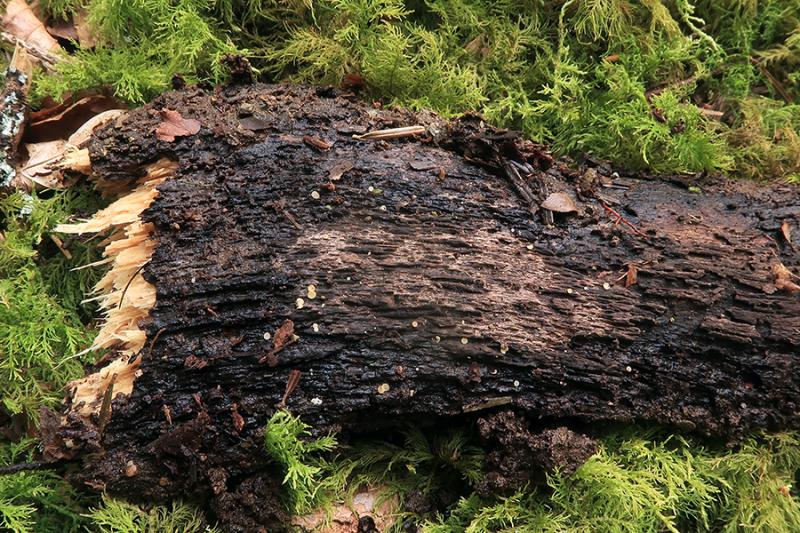
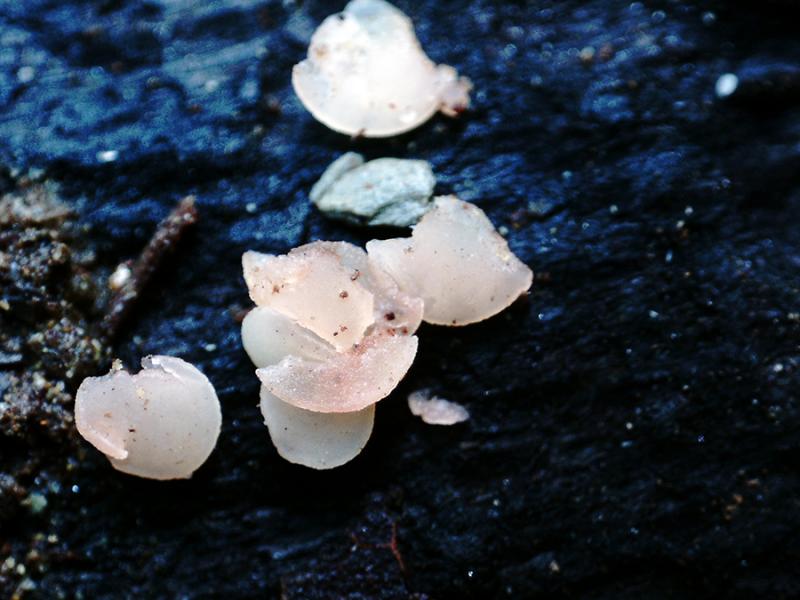
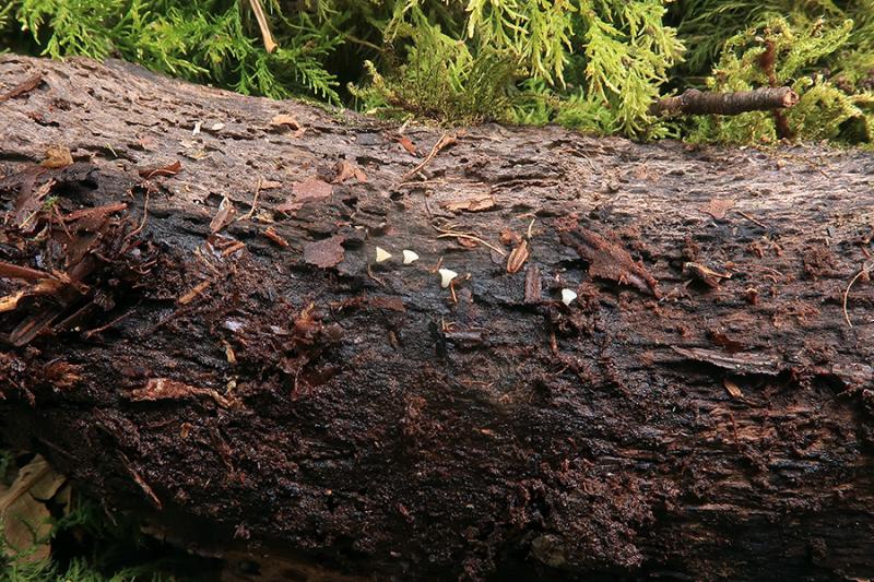
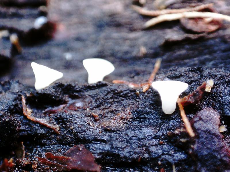
Tiny pinkish discomycetes, photographed and collected on August 17, sprouting scattered on the surface of a fallen spruce (Abies alba) trunk.
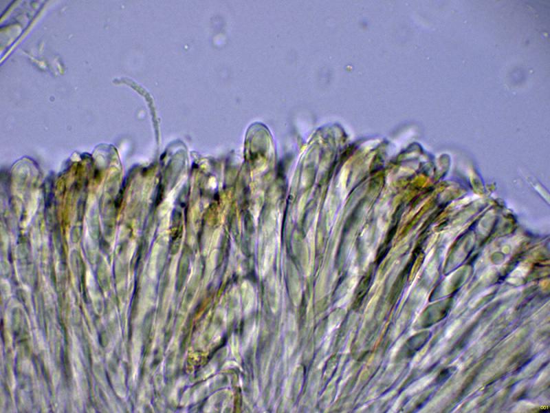
Apothecia attached to the substrate only by the central part, without a distinct stem, with a diameter between 1.17 and 1.75 mm, whitish with pinkish tinges.
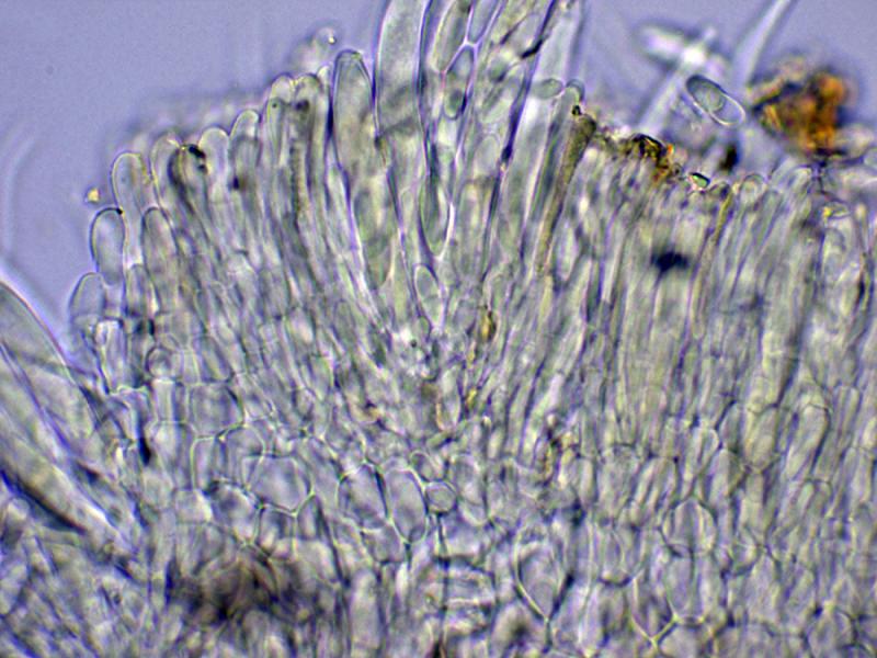
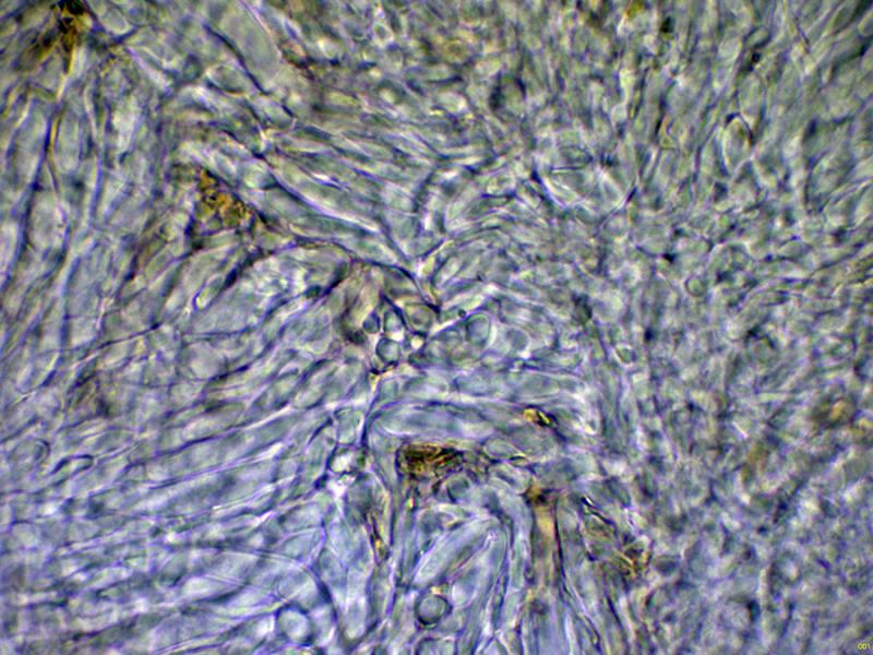
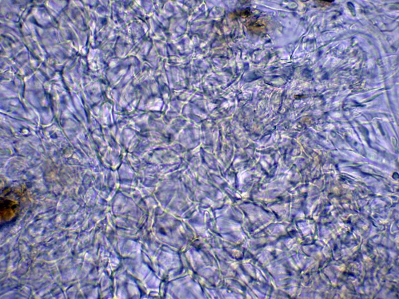
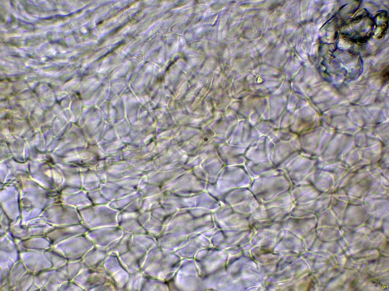
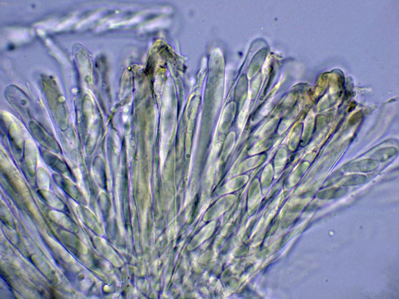
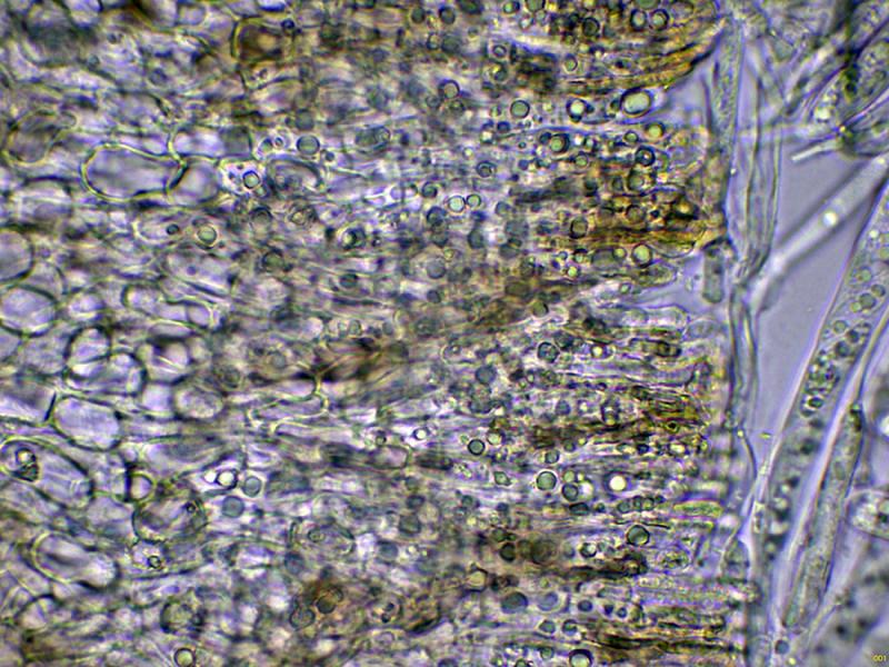
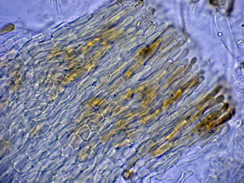
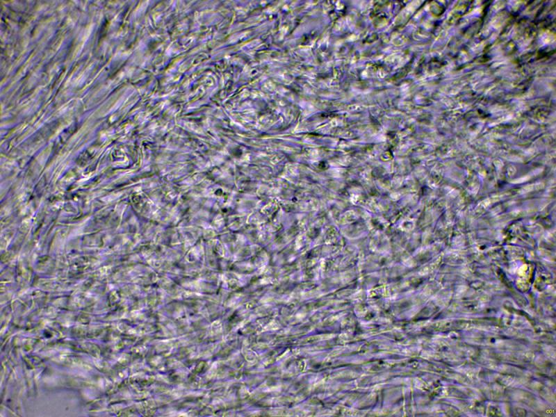
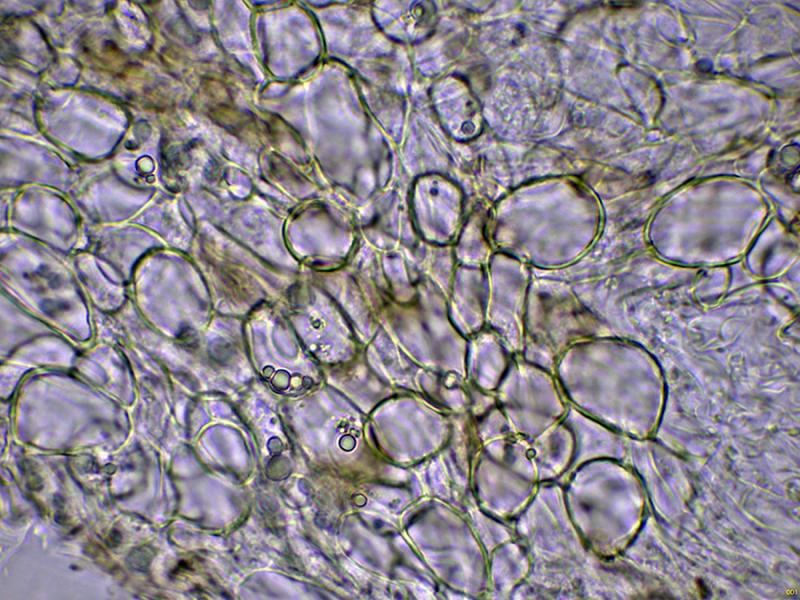
The medullary exciple is filamentous, with an intricate texture, and the ectal exciple has a globose-angular texture.
Short, septate marginal hairs with rounded ends.
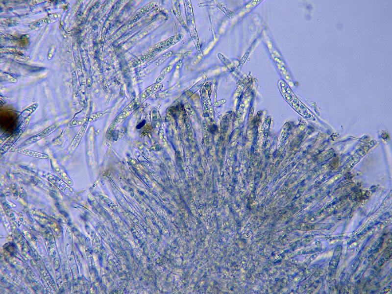
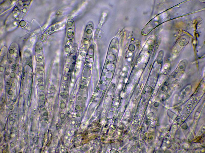
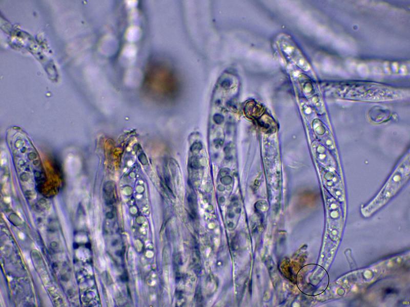
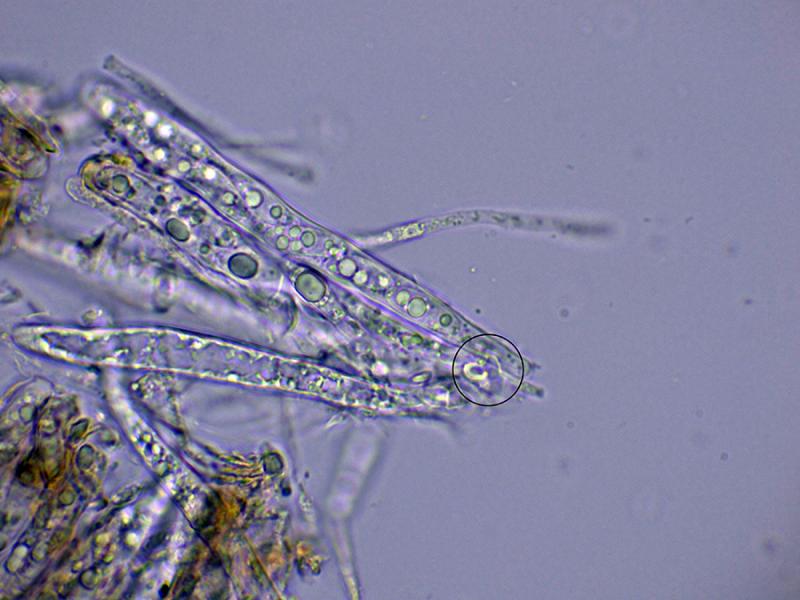
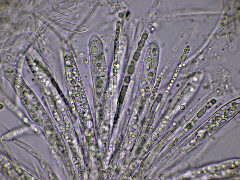
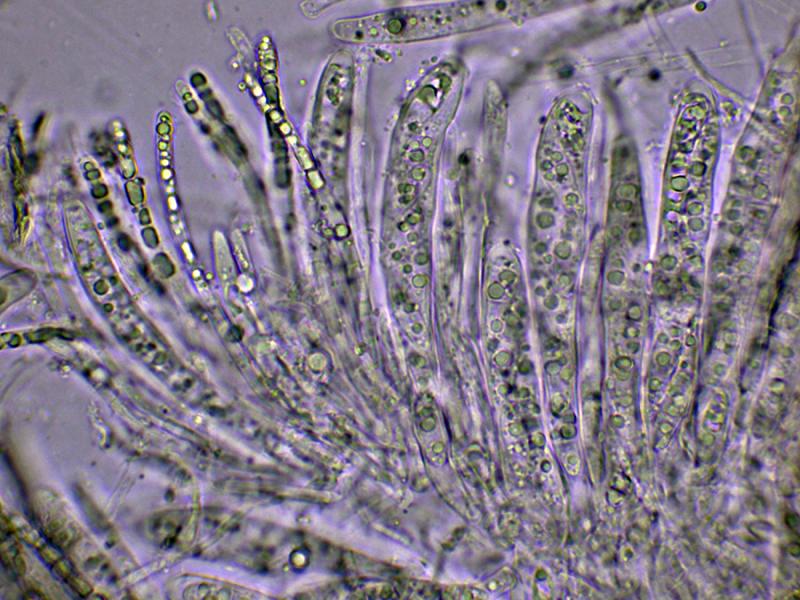
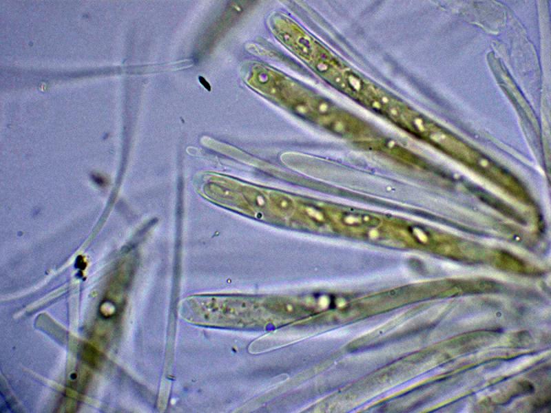
Octosporic asci, in some cases with uncinules.
The paraphyses are septate, filiform, unbranched, and protrude very slightly above the level of the asci.
No significant reaction of the asci with Melzer's reagent has been observed.
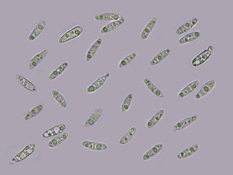
Cylindrical ascospores, with one or two large lipid droplets inside and other smaller ones scattered throughout. Between one and three septa can be discerned in some of these ascospores, with measurements in water of:
(11.5) 12.5 - 15.2 (15.6) × (3.8) 4 - 4.8 (5) µm
Q = (2.4) 2.9 - 3.6 (4) ; N = 30
Me = 13.9 × 4.3 µm ; Qe = 3.2
These specimens were found very close to what appears to be Hymenoscyphus imbrebis (there were three specimens on a nearby trunk), so at first I thought they were probably the same, although the microscopy doesn't match at all. Based on its microscopic characteristics, I'm thinking of Calycellina, and within Calycellina, the best fit is Calycellina subtrabinella, but there are too many conflicting characteristics, such as the host, which I haven't been able to find information on whether it can grow on coniferous wood, and the lack of reaction to Melzer's, as well as the spore width, don't agree.
Any feedback from you would be welcome.
Thank you in advance.
Best regards.

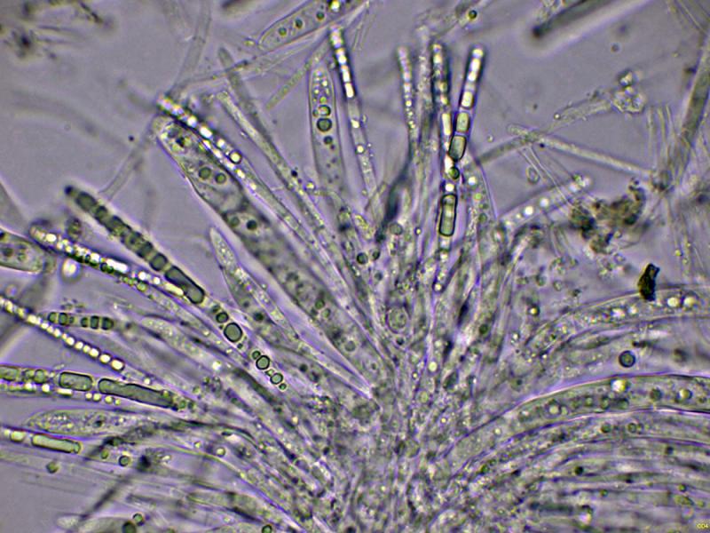
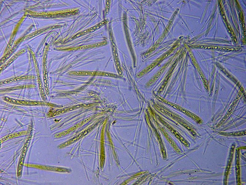
I mentioned in my thread that these apothecia sprouted right next to what I considered the typical Hymenoscyphus imberbis.
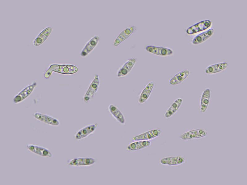
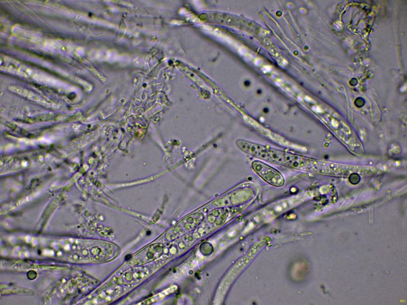
I'm adding a few images from my study of what I believe to be Hymenoscyphus imberbis, growing right next to it. In this case, a faint amyloid reaction of the apical apparatus is visible, along with many paraphyses with contents, and the uncinules at the base of the asci.
In my study, the spores measured 13.1 - 16.05 (16.1) × (5.1) 5.15 - 6.3 µm, with a width well above the previous ones (3.8) 4 - 4.8 (5) µm. It is because of this data that I have come to doubt that it was the same thing.
I'll leave both studies as Hymenoscyphus imberbis sl.
Thank you again for your opinion.
Best regards,
Josep.
So, if it's not Hymenoscyphus imberbis or Hymenoscyphus kathiae, is there another option within Hymenoscyphus that fits my proposals? Or should I just file it as Hymenoscyphus sp.?
Best regards.
I'll try to recover the exsiccata and gather new data. My mistake was that, since it grew on practically the same trunk, I assumed it was probably the same thing, at different stages of development.
Best regards.
Sorry for the delay. Since I couldn't obtain spores, I sent one of the specimens for sequencing. The ITS gene result determined that, just as I suspected, it was a 100% match for Hymenoscyphus kathiae.
Best regards.
You already have everything in your email.
Best regards.