
19-04-2026 21:23
Steve ClementsBonjour, I found this anamorphic fungus on old pl
19-04-2026 20:46
Steve Clements1 mm diameter approx spherical conidiophores on pl
12-04-2026 17:56
 Hardware Tony
Found on dead stems in February earlier this year
17-04-2026 19:16
 Enrique Rubio
Hi to everybodyI would appreciate any assistance r
14-04-2026 05:32
Ethan CrensonHi all, A few weeks back a friend pointed out som
17-04-2026 15:14
 Bruno Coué
Bonjour.Récoltes du 16/04/2026, sur feuilles mort
12-04-2026 15:52
Gernot FriebesHi,I'm looking for help with this anamorph collect
14-04-2026 21:52
Gernot FriebesHi,found on dead leaves of Carex elata. Conidia: 4
16-04-2026 22:09
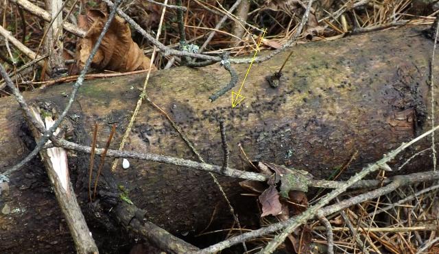
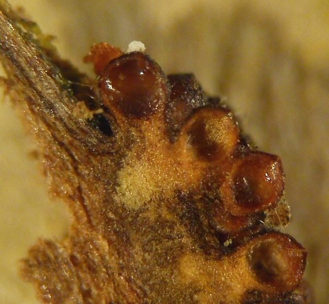
Buckwheat PeteHello, I'd like to ask about this older specimen:
15-04-2026 19:33
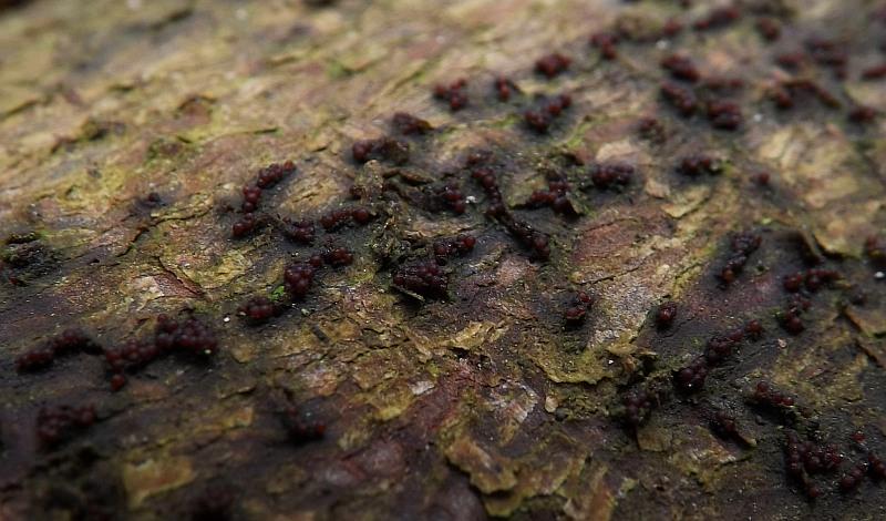
Fátima Durán ManzanequeHi!! I need help, I found this Ascomycete but I d
Will someone help me identify?
Thank you in advance.
Regards
Mirek


A very hard topic for such a novice in this topic as me.
I had to review everything that is available on the internet before I came to any conclusions.
Initially, I tried to compare my collection to T. fuckeliana. However, the features did not suit me at all, although on the asco-sonneberg website I found collections identical to mine, signed just as T. T. fuckeliana;
http://asco-sonneberg.de/pages/gallery/nectria-fuckeliana-100325-mcol-0123451.php?group_id=7071&position=16
However, I measured the spores visible in the pictures themselves and their size is rather very similar to mine and not as stated in the description so I gave up this option.
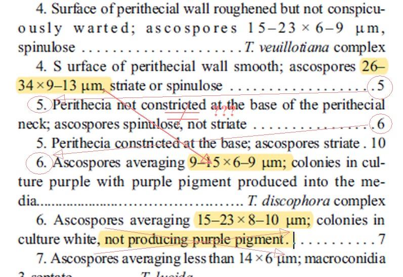
Then I used the work "The genus Thelonectria (Nectriaceae, Hypocreales, Ascomycota) and closely related species with cylindrocarpon-like asexual states - 2016". I may be wrong but it seems to me that it is written with errors. There are large inaccuracies in the key (see scan No. 01).
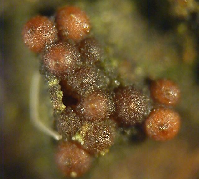
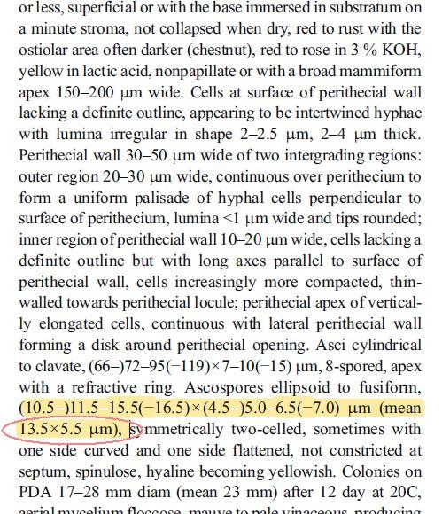
Yesterday I came to the earlier work of the same authors and according to her my collection is the closest to Thelonectria discophora;
"Phylogeny and taxonomic revision of Thelonectria discophora
(Ascomycota, Hypocreales, Nectriaceae) species complex - 2013 ".
(See scan 02)
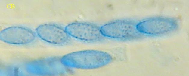
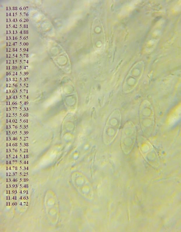
Today I have measured a greater number of spores and their dimensions are practically perfectly consistent with this description!
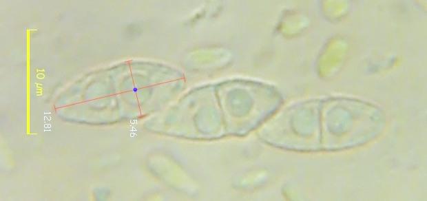
(11.4) 11.9 - 15.1 (16.2) × (4.6) 4.9 - 5.9 (6.2) µm
Q = (2.1) 2.2 - 2.8 (3); N = 34
Me = 13.4 × 5.5 µm; Qe = 2.5
Individual dimensions of the spores are given in the picture nr. 03
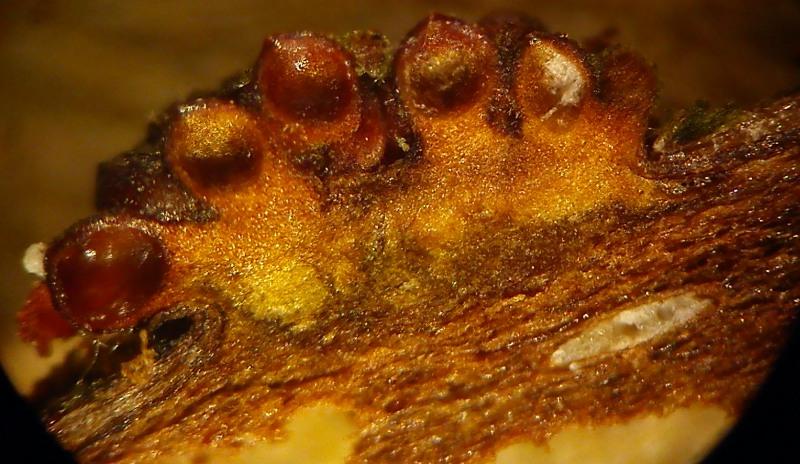
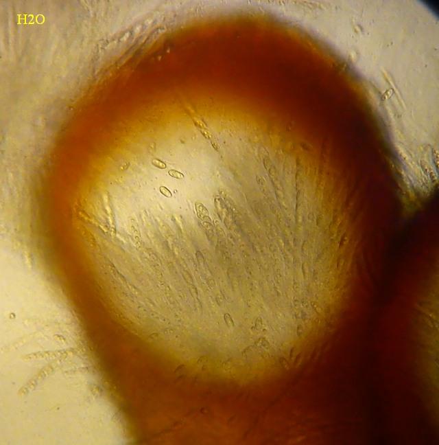
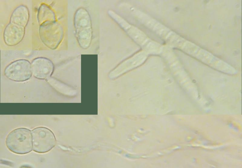
Christian, fruiting bodies are not overripe. I showed germs germinating but there were very few. In my opinion, the fruiting bodies are of the perfect age for microscopy. In my collection there are completely immature spores and free spores that are already germinating. However, the vast majority of spores are moderately mature, with ornamentation already formed. This time I have measured just such.
I compared other species but in their case the size of the spores is not compatible with mine!
Thank you for the hint!
You'll agree with me?
Regards
Mirek
There is no experience with this type.
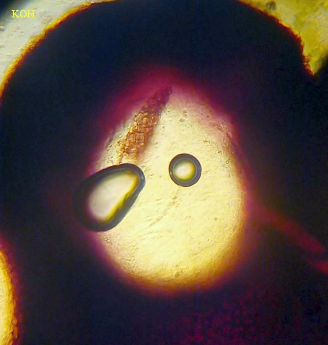
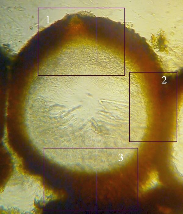
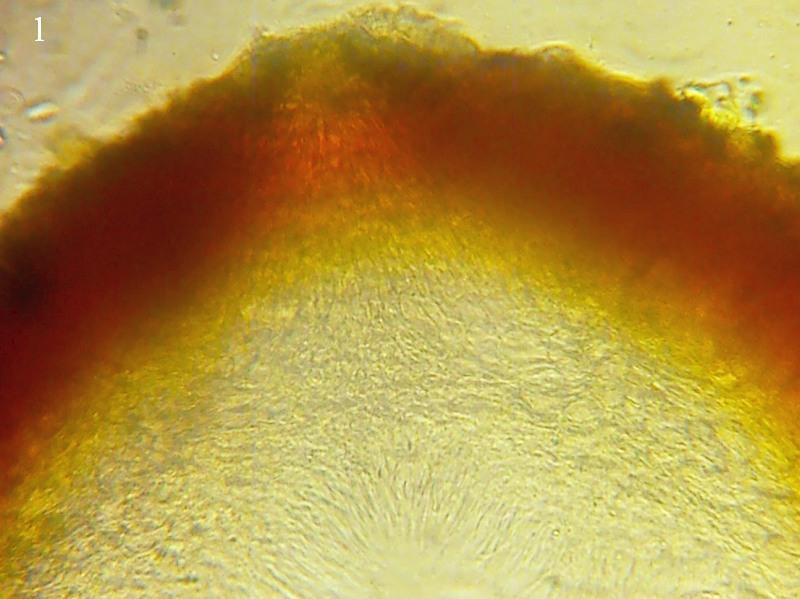
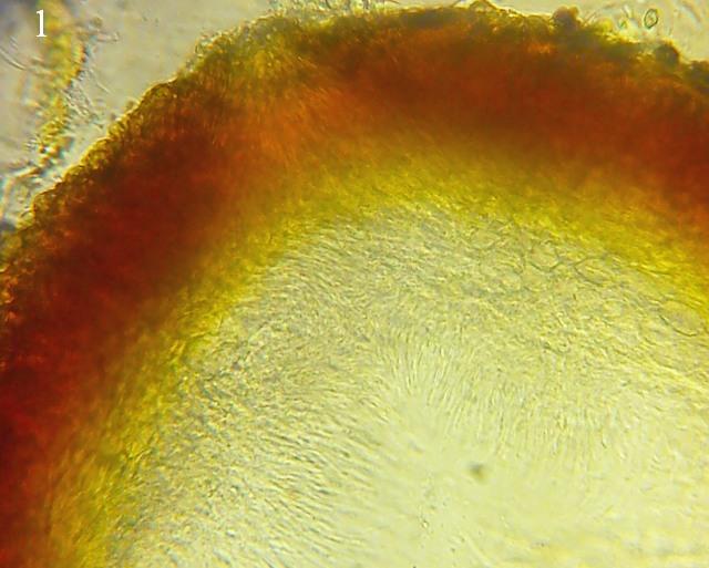
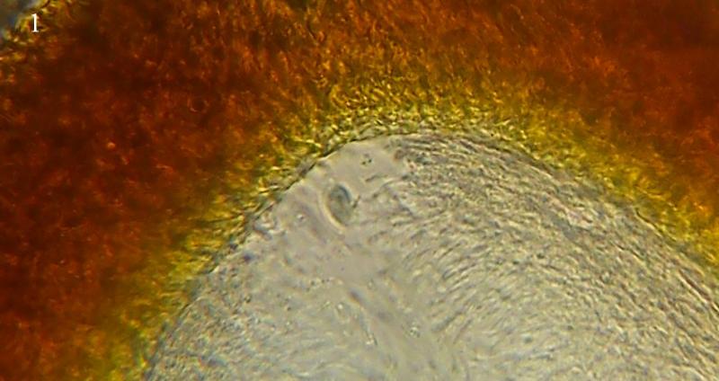
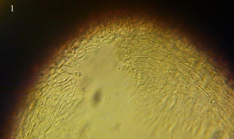
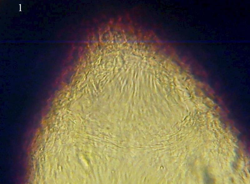
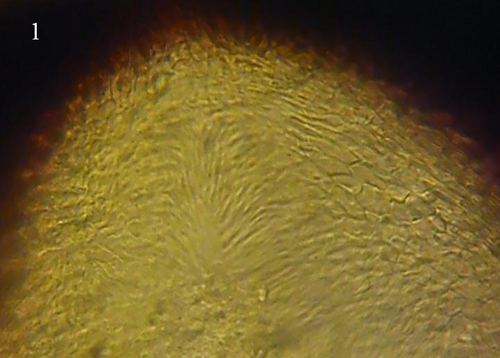
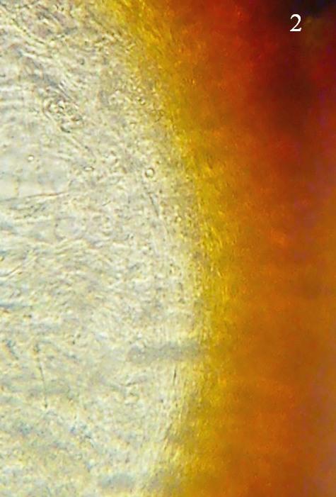
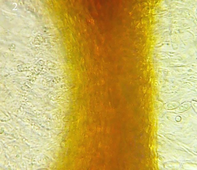
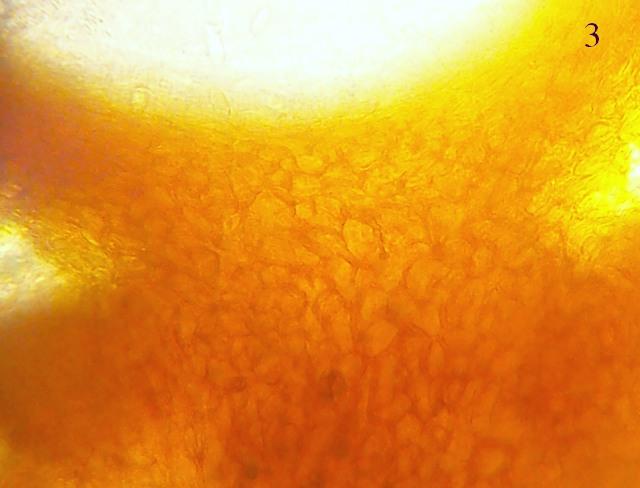
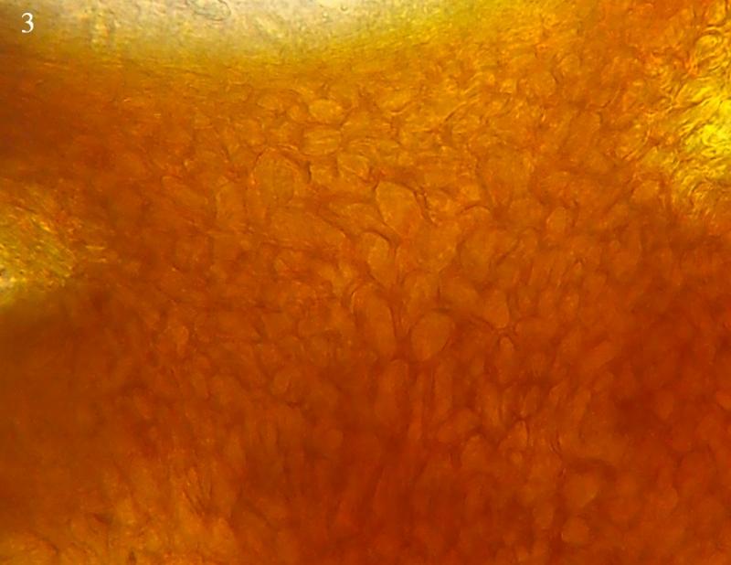
I prepared some photos. Maybe you can read the necessary qualities from them?
Mirek
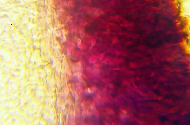
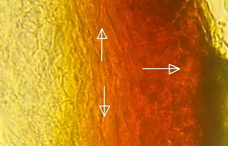
http://www.centrodeestudiosmicologicosasturianos.org/?p=15486
It is true that my photo is not as perfect as Enrique but you can see sufficient arrangement of the cells.
Is this how it was supposed to look like?
Regards
Mirek
Your help was priceless!
Mirek