12-07-2015 00:05
 Nedim Jukic
This one from the same locality as the previous on
12-11-2019 10:32
 Miguel Ángel Ribes
Hi againExactly at the same place than my previous


30-05-2026 21:12
Philippe PELLICIERSur branche de mélèze (Larix) près de la neige,
31-05-2026 10:35
Hulda Caroline HolteHello,I collected this species growing on a rather
25-05-2026 16:35
 Bernard CLESSE
Bonjour à toutes et tous,J'ai trouvé récemment,
29-05-2026 15:35
daniel FERREBonjour à tous,Je voudrais votre aide pour cette
28-05-2026 16:15
James MitchellHello,Does anyone have the original publication of
28-05-2026 11:06
Thomas Læssøehttps://svampe.databasen.org/observations/10596750
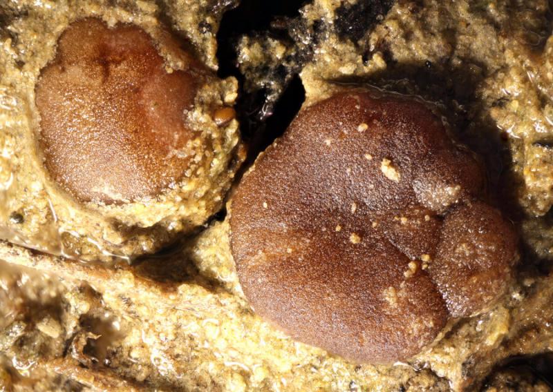
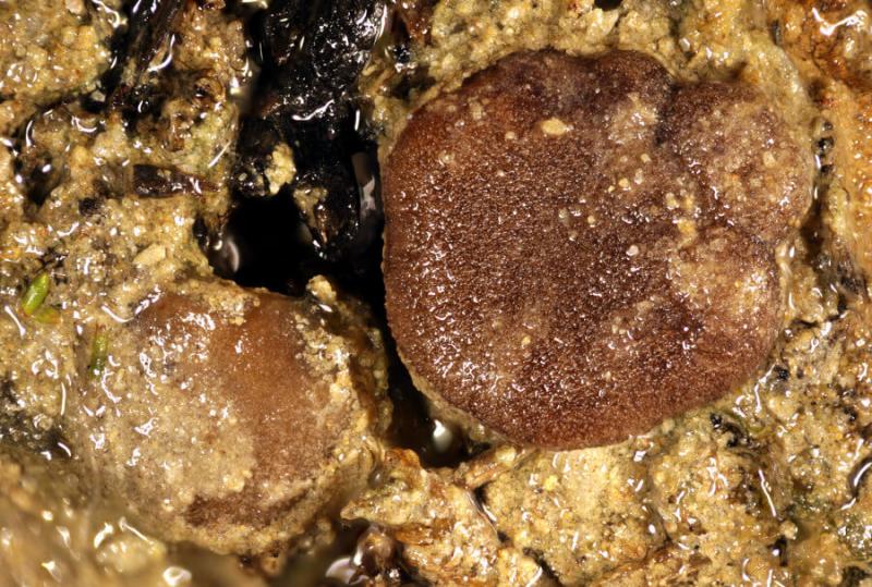
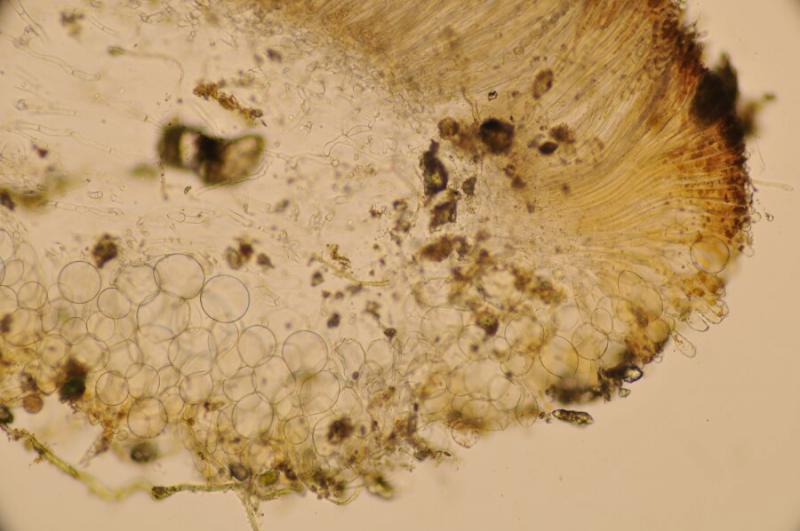
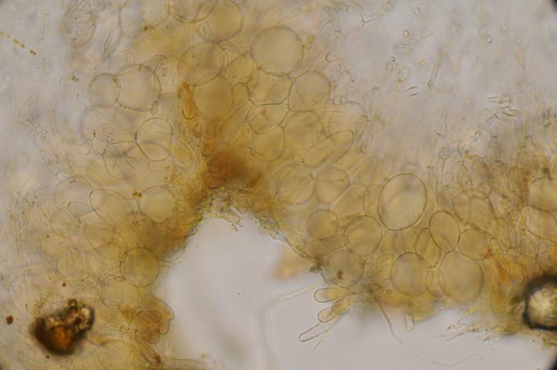
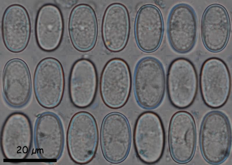
Hi againExactly at the same place than my previous species (Aragüés del Puerto) that I just sent, even in the same square centimeter, and with an almost identical macro appearance, this Pachyella grew. At first glance it is impossible to differentiate them, but the micro does not lie.
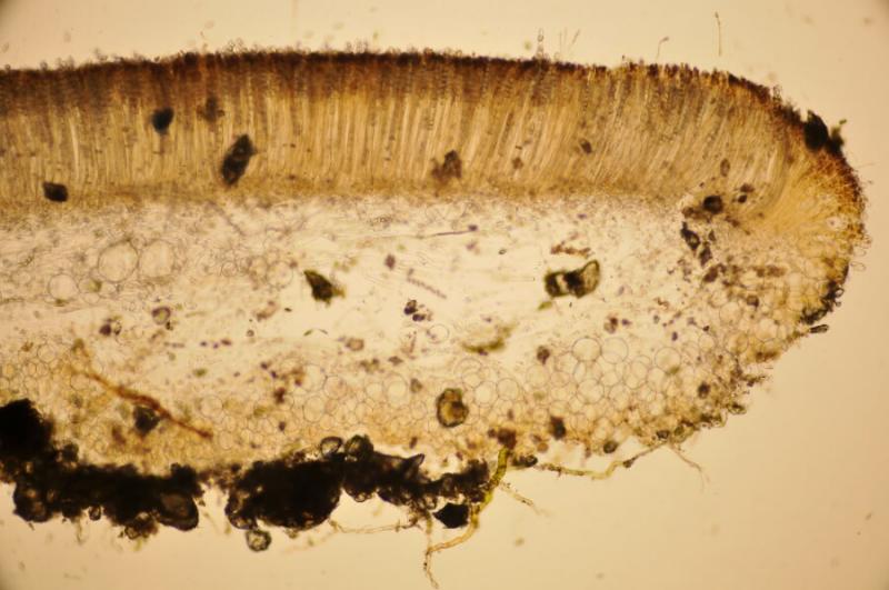
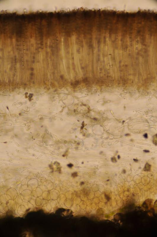
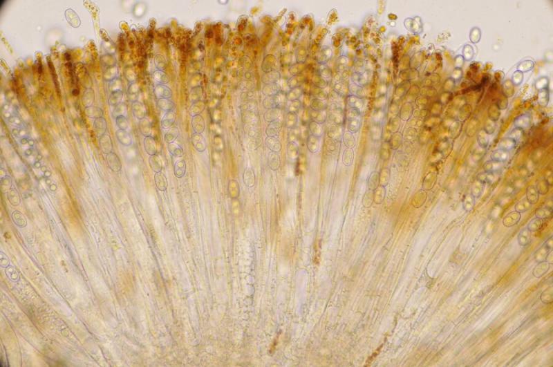
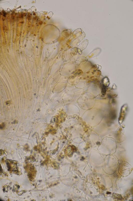
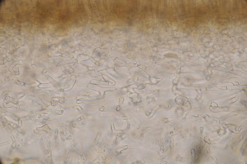
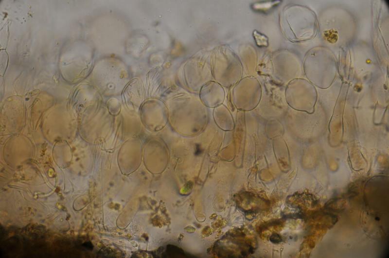
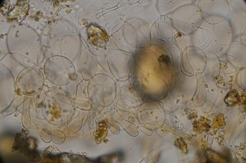
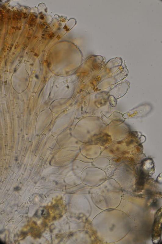
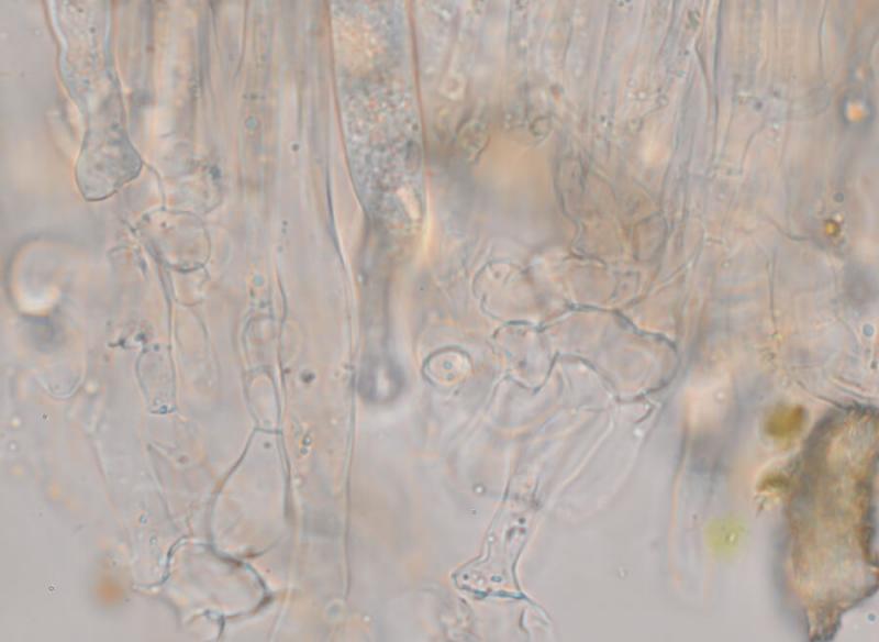
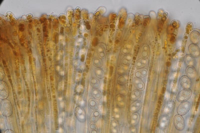
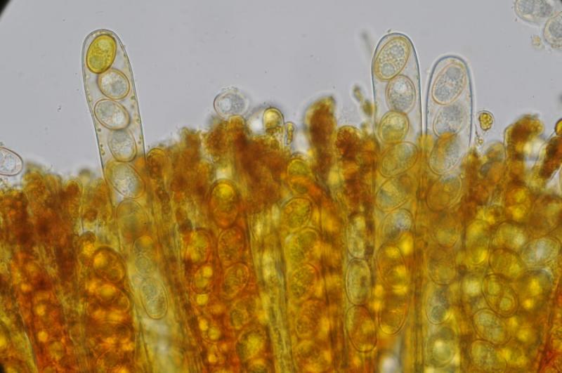
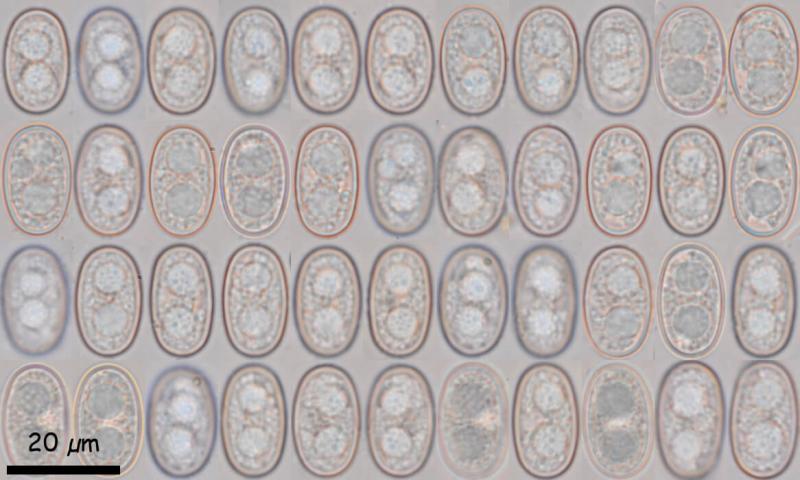
Exctal excipulum with textura globulosa finished in a cylindrical-claviform appendix. Margin with chains of 2-3 subglobose cells and cylindrical-claviform appendix. Medullary excipulum with very lax (gelled) intricate texture. Paraphysis with large brown pigment gutules, slightly thickened at the apex. Uniseriate octosphoric asci, with croziers and IKI -. Ellipsoidal spores, with 2 large LBs, apparently finely rough in water, but virtually imperceptible in cotton blue, of (18.3) 18.8 - 20.1 (20.8) × (10.8) 11.2 - 12.1 (12.3) µm; Q = 1.6 - 1.78 (1.8); N = 50; Me = 19.5 × 11.6 µm; Qe = 1.7
With this spore size, in the literature I only see Pachyella adnata, but the spores have large spines. Macroscopically reminds me to P. celtica, but paraphysis, asci, spore size, etc. do not fit. It also looks like Peziza subisabellina, but the micro has nothing to do with it. Perhaps a simply P. babingtonii growing on land?
Thank you.