22-04-2026 20:17
 Marian Jagers
Is anyone familiar with the Hyphomycetes genus Pse
22-04-2026 20:54
 Enrique Rubio
Hi to everybody.This Pyrenopeziza grew in moist le
22-04-2026 01:06
 Richard VALERI
Bonjour à tous.Je vous présente cette Nectria s.
21-04-2026 13:36
Gernot FriebesHi,I am out of ideas for this one. I collected Sal
21-04-2026 13:19
Gernot FriebesHi,this Lophodermium on Typha has ascospores measu
21-04-2026 13:05
Gernot FriebesHi,this hyphomycete feels familiar but I was not a
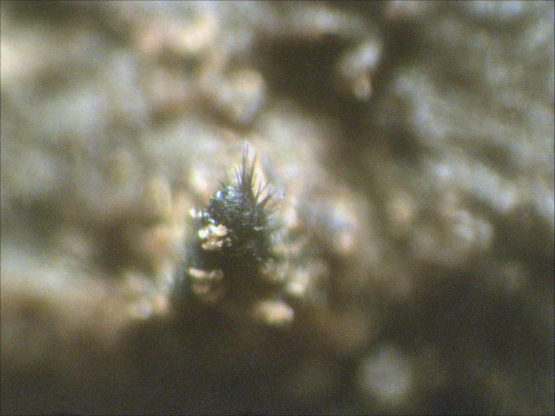
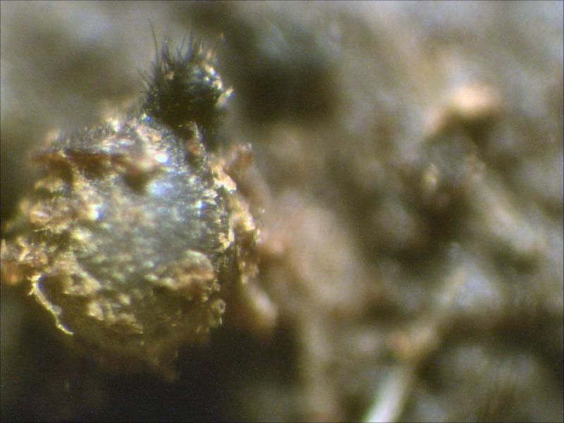
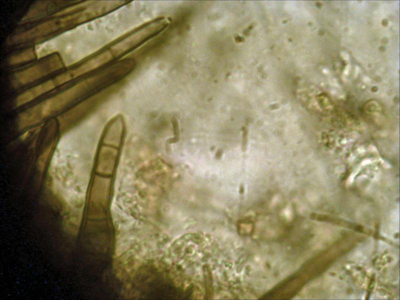
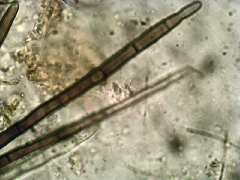
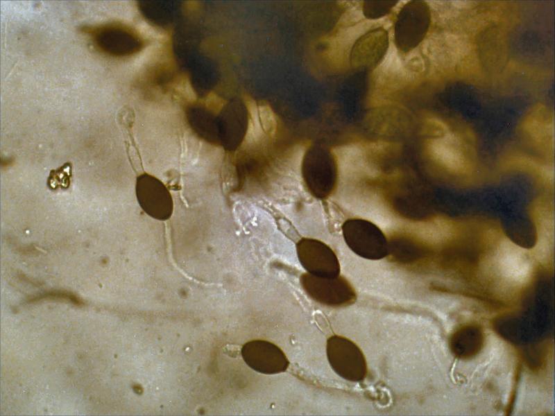
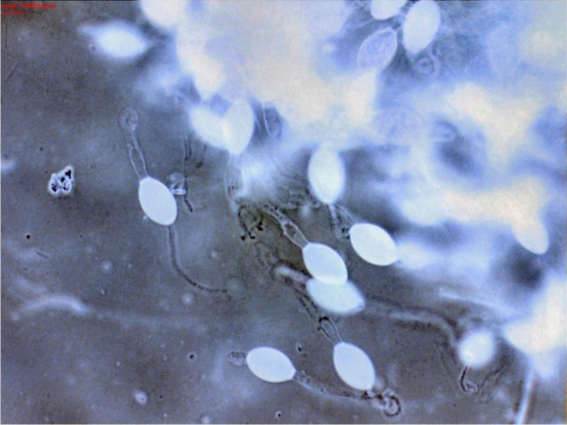
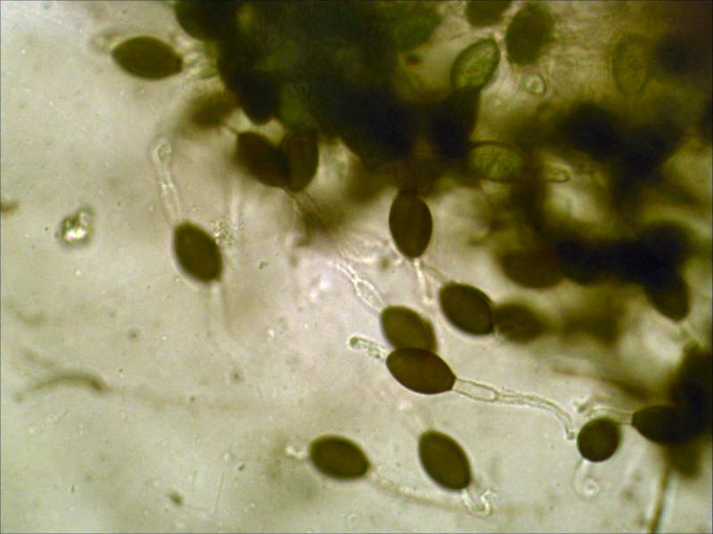
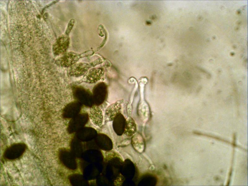
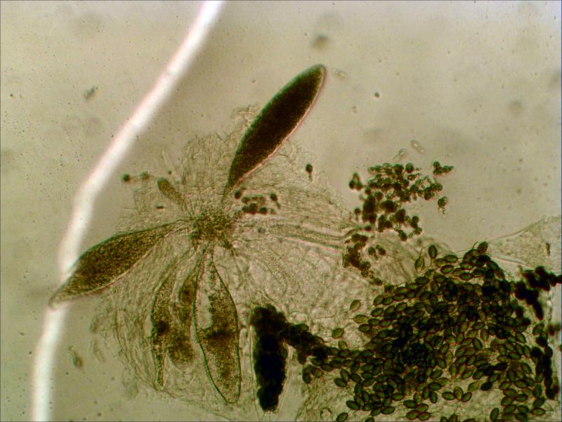
 Found on cow dung.
Measurements of the perithecia and asci do not represent the mentioned values in the documentation because it was not a fully mature species.
Perithecia: 493x458 um with a neck of 168x162 um; sporadically covered with flexuous, septated hairs 2.0-2.5 um wide; the neck is covered with straight thick walled, constricted, multiple septated and hyaline-tipped hairs 87-132.5x4.3-5.6 um with a wall of 0.8-0.95 um.
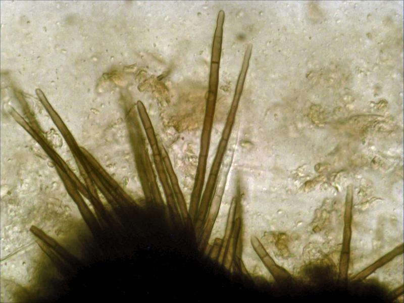
Ascus: 512 spores; 275x58 um with a stolk of 43.5 um.
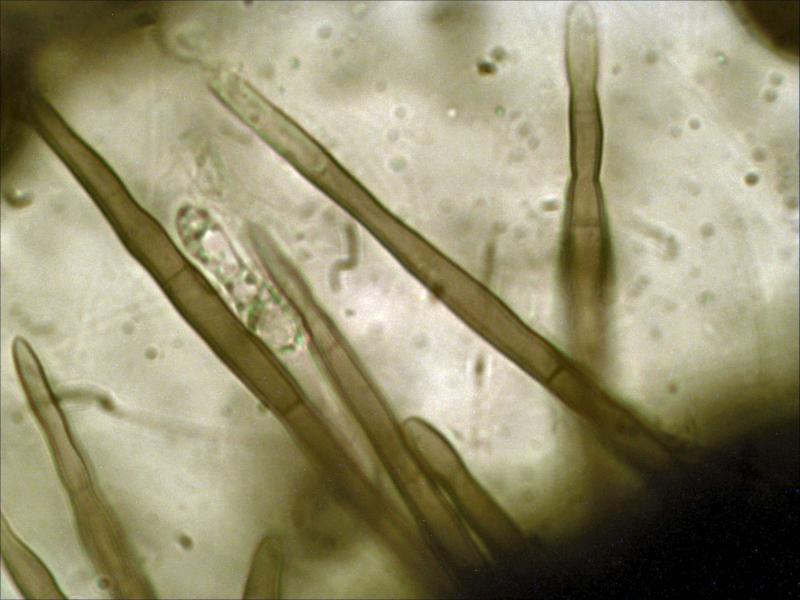
Spores: 21-25x13-16 um; pedicel 12-16x5.0-6.5 um; upper cauda probably consisting of agglutinated filaments 38.5-40.0x3.4-5.2 um; secondary cauda consisting of two to 4 filaments connected to the side of the pedicel, at first separated but half way agglutinated and finally curved whereby the two main filaments are clearly visible by means of two bright spots.
Filaments and the top of the pedicel are covered with granules but they are hard to detect and certainly not with mature spores.
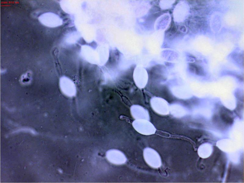
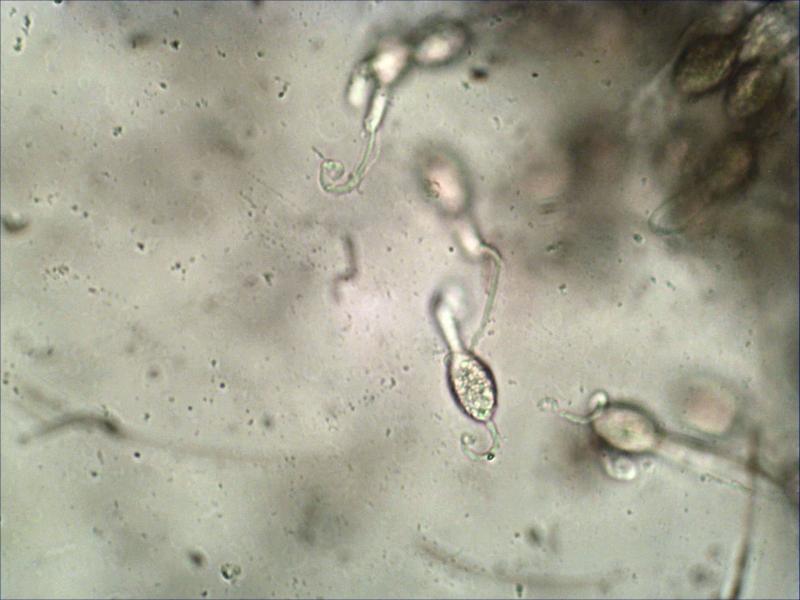
Photos 11&12 show the filaments attached to the side of the pedicel and in my opinion they agglutinate halfway.
The white spots at the and of the caudea clearly show that two main filaments are present probably surrounded by others.
Photo 9 show two small white dots on the pedicel of the most left spore indicating the connection to the pedicel.
To me it looks like the agglutinated filaments of the secondary caudea bend down like the pistil of a flower when they curl.
The upper caudea do not have that, despite the fact that they are already agglutinated and also do curl at the end.
Photo 8 is a negative from the previous one showing the granules at the end of the pedicel and covering the filaments, they are not visible on a normal coloured photo.
I used a 40x objective because a 100x objective blurred the photos so the granules were not clearly visible.
Joop