
24-04-2026 03:16
 David Chapados
Found while looking at something else from wood in
22-04-2026 20:54
 Enrique Rubio
Hi to everybody.This Pyrenopeziza grew in moist le
22-04-2026 01:06
 Richard VALERI
Bonjour à tous.Je vous présente cette Nectria s.
22-04-2026 20:17
 Marian Jagers
Is anyone familiar with the Hyphomycetes genus Pse

21-04-2026 13:36
Gernot FriebesHi,I am out of ideas for this one. I collected Sal
21-04-2026 13:19
Gernot FriebesHi,this Lophodermium on Typha has ascospores measu
Mycoarachis inversa
Joop van der Lee,
24-11-2018 19:58
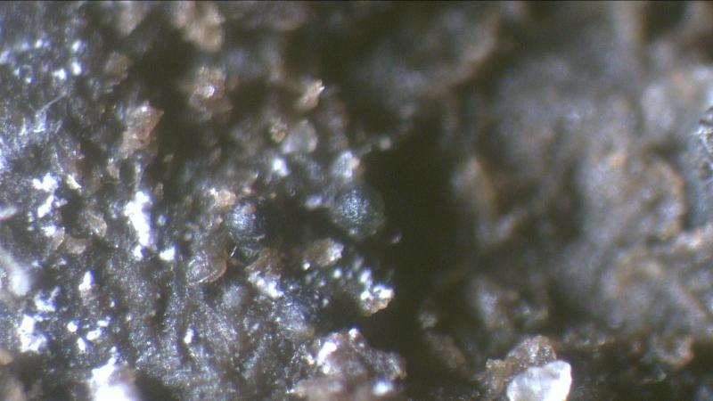
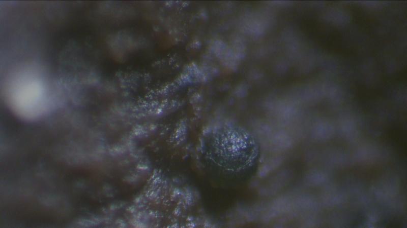
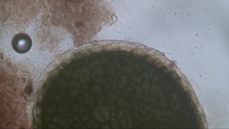
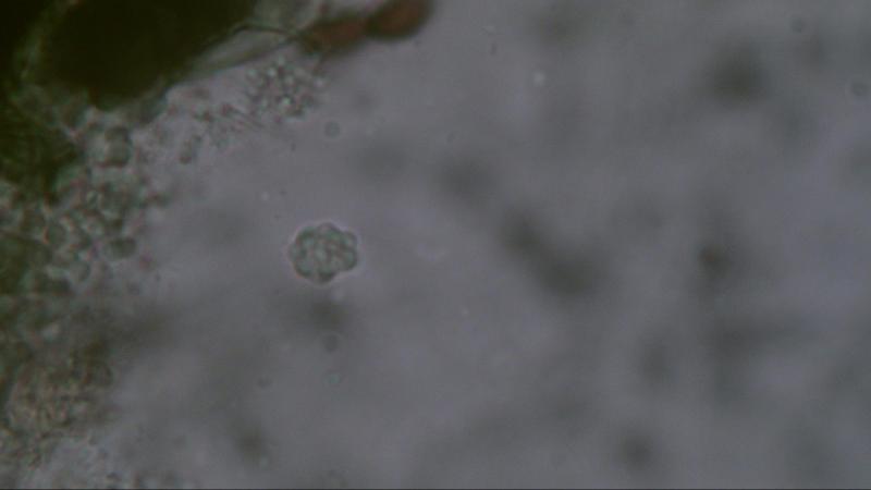
 Found on cow dung, most of the time in the visinity of Schizothecium species.
Fruitbody: Round 170,2-173 um in diameter, surrounded by a gelatinous layer approx. 60 um thick, dark green in colour.
Spores: Round and/or pointed 6.8-7.3 um covered with round warts 2.2-2.5 um.
The first time spores were measured when in water but missing the warts so the second time measurement was performed in Melzer.
David Malloch,
25-11-2018 01:41

Re : Unknown asco
Hello Joop,
That is a very interesting fungus. Do the fruiting bodies have an opening of some kind? The round asci look like the kind you find in a cleistothecium but your pictures suggest that maybe there is an ostiole. Also, do the asci contain 8 ascospores or are there more than that?
Regards,
David
That is a very interesting fungus. Do the fruiting bodies have an opening of some kind? The round asci look like the kind you find in a cleistothecium but your pictures suggest that maybe there is an ostiole. Also, do the asci contain 8 ascospores or are there more than that?
Regards,
David
Michel Delpont,
25-11-2018 11:34

Re : Unknown asco
Hello !
What you show on all your photos are in my opinion asci and it is very difficult to see the spores alone. Are there any hairs? Afraid to be could you look for the genera Lophotrichus, Kernia. There is also the genus Orbicula but in the latter the asci are cylindrical.
Michel.
Sven Heinz,
25-11-2018 17:44

Re : Unknown asco
Hello Joop,
your fungus reminds me of Pithoascus nidicola.
Greetings Sven
Joop van der Lee,
26-11-2018 20:39
Re : Unknown asco
Hello Sven,
Can you provide me the following article.
Pithoascus nidicola (Massee & E.S. Salmon) Arx, Proceedings van de Koninklijke Nederlandse Akademie van Wetenschappen Section C 76 (3): 292 (1973) [MB#320551]
Regards,
Joop
Can you provide me the following article.
Pithoascus nidicola (Massee & E.S. Salmon) Arx, Proceedings van de Koninklijke Nederlandse Akademie van Wetenschappen Section C 76 (3): 292 (1973) [MB#320551]
Regards,
Joop
Sven Heinz,
26-11-2018 21:02
Re : Unknown asco
Hello Joop,
unfortunately, I do not own this article!
Greetings Sven
Björn Wergen,
26-11-2018 21:26

Joop van der Lee,
27-11-2018 07:48
Re : Unknown asco
Hello Sven,
I will try to get the article in the library of Naturalis when I can find the time to do so.
If it is succesful I will send you a copy.
Joop
Joop van der Lee,
27-11-2018 07:48
Re : Unknown asco
Thanks Bjorn I will have a look.
Joop
Joop
Joop van der Lee,
27-11-2018 07:57
Re : Unknown asco
Hello David,
In my opinion it is a single ascus species like Thelebolus stercoreus containing hundreds of spores.
I did not find any ostiole but maybe that is possible when these species are ripe.
It is typical that these species were found together with Schizothecium conicum species.
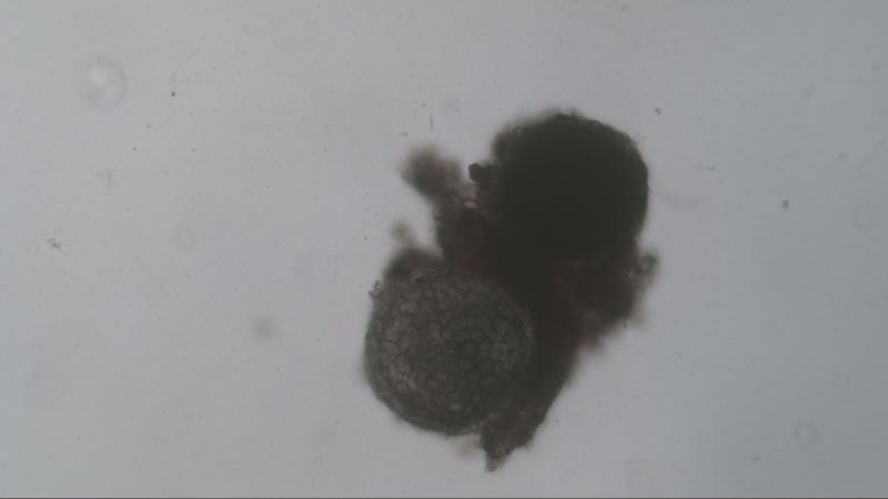
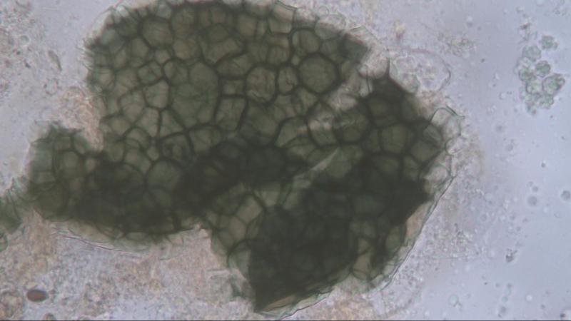
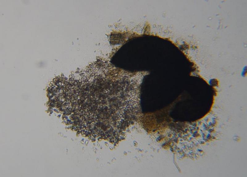
When putting pressure on the cover glass the species bursts open (photo #4).
Joop
In my opinion it is a single ascus species like Thelebolus stercoreus containing hundreds of spores.
I did not find any ostiole but maybe that is possible when these species are ripe.
It is typical that these species were found together with Schizothecium conicum species.
When putting pressure on the cover glass the species bursts open (photo #4).
Joop
Joop van der Lee,
01-12-2018 12:37
Re : Unknown asco
Hello Sven,
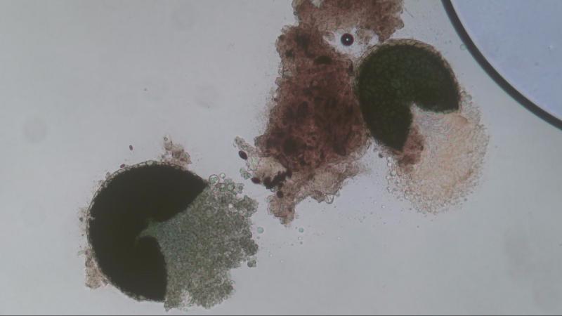
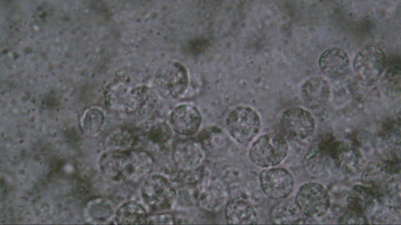
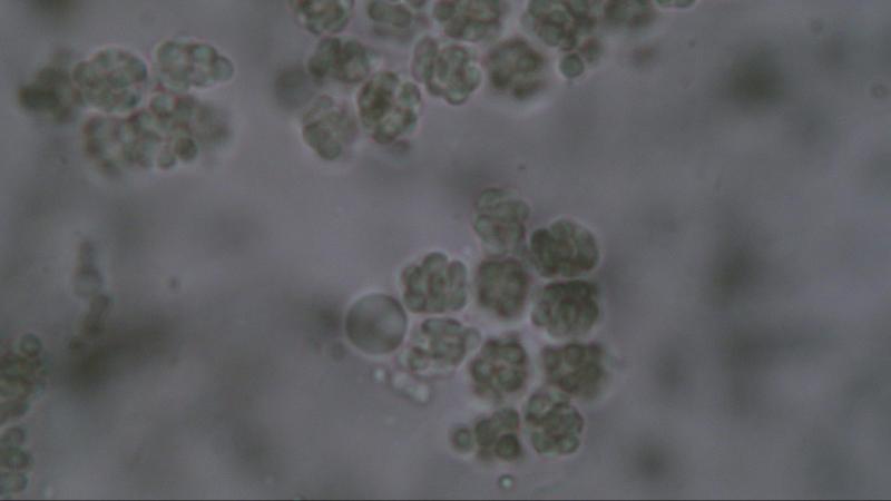
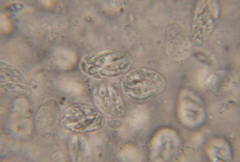
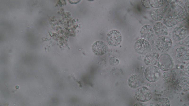
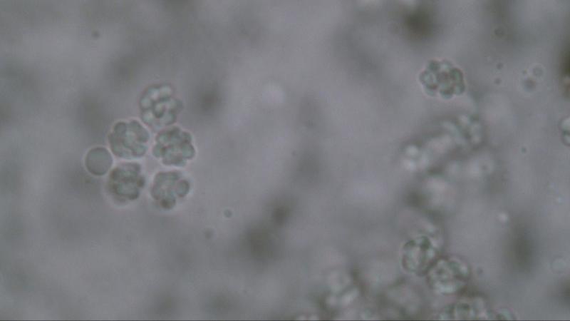
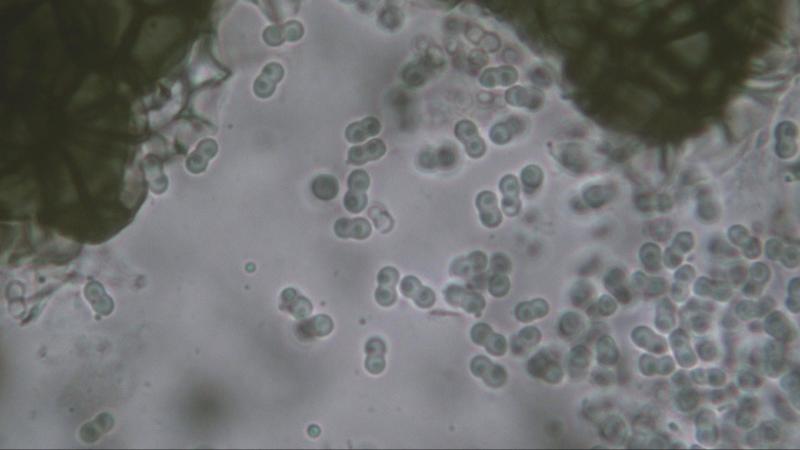
I collected some more information about this species and it will present different shapes of spores when using different fluids.
Photo #1 when using water.
Photo #2 when using Melzer
Photo #3&4 when using Congo Red.
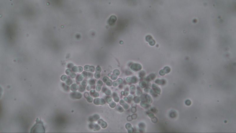
It seems to me that the presentation on #2 show the spores as seen in #3&4 clinging to each other. Whereby at first I thought that they were warts.
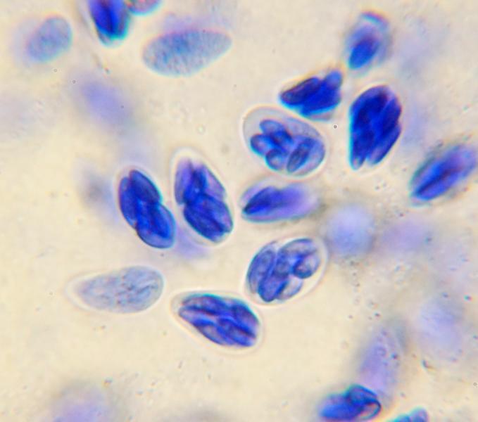
The spores consist of 2 cells, each cell measures 2.45 um in diameter, the total of 2 cells combined is 4.9 um.
Each cell is filled with a "the bary bubble".
When measuring the cells as presented in #2 (or other photos I made) the result will be the same as in #3&4 namely 2.45 um.
Greetings,
Joop
I collected some more information about this species and it will present different shapes of spores when using different fluids.
Photo #1 when using water.
Photo #2 when using Melzer
Photo #3&4 when using Congo Red.
It seems to me that the presentation on #2 show the spores as seen in #3&4 clinging to each other. Whereby at first I thought that they were warts.
The spores consist of 2 cells, each cell measures 2.45 um in diameter, the total of 2 cells combined is 4.9 um.
Each cell is filled with a "the bary bubble".
When measuring the cells as presented in #2 (or other photos I made) the result will be the same as in #3&4 namely 2.45 um.
Greetings,
Joop
David Malloch,
01-12-2018 15:03
Re : Unknown asco
Hello Joop,
I believe this fungus is Mycoarachis inversa, a species characterized by two-celled peanut-shaped spores and a cleistothecial peridium with the hyaline layers on the outside (hence "inversa").
David
I believe this fungus is Mycoarachis inversa, a species characterized by two-celled peanut-shaped spores and a cleistothecial peridium with the hyaline layers on the outside (hence "inversa").
David
Joop van der Lee,
01-12-2018 23:25
Re : Unknown asco
Thanks David.
Joop
Joop
Norbert Heine,
05-12-2018 15:05

Re : Unknown asco
This is a very nice found, Joop!
Thanks to all for this interesting discussion.
I didn't know this genus before.
I think that this is exactly the species, descriped by Dave & R.F.Cain, 1970, as Mycoarachis inversa.
At the fotos you can see the hyaline outer layer of the peridium and the unique, peanut shaped ascospores.
In the article by Melo et al. 2017 you can find also a description with some nice pics. Plate 1, figs. 17-21
It seems that this species until now is only known from North and South America and Africa.
Best regards, Norbert
Michel Delpont,
05-12-2018 16:44
Re : Unknown asco
Joop avait déjà trouvé cette espèce en 2015 !
Michel.
Norbert Heine,
05-12-2018 17:00
Re : Unknown asco
Thank you, Michel!
I think that I overlooked this.
Nevertheless it seems to be a nice and rare species!
Regards, Norbert