

10-06-2026 12:54
Steve ClementsBonjour encore, Pouvez-vous m'aider, s'il vous pl
09-06-2026 18:32
Camille MertensSur morceau de roseau immergé 0,5 - 0,7 mm de dia
10-06-2026 21:16
 François Freléchoux
Bonsoir,Le dernier du jour, en attendant votre avi
10-06-2026 21:07
François Freléchoux
Toutes les tiges de gentianes jaunes de l'an pass�
10-06-2026 13:41
François Freléchoux
Bonjour à nouveau, Voici une trouvaille d'hier.
10-06-2026 11:53
Steve ClementsBonjour, This disco is abundant on dead stems of
10-06-2026 10:45
François Freléchoux
Bonjour à nouveau, Encore une détermination qui
08-06-2026 10:16
 Spooren Marco
I don`t have a clou about this fungus,it is not in
10-06-2026 09:24
François Freléchoux
Bonjour, J'imagine que cette détermination ne do
Nitschkia parasitans on Graphostroma?
Gernot Friebes,
24-03-2015 11:31
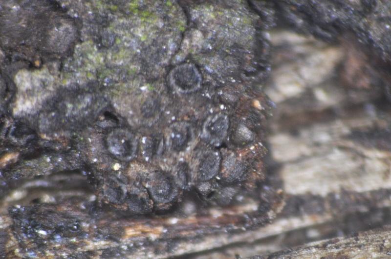
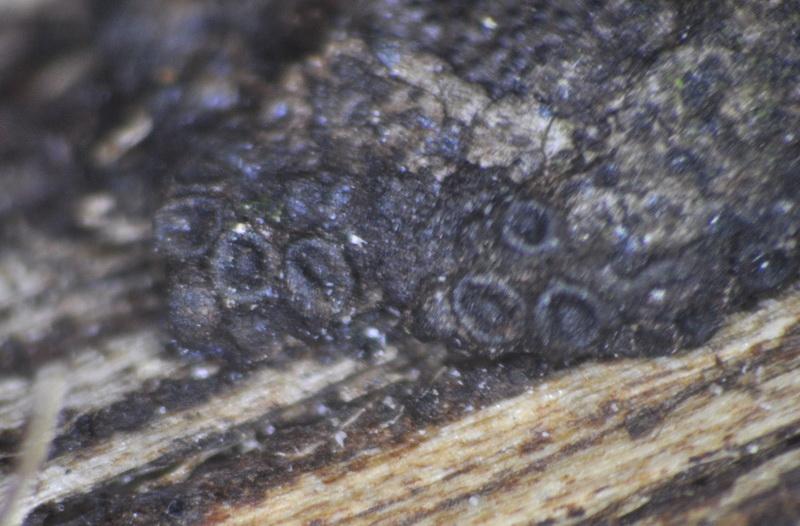
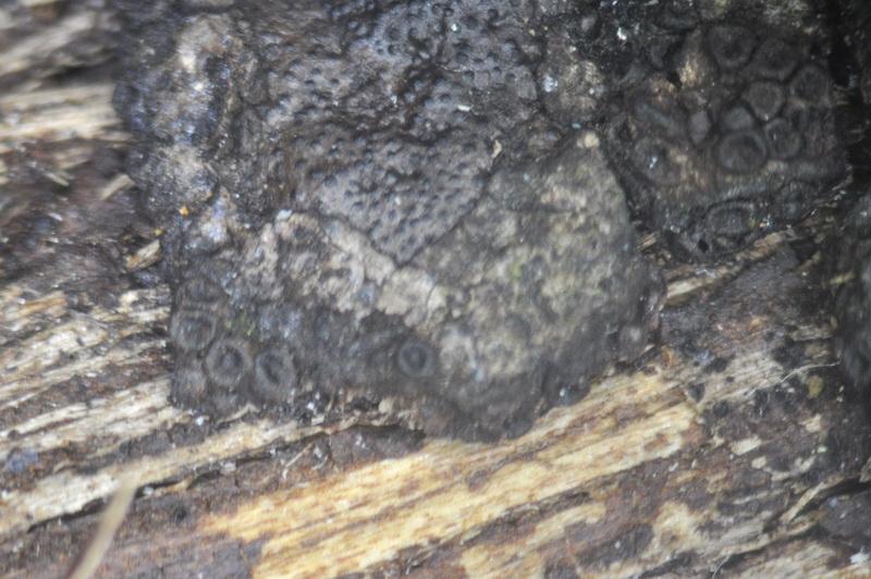
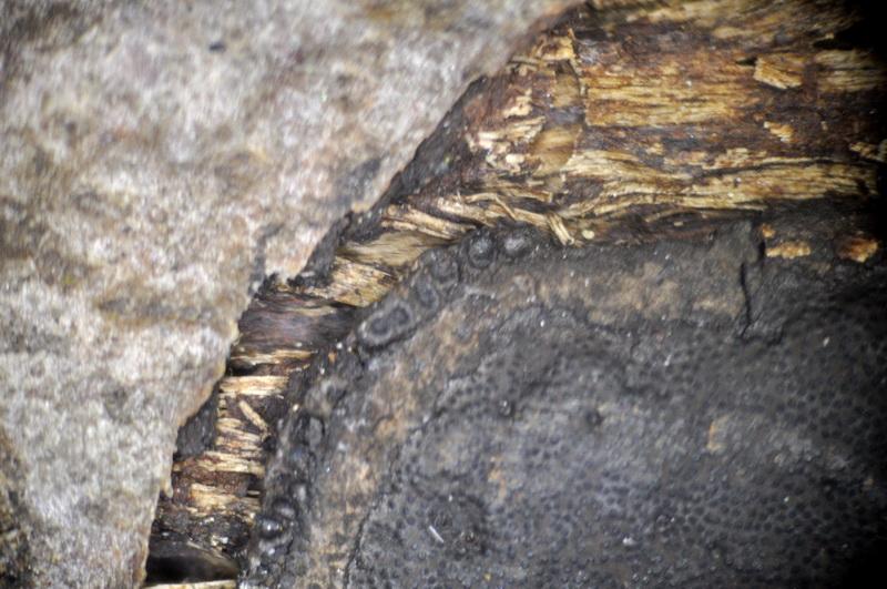
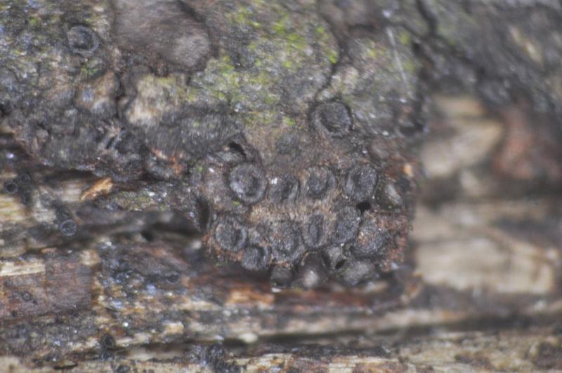
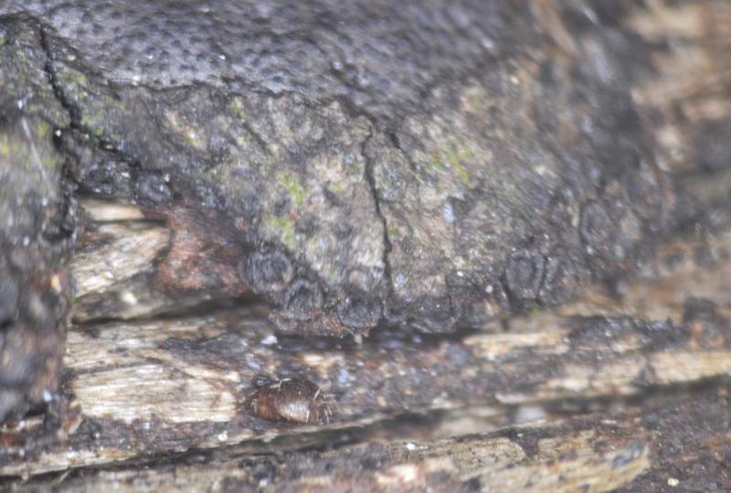
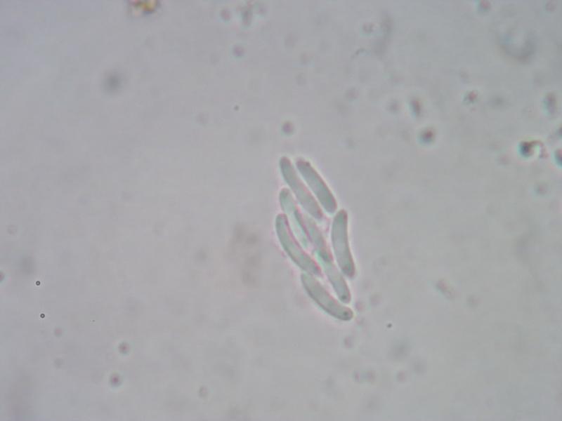
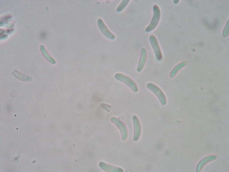
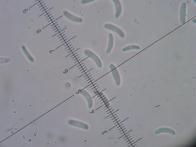
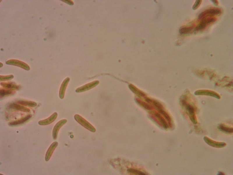
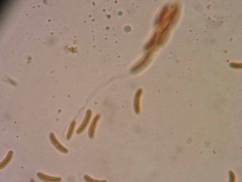
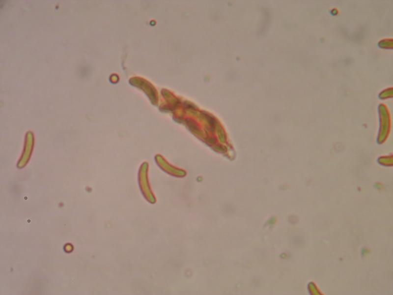
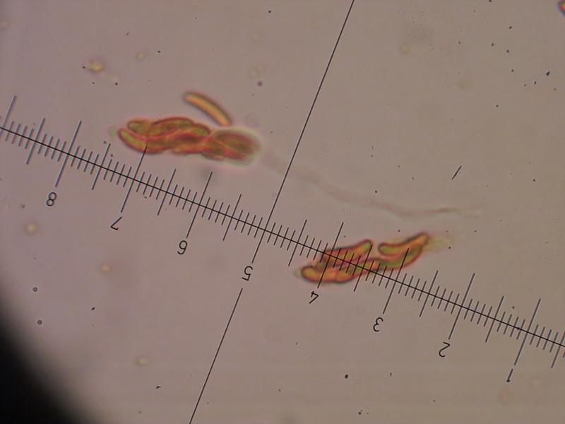
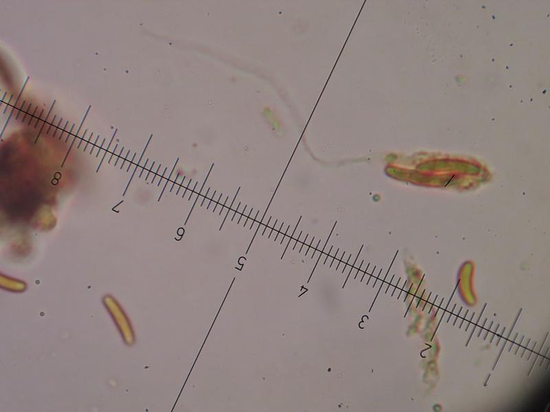
as far as I'm aware Nitschkia parasitans is always described as growing on stromata of Nectria cinnabarina. However, I have received a collection that comes close to N. parasitans microscopically but grows on the margin of stromata of Graphostroma platystoma (on Castanea sativa). At least I don't know any other Nitschkia species with 8-spored asci and allantoid, hyaline ascospores which in the present fungus measure 9–11 x 2–2.5 µm. Maybe there is one that I overlooked? Or does N. parasitans indeed grow on fungi other than N. cinnabarina occasionally?
The long stipe of the dead asci is also quite remarkable. Munk pores are frequent and encircled by the dark, thickened cell wall. The macroscopic appearance is also a bit different from typical N. parasitans I think, because that species usually grows more densely fasciculate.
On a side note: it was very interesting to observe the ascomata upon rehydration because some of them quickly ejected a whitish conical "body" from which again the ascospores where visibly ejected under the stereo microscope. This must have been the "Quellkörper" which I have never seen in action before. Unfortunately I was at a microscope without a chance to take photos...
Best wishes,
Gernot
PS: The attached photos are not mine.
Jacques Fournier,
24-03-2015 14:52

Re : Nitschkia parasitans on Graphostroma?
Hi Gernot,
I don't know your fungus but when run through Huhndorf and Mugambi's key (Mycologia, 102(1), 2010, pp. 185–210) it comes to the genus Coronophorella, with C. chaetomioides as the only species. Hope it helps...
Cheers,
Jacques
I don't know your fungus but when run through Huhndorf and Mugambi's key (Mycologia, 102(1), 2010, pp. 185–210) it comes to the genus Coronophorella, with C. chaetomioides as the only species. Hope it helps...
Cheers,
Jacques
Gernot Friebes,
24-03-2015 21:08
Re : Nitschkia parasitans on Graphostroma?
Hi Jacques,
thanks for the suggestion. Nannfeldt describes the ascospores as shorter and wider than in this collection (6–8 x 2–3 µm; as "Nitschkia chaetomioides") and in the following link the ascospore size is also considerably shorter and wider (as "Scortechinia chaetomioides"): http://www.bcrc.firdi.org.tw/fungi/fungal_detail.jsp?id=FU200802050069. The description here: https://www-s.life.illinois.edu/pyrenos/records/show_by_page?page=114 fits better but unfortunately the images are not available. I think for now this fungus has to stay without a full name...
Best wishes,
Gernot