02-06-2026 14:33
 Nicolas VAN VOOREN
Hello.I'm searching for a PDF copy of the followin
02-06-2026 17:58
Louis DENYBonjour forum, Sur feuille de Populus tremula, en

28-07-2011 18:31
 Alex Akulov
Dear FriendsToday I made the pdf file of Velenovsk
11-09-2025 16:57
 Jason Karakehian
Our revision of Marthamycetales (Leotiomycetes) is
12-11-2019 10:32
 Miguel Ángel Ribes
Hi againExactly at the same place than my previous
25-12-2019 17:54
Valencia Lopez Francisco JavierHola a todos/asEstas supuestas pezizas estaban en
12-07-2015 00:05
 Nedim Jukic
This one from the same locality as the previous on

30-05-2026 21:12
Philippe PELLICIERSur branche de mélèze (Larix) près de la neige,
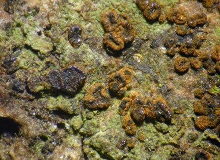
 Hi everyone,
I got a fungus on bark from southern Italy, seemingly with no connection to the surrounding lichens. If it were lichenicolous, I would presume it to be a Buelliella. Has anybody any idea what this could be?
Ascomata sessile, constricted below, initially closed, later apothecioid, up to 0.5 mm diam., irregularly roundish, disc black but covered with a rusty pruina, margin prominent, densely rusty pruinose.
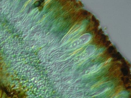
Hypothecium brownish, 55 µm high, hymenium hyaline below, brownish above, 120 µm high, epithecium brown, covered with dark brown granules, excipulum dark brown, up to 50 µm thick.
Paraphyses septate, sparsely ramified, 2–2.5 µm wide, hyaline below, brownish above, the upper cell sometimes enlarged up to 4 µm, brown in the upper half.
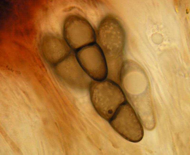
Asci clavate, 80–95 × 15–26 µm, apically thickened, with an internal beak, with a long stalk, 4–8-spored.
Ascospores 1-septate, grey, the upper cell rounded or slightly attenuated, the lower attenuated, narrower than the upper one, constricted at the septum, with one big guttule in each cell, surface ± smooth but appearing foveate, (20–)20.8–23.1(–24) × (8.5–)9–9.9(–10) µm, l/b = (2–)2.2–2.5(–2.6) (n = 20).
Pruina K+ violet, not dissolving. Hymenium above K+ grey, I+ reddish. Asci externally I/KI+ pale blue, no I/KI reacting apical structures.?