
25-05-2026 16:44
François BartholomeeusenHi forum members,During an excursion organised by
25-05-2026 16:35
 Bernard CLESSE
Bonjour à toutes et tous,J'ai trouvé récemment,
22-05-2026 13:29
Gernot FriebesHi,I am curious to hear your opinion on this mater
23-05-2026 11:44
 Charles Grapinet
Hello, I am having trouble identifying this copro
23-05-2026 18:57
Sylvie Le GoffBonjour à tousRécolté sur une branchette de Sal
22-05-2026 14:44
 Lothar Krieglsteiner
in unripe condition citrine yellow, then soon fadi
22-05-2026 21:35
Steve ClementsBonjour, I expected this find on old wood on our
22-05-2026 18:12
Lothar Krieglsteiner
... in moist chamber from Portugal.As the fungus s
22-05-2026 20:08
Ethan CrensonHello all, Yesterday in NYC I was visiting an e
Niptera / Mollisia indet.
Uwe Lindemann,
03-12-2009 18:54
 Hello Forum,
 recently Dirk and I found a little inoperculate discomycet which we can't determine.
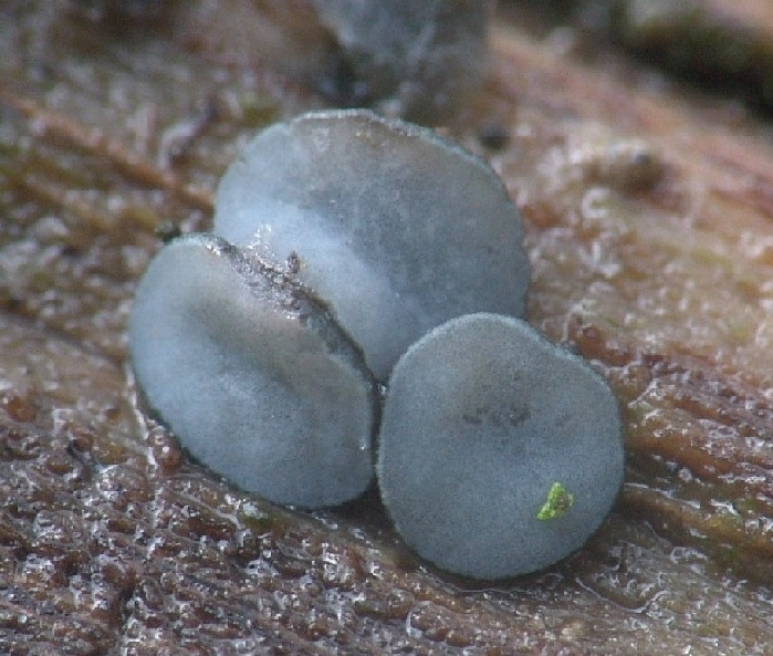
Macroscopic features:
Apothecia sessile, 0,3-0,4 mm diam., greyish-blueish (see photo)
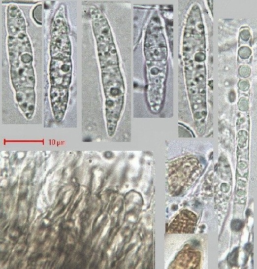
Microscopic features:
Asci arising from croziers, 8spored, 125-130 x 11,5-12 µm, mostly biserate, IKI+ (blue).
Ascospores hyalin, fusiform with (1-)3(-4) septa, with many little to medium guttules, 25,5-34,5 x 5,2-6,5 µm; spores not septate in the asci.
Paraphyses filiform, with many little “oildrops”/vacuoles, up to 3,5 µm wide, not thickened at the apex; in KOH not yellow.
”Hairs” brownish, smooth, up to 4,5 µm wide, up to 25 µm long
Ectale excipulum brownish cells, textura angularis/textura prismatica
Medullary excipulum brownish cells, textura angularis
The substrate is Rubus fruticosus s.l.
Dirk and I thought first of Niptera/Mollisia pilosa or Niptera/Mollisia pulla, but size of the spores, the blue porus-reaction and the substrate are not fitting well to pilosa or pulla.
What do you think about it?
Best
Uwe and Dirk
Uwe Lindemann,
03-12-2009 18:54
Re:Niptera / Mollisia indet.
Micro
Hans-Otto Baral,
03-12-2009 21:33

Re:Niptera / Mollisia indet.
Hi Dirk and Uwe
your fungus is very interesting. Looks like a Mollisia but from the paraphysis content it cannot be. Your micros have a strong punctation which makes the structure of the amyloid rings difficult to see (do you use a too long exposure?). It reminds me a bit of a type similar as in Tatraea and Phaeohelotium fulvidulum.
I assume the drops in the paraphyses are vacuoles (you could verify this with Cresylblue which stains them turquoise in the living state). They would better fit to a Helotiaceae. Did you see them like this in many paraphyses? You can also test the spore surface with Cresylblue if there is some gel.
The asci: are the spores inside them really aseptate? Do you have a photo?
A section through the apo would also be informative. A medulla of brownish angularis would be very unusual for Mollisia s.l. My database contains nothing which fits to Rubus and this spore size.
Uwe Lindemann,
04-12-2009 00:22
Re:Niptera / Mollisia indet.
Hello Zotto,
thank you for your fast reply and your very interesting remarks! Yes, you are right: when we look at the really bad pictures of the outer layer of the excipulum we think it is gelatinized. Concerning you question about the the asci: yes, it's an error in our description. The spores are septate in the asci.
I send to you some pictures of the micros with a better (high..) solution to your private email-address. I hope that you can see better what we describe.
Thank you!
Best Uwe and Dirk
Hans-Otto Baral,
04-12-2009 12:41
Re:Niptera / Mollisia indet.
Hallo Uwe
The apical rings actually look something like Hymenoscyphus s.lato, not s. stricto. There exists a Mollisia spectabilis, of which I have no picture and their are spores only up to 17 microns, also the ring is of the typical Hymenoscyphus type.
Perhaps your mushroom is next to Hymenoscyphus fulvidulus. There, the spores go up to 27 microns and also the apical rings agree. However, the asci are always without croziers. A similar species, which I call griseobrunneus, would have croziers, but is only very weakly amyloid, and has much smaller spores (17-22 x 4-5). The Apos indeed recall a Mollisia.
The spores in you find could well be unicellular, when inside the living ascus. Those on your photo, unfortunately, are already dead. The find is probably slightly overripe, therefore the spores are so large and septate.
Anyhow, if you manage a photo of a section this could help in confirming my idea.
Zotto