Accès membres

Mot de passe perdu? S'inscrire

28-04-2024 18:05

Bernard CLESSE Bernard CLESSE

Bonsoir à toutes et tous,J'ai trouvé ce matin ce

28-04-2024 13:30

Juuso Äikäs

On Friday I found these pale, hairy little discos

24-03-2024 08:27

Thierry Blondelle Thierry Blondelle

HiOn Hedera helix fallen branchEcological habitat:

26-04-2024 10:07

Mathias Hass Mathias Hass

Hello, Does anyone know what this is? Found on J

24-04-2024 21:54

éric ROMERO éric ROMERO

Bonjour, J'ai trouvé ce Lasiobolus sur laissées

23-04-2024 15:18

Lothar Krieglsteiner Lothar Krieglsteiner

... but likely a basidiomycete. I hope it is o.k.

23-04-2024 13:17

Edouard Evangelisti Edouard Evangelisti

Bonjour à tous, Je viens de récolter ce que je

23-04-2024 21:49

Ethan Crenson

Hello all, A friend recently found this orange as

22-04-2024 11:52

Zuzana Sochorová (Egertová) Zuzana Sochorová (Egertová)

Hello,I made a loan of a collection of Microstoma

11-01-2022 16:36

Jason Karakehian Jason Karakehian

Hi does anyone have a digital copy of Raitviir A (

« < 1 2 3 4 5 > »
Ionomidotis fulvotingens group
Danny Newman, 03-02-2024 07:18
Danny Newman
observation with full resolution images available at https://www.inaturalist.org/observations/193075223

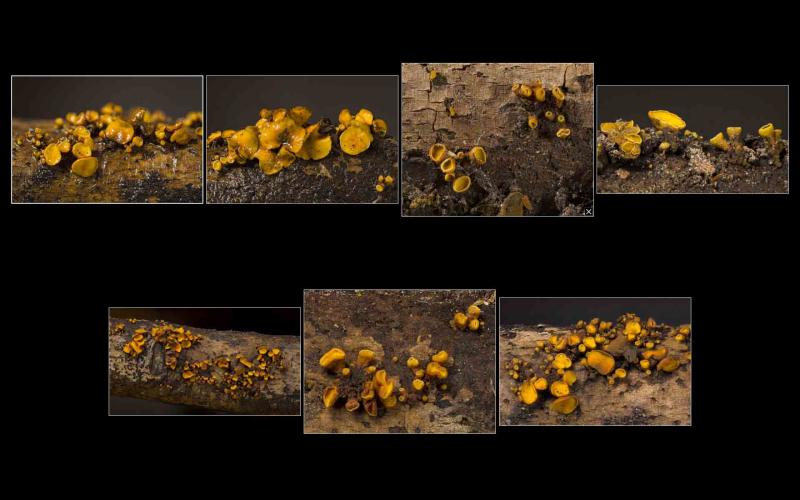
Loc: Plantation Trail, Jean Lafitte National Historical Park and Preserve, Louisiana, USA
Coll: D. Newman, R. Cronce & K. Thorstad
Substrate: on dead, downed, corticate, firm/undecomposed, hardwood stick


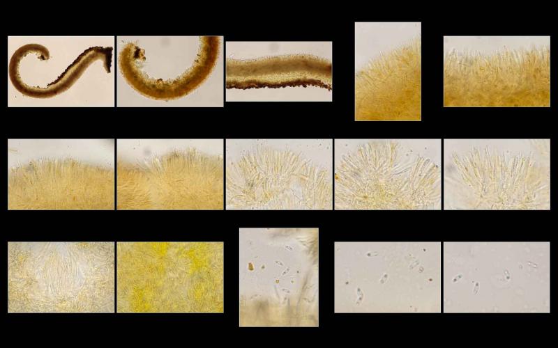
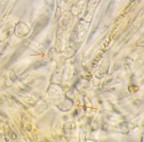
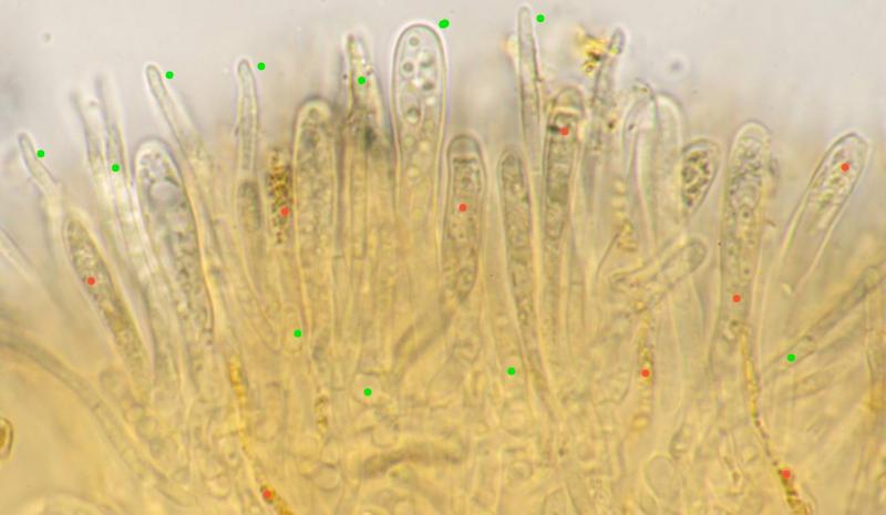
all mounts made in KOH. excipulum composed of irregularly inflated/swollen chains of cells (asexual propagules as in I. irregularis?); context composed of long, slender, even hyphae, possibly embedded in a gelatinous matrix; paraphyses filiform and short, only slightly exceeding asci; both hymenium and abhymenium appear to contain ionomidotic granules/contents; asci biseriate; spores bi-guttulate and (probably) aseptate.
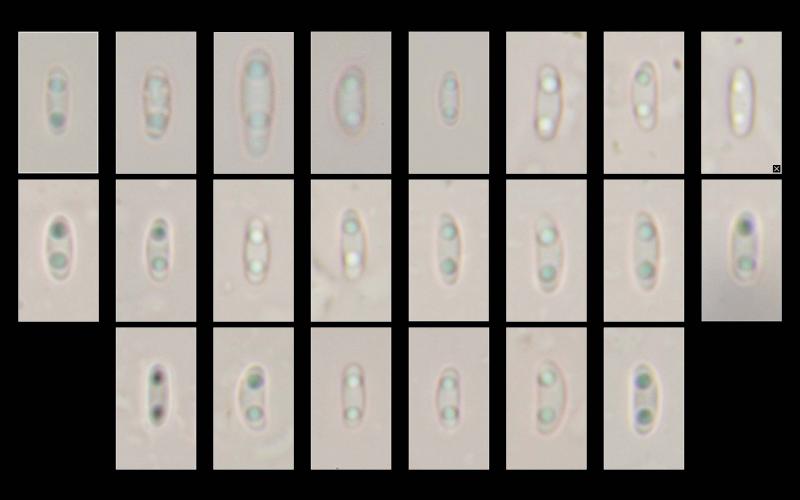


Spores:
(5.1) 5.3 - 6.5 (6.8) × (2.1) 2.2 - 2.6 (2.7) µm
Q = (2) 2.2 - 2.9 (3) ; N = 20
Me = 5.9 × 2.4 µm ; Qe = 2.5


GIF of ionomidotic reaction available at https://inaturalist-open-data.s3.amazonaws.com/photos/347983283/original.gif

  • message #78127
  • message #78127
Hans-Otto Baral, 03-02-2024 10:24
Hans-Otto Baral
Re : Ionomidotis fulvotingens group
Your above link leads to another fungus which is a Strossmayeria as far as I can see.

Without seeing the micros, I can say that this is a Ionomidotis in the current sense, which is microscopically very similar to I. fulvotingens but differs merely in having an ascus apical thickening (in dead asci) and of course by yellow-ochre apothecia.

Its rDNA is very distant, however.

Kadri and I consider this to be the old Cenangium collemoides (Rehm) Rehm.

So far we appear to have only European sequences and I am not sure if they are all the same. An American DNA would be great to compare.

In which genus I. fulvotingens and relatives finally will end up is as yet unclear.


Danny Newman, 03-02-2024 16:34
Danny Newman
Re : Ionomidotis fulvotingens group
Thank you for the prompt and informative reply, and please forgive the link mixup.  The correct one has been added.  Sequencing this has been a priority practically from the moment it was collected.  If there are fresh characters (such as ascus apex thickness compared to dry/dead tissue) which would be important to record, I am returning to the collection site today and can keep an eye out for more.
Hans-Otto Baral, 03-02-2024 17:15
Hans-Otto Baral
Re : Ionomidotis fulvotingens group
The ascus photos are splendid, they show well the thickening, most pronounced in immature asci. But I would appreciate living spores, and also a lok at the ascus base for croziers. Spore measurements of course. The diameter of the apos is up to ?5 mm?
Danny Newman, 05-02-2024 02:16
Danny Newman
Re : Ionomidotis fulvotingens group
Zotto,

Please find two thumbnail plates attached showingn the requested information.  Full resolution versions of these images are available on the iNaturalist observation.  Once I replace my lost stage micrometer I will have measurements to provide. In the meantime, croziers are something of a challenge for me to determine the presence or absence of with certainty.  I have tried to clearly illustrate many ascus bases so that someone with a keener eye (ie: yourself) might make that determination.

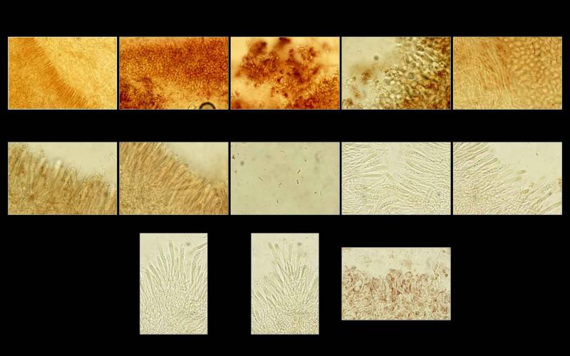
Two new things I've noticed in these recent mounts: 1: the paraphyses are multi-septate, and 2: many contain bright yellow, refractive contents.  I had observed it rather vaguely in the hymenium before, but not inside the paraphyses.

I'm must look further into this Cenangium collemoides (Rehm) Rehm which you reference.  I am equally eager to have this species sequenced, along with a wide variety of other charismatic but neglected North American ascomycetes.  Even in 2024, these fungi often evade attention, collection and study by most people here.
  • message #78157
  • message #78157
Kadri Pärtel, 05-02-2024 07:47
Re : Ionomidotis fulvotingens group
Dear Danny, 

Thank you for sharing these high-value images. I would like to sequence this specimen, it would help to clear the mysterious taxonomy in this group...

If you like to send a piece of specimen to our lab, please, use this address:

Kadri Pärtel
Chair of Mycology
Department of Botany
Institute of Ecology and Earth Sciences
University of Tartu
Oecologicum
J. Liivi St. 2
50409 Tartu
Estonia
kadri.partel@ut.ee
Phone +372 5226179

THANK YOU FOR COOPERATION!
Hans-Otto Baral, 05-02-2024 10:09
Hans-Otto Baral
Re : Ionomidotis fulvotingens group
Very good images! And very instructive for explaining vital taxonomy.

1. croziers are clearly seen. The V-shaped ascus base is necessary to see, it represents the two septa at the connection to the crozier cell.

2. Paraphyses are always septate, but in killing agents they often get invisible (unlke in multiguttulate ascospores where septa are much better seen in the dead state).

3. The yellow contents are only present in dead paraphysis cells.

green = living, red = dead

So far you did not give us any measurements. Full resolution is nice, but a scale would be important. Apo size and spore size at least.

I did not remember that we have already an American sequence. KL348B (coll. Jason Karakehian) falls to European sequences (not sure how close).
  • message #78159
  • message #78159
Khomenko Igor, 05-02-2024 17:22
Khomenko Igor
Re : Ionomidotis fulvotingens group
I also found this species 3 times around Ottawa, ON Canada on hardwood, Acer saccharum branch and ?Tilia log, so it is not that uncommon here.
https://inaturalist.org/observations?place_id=any&q=Ionomidotis%20cf.%20fulvotingens&search_on=tags&user_id=ikhom&iconic_taxa=Fungi
I kept the first two speciments.

I wonder about I. fulvotingens that grows on Pinus strobus. Has it been ever sequenced? I found a specimen in Feb 2020 and I kept it. (https://inaturalist.org/observations/38581250)

Kadri Pärtel, 09-02-2024 08:01
Re : Ionomidotis fulvotingens group
Dear Igor,
Thank you for sharing your found.

I checked my data: all specimens we have studied and sequenced are on hardwood. It would be intresting to add one from a conifer.

Best regards,
Kadri
Danny Newman, 19-02-2024 01:16
Danny Newman
Re : Ionomidotis fulvotingens group
Spores:
(5.1) 5.3 - 6.5 (6.8) × (2.1) 2.2 - 2.6 (2.7) µm
Q = (2) 2.2 - 2.9 (3) ; N = 20
Me = 5.9 × 2.4 µm ; Qe = 2.5
Hans-Otto Baral, 19-02-2024 09:01
Hans-Otto Baral
Re : Ionomidotis fulvotingens group
This is exclusively in water?
Danny Newman, 19-02-2024 09:19
Danny Newman
Re : Ionomidotis fulvotingens group
yes, tap water