07-06-2026 15:10
 William Slosse
Hello everyone,On 05-06-26, I found following asco
07-06-2026 12:09
 François Freléchoux
Bonjour, Voici une brève description de ce qui m
05-06-2026 11:02
Thomas Læssøehttps://svampe.databasen.org/observations/10596691
07-06-2026 12:43
Steve ClementsBojour. This was a strange find on a stick on my
12-07-2015 00:05
 Nedim Jukic
This one from the same locality as the previous on
06-06-2026 17:44
Steve ClementsBonjour, This disco was on planed wood 3 x 1.5 cm
14-08-2016 23:15
 Alex Akulov
Dear friendsCan you help me to find the descriptio

04-06-2026 11:36
Gernot FriebesHi,found on Vaccinium myrtillus.Asci: IKI –, 8-s
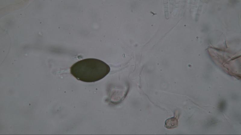
Schizothecium tetrasporum
Joop van der Lee,
07-04-2019 10:18
 Found on deer dung,
The fact that pedicel and upper cauda are covered with a gelatinous layer does not heve much attantion in documentation. In my opinion it is best described in "Coplrophilous fungi in New Zealand. I. Podospora species with swollen agglutinated perithecial hairs" Mycologia 87(3) 1995 pp. 375-396. Under Podospora tetraspora page 393.
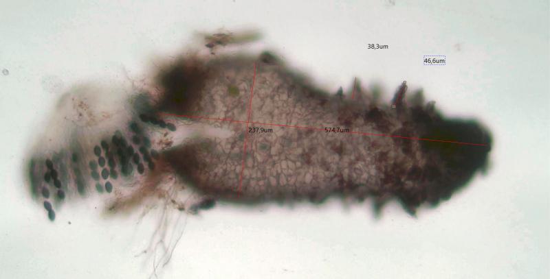
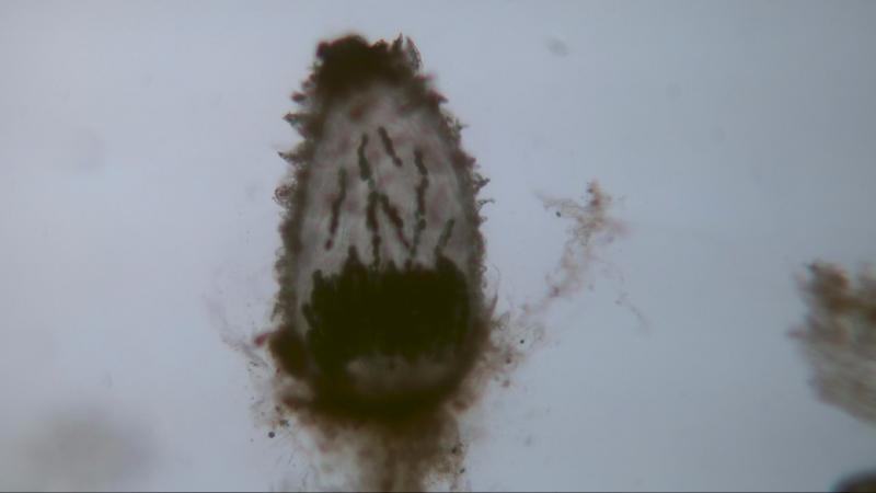
Perithecia: 574x237 um; neck and area just below the neck covered width short hairs; one third of the body covered with agglutinated hairs 38-46 um.
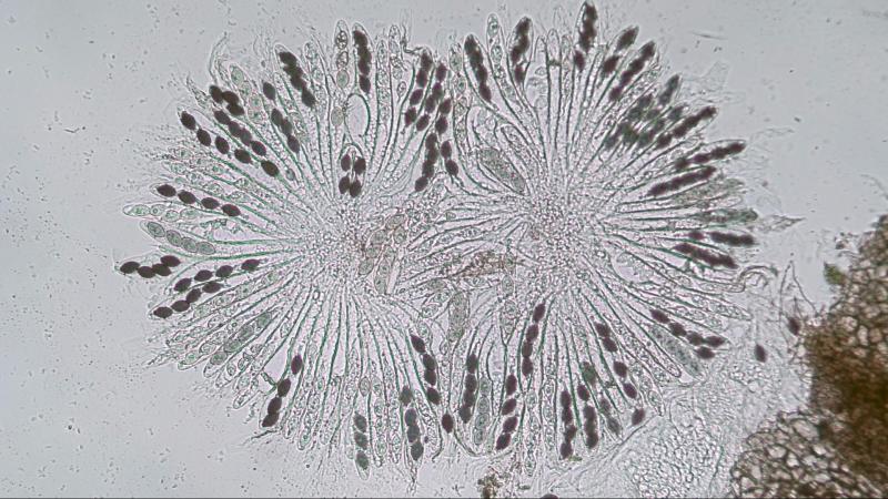
Asci: 81-spored; 196-204x22-24 um
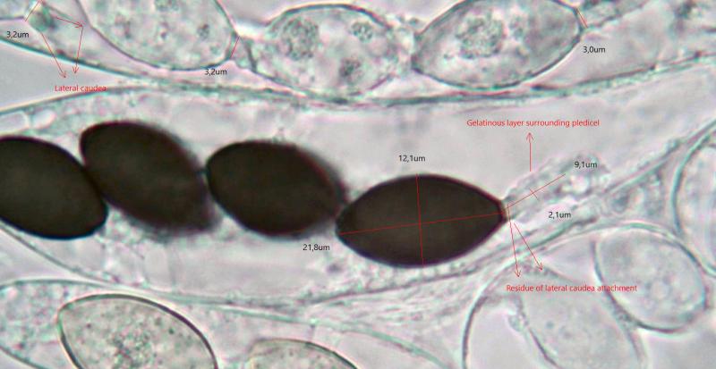
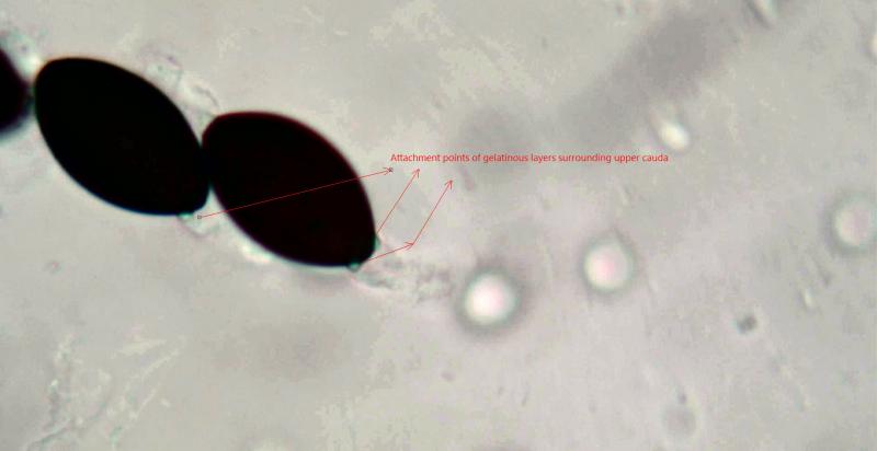
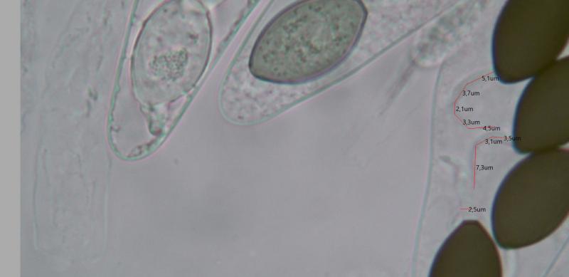
Spores: 21.8x12.1 um; pedicel 8.2-9.1x2.1-2.5 um, at least two lateral cauda at the base of the pedicel, pedicel covered with a gelatinous layer; upper cauda 13-15x1.1-1.6 um, cauda covered with gelatinous layer originating on both sides of the germ pore.
Residue of lateral caudea on base of the pedicel is visible by means of black or lighted spots.
The same is visible with the gelatinous layer around the upper cauda originating just beside the germ pore.
It is exeptional to see that the width of the pedicel is greater with immature spores than with mature spores. 3.2 um against 2.3 um.
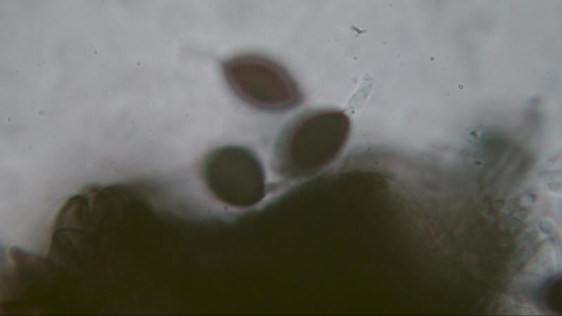
Photos 7-9 are from a S. tetrasporum with a smaller spore size 15.3-18x8.2-9.2 um and found on rabbit dung.
Perithecia: 398x215 um.
Photo 8 shows the gelatinous layer around the pedicel.
Photo 9 shows the gelatinous layer around the upper cauda originating on both sides of the germ pore.
Joop