
27-04-2026 20:52
 Lothar Krieglsteiner
Found on hanging tiwg of Olea europaea in dried-ou
27-04-2026 18:48
Tony MoverleyCollected 23rd April 2026, Norfolk, EnglandSwarms
27-04-2026 17:41
Lothar Krieglsteiner
.. Algarve, same leaf than the last post. The con
27-04-2026 18:05
Lothar Krieglsteiner
... still attached at standing tree. The green con
27-04-2026 17:16
Lothar Krieglsteiner
.. Algarve, moist lying.The conidiomata look like
27-04-2026 12:54
Steve ClementsBonjour. Ce petit champignon blanc résupiné et
27-04-2026 09:59
Pauline. PennaBonjour Can anyone advise me on these pycnidia fo

22-04-2026 20:54
 Enrique Rubio
Hi to everybody.This Pyrenopeziza grew in moist le
Hyphodiscus or Cistella?
Uwe Lindemann,
14-11-2009 01:37
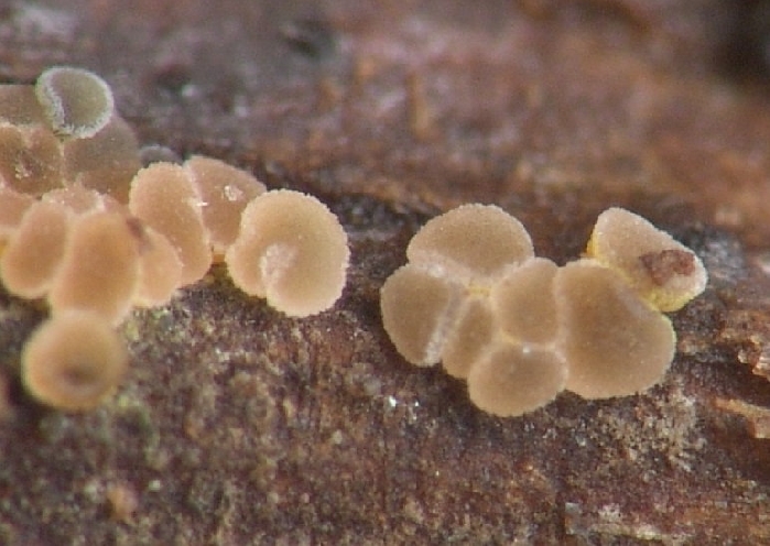
 Hello Forum,
I recently found a little discomycet which I can't determine.
Macroscopic features:
Apothecia gregarious, sessile; Disc 0,1-0,3 mm, young pale beige, old greyish and brownish with a greenish tint (see photos)
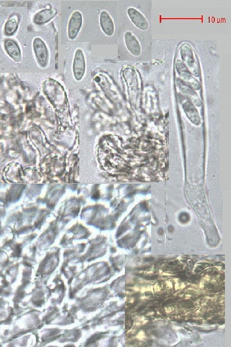
Microscopic features:
Asci arising from croziers, 8spored, 45-55 x 6-7 µm, mostly biserate, IKI- and MLZ- !
Spores ellipsoid, hyaline, aseptate, with two polar guttules and/or some small guttules, 6-7,3(9) x 2,7-3(3,5) µm
Paraphyses filiform, 2 µm wide, sometimes slightly thickened at the apex
Hairs hyaline, thin to thick walled, coarsely warted, up to 5 µm wide; in some areas of the excipulum they are yellowish caused by a yellowish "exudate" which covered the outside of the excipulum.
Ectale excipulum: textura prismatica, mostly getalinous
It grows on Rubus.
My first guess is Hyphodiscus hymeniophilus but the size of the spores and the substrate are not fitting well.
What do you think about it? I would be very happy if you can help me!
Best Uwe
Uwe Lindemann,
14-11-2009 01:38
Re:Hyphodiscus or Cistella?
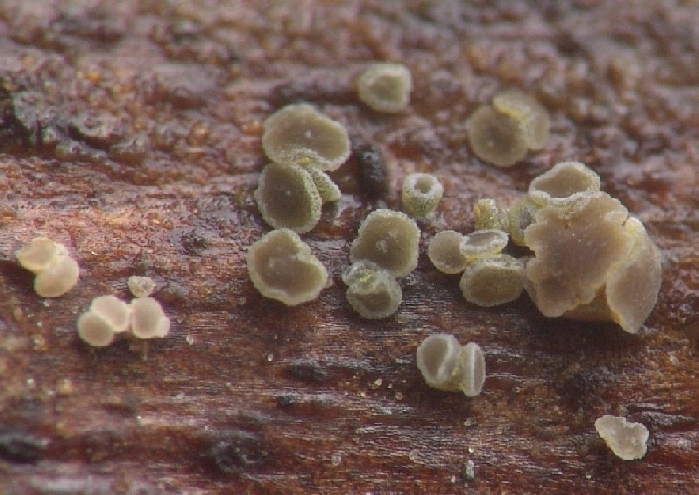
Makro2
Uwe Lindemann,
14-11-2009 01:38
Re:Hyphodiscus or Cistella?
Mikro
Hans-Otto Baral,
14-11-2009 10:05

Re:Hyphodiscus or Cistella?
Hi Uwe
this is 100% a Hyphodiscus, because of brownish colour and yellow exudate (which is onoly inconsistently present in Hyphodiscus but never in Cistella). The strongly gelatinized excipulum is also typica.
I would also say H. hymeniophilus, spore size , shape and gutules would fit quite well. But if you are right that the asci are inamyloid (in Lugol) then this would be the first time. I suggest you try pretretment with KOH and then test IKI or MLZ again. Hyphodisci are often hemiamyloid, so a clear blue is only obtained after KOH, and the IKI-red reaction sometimes overlooked.
My only collection with sch elongate spores and inamyloid asci was on Alnus viridis. Regrettably it was in bad shape, so I had no free spores:
H. cf. hymeniophilus. Asci IKI-/K+IKI-. Sp. *5-6(-8) x 2-2.2 µm, Ölm.1: 2-3 minute LBs in each end.
Was it Rubus idaeus or fruticosus-group?
Zotto
Uwe Lindemann,
14-11-2009 11:02
Re:Hyphodiscus or Cistella?
Hi Zotto,
yes, the porus is hemiamyloid! After the pretreatment with KOH I can see the IKI-reaction (blue) very well. Thank you for your help!
An the substrate is of the rubus fruticosus-group (Brombeere).
All my best,
Uwe
yes, the porus is hemiamyloid! After the pretreatment with KOH I can see the IKI-reaction (blue) very well. Thank you for your help!
An the substrate is of the rubus fruticosus-group (Brombeere).
All my best,
Uwe