
08-04-2026 10:39
FRANCIS FOUCHIERBonjour , je recherche en pdf cet article: KORF R

06-04-2026 21:36
 Viktorie Halasu
Hello, could anyone please send me the article wi
06-04-2026 19:40
 David Gibbs
Help with this one much appreciated, on rotting Fa
06-04-2026 11:07
Louis DENYBonjour forum, Trouvé sur bois de feuillu très d
06-04-2026 16:24
Juuso ÄikäsLast Tuesday I found some tiny white Helotiales gr
05-04-2026 20:40
 Robin Isaksson
Hi!Found i Japan on bark of Abies sp. Spores 35-4
Discomycete on leaves
Josep Torres,
02-10-2025 09:16
 Hello.
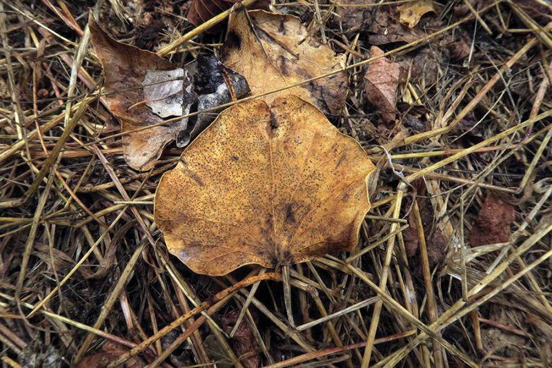
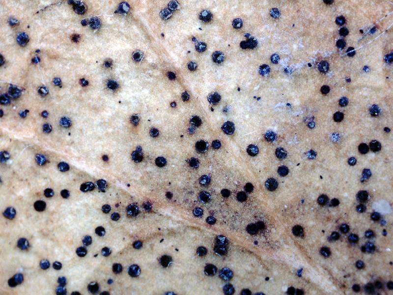
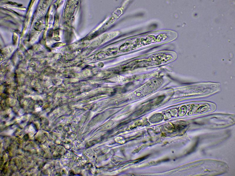
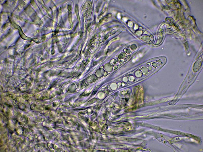
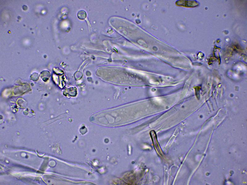
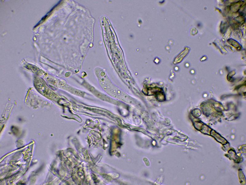
Some tiny apothecia sprouting in a scattered but abundant manner on decaying leaves in the riverine forest under poplars (Populus).
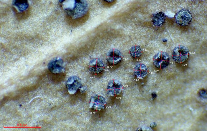
The apothecia are blackish, only 0.2 to 0.3 mm in diameter, without a clearly distinct margin.
Under microscopy, I couldn't see any structures that could correspond to marginal hyphae; if they were present, they would be very few.
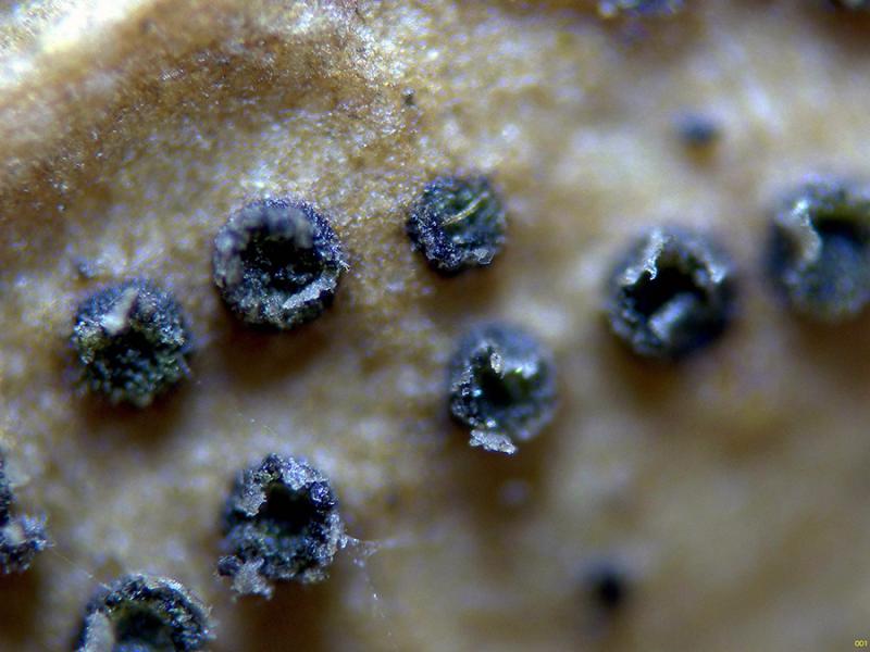
The excipule is very sparse, globose in texture, angular.
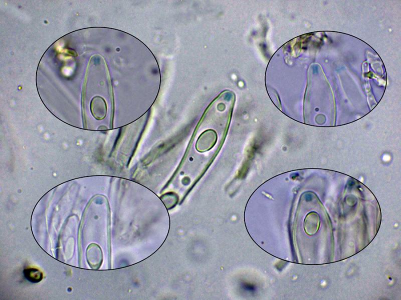
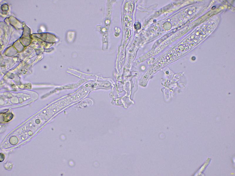
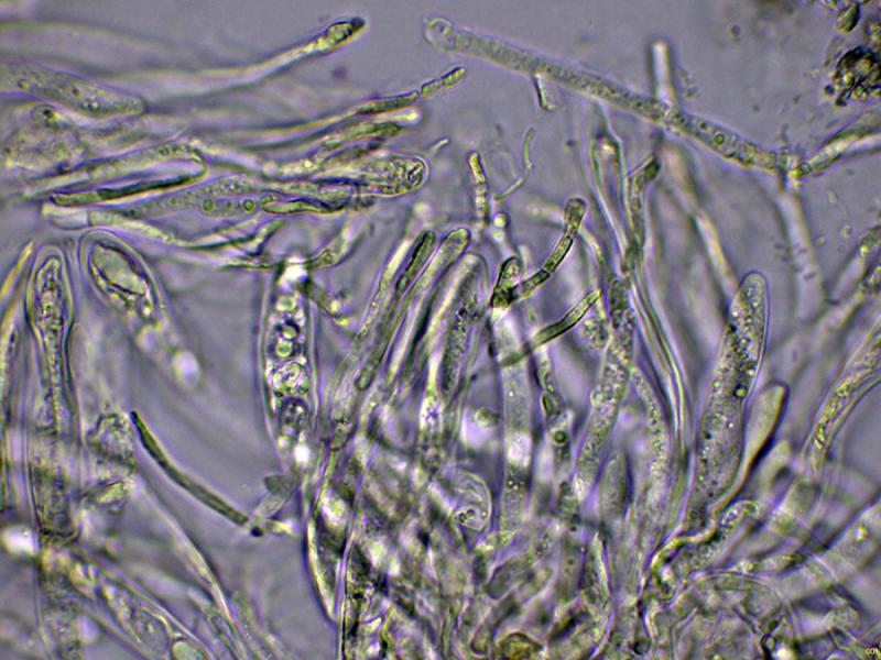
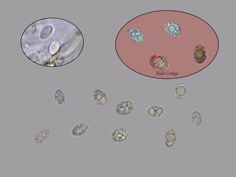
Octosporic asci, uniseriate, thick-walled, apparently without clear uncinules at their base, with measurements in water of (50.1) 56.1 - 70.8 (79.2) × (8.6) 9.3 - 11.4 (11.6) µm., Me = 64.9 × 10.4 µm., and with an amyloid apical apparatus to Melzer, cylindrical to slightly widened at its upper part, with measurements of 2.6 - 3.1 (3.4) × (2.1) 2.12 - 2.29 (2.3) µm. As a curiosity, I could not observe this apical apparatus in the preparations with Lugol. The paraphyses are filiform, septate, with a slight widening at the apex, protruding above the level of the asci.
Ellipsoidal ascospores, with one or more lipid droplets inside, inamyloid in the Melzer test, and with measurements in water of (8.2) 8.8 - 11.2 (11.4) × (5.4) 5.9 - 8 (8.4) µm, Me = 10 × 6.8 µm; Qe = 1.5.
Based on microscopy and the amyloid reaction of its apical apparatus, the closest I could find would be Drepanopeziza, but its appearance doesn't match.
Any feedback from you would be welcome.
Thank you in advance.
Best regards.
Josep Torres,
02-10-2025 09:18
Re : Discomycete on leaves
The rest of the images.
Hans-Otto Baral,
02-10-2025 09:23

Re : Discomycete on leaves
You have Trochila craterium on Hedera leaves I think, unless these are not Ilex leaves.
Josep Torres,
02-10-2025 09:26
Re : Discomycete on leaves
Thank you, Zotto, for your prompt response. Ivy (Hedera helix) is abundant in the area, and it's most likely the Trochila craterium you're suggesting.
Best regards.
Best regards.