
27-04-2026 17:41
 Lothar Krieglsteiner
.. Algarve, same leaf than the last post. The con
27-04-2026 17:20
Lothar Krieglsteiner
.. Algarve, moist lying.The conidiomata look like
27-04-2026 17:17
Lothar Krieglsteiner
.. Algarve, moist lying.The conidiomata look like
27-04-2026 17:16
Lothar Krieglsteiner
.. Algarve, moist lying.The conidiomata look like
27-04-2026 17:16
Lothar Krieglsteiner
.. Algarve, moist lying.The conidiomata look like
27-04-2026 12:54
Steve ClementsBonjour. Ce petit champignon blanc rÃĐsupinÃĐ et
27-04-2026 09:59
Pauline. PennaBonjour Can anyone advise me on these pycnidia fo

22-04-2026 20:54
 Enrique Rubio
Hi to everybody.This Pyrenopeziza grew in moist le
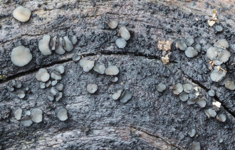
 Good afternoon
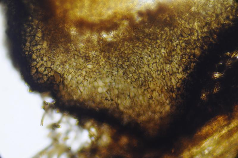
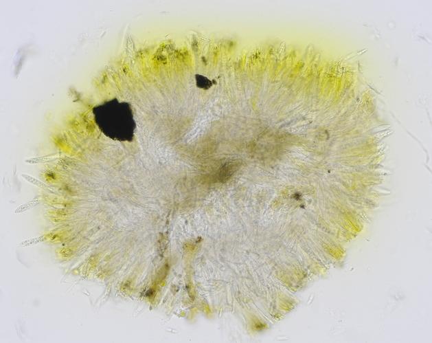
This 1-2 mm Mollisia was growing on Cistus ladanifer wood and I can't find any that look like it.
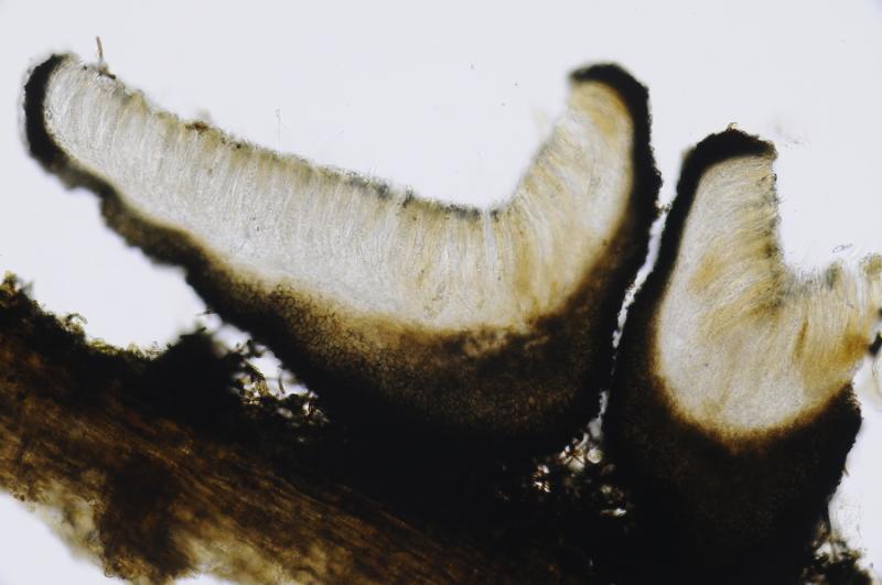
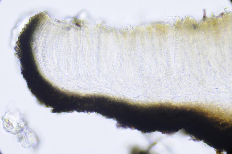
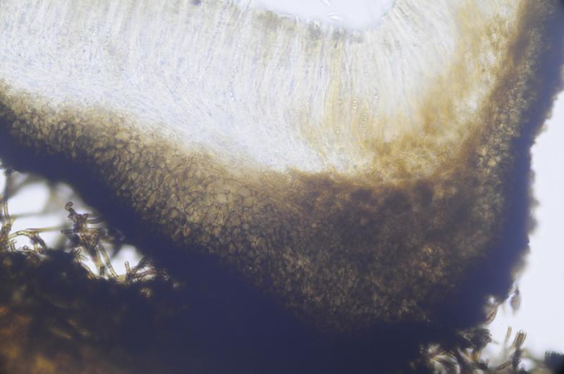
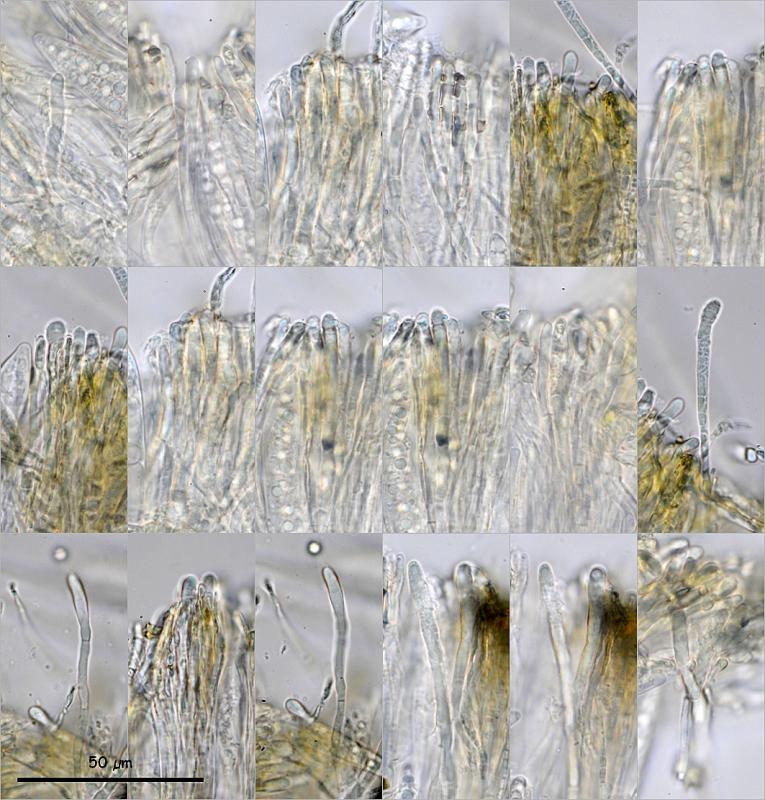
Ectal excipulum brown with textura prismatica to angularis, with claviform terminal cells. Medullary excipulum hyaline with textura prismatica to intricata.
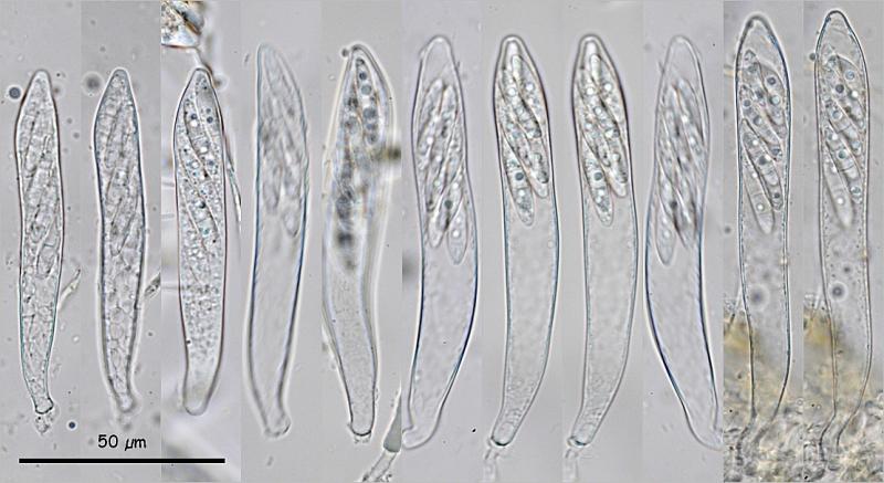
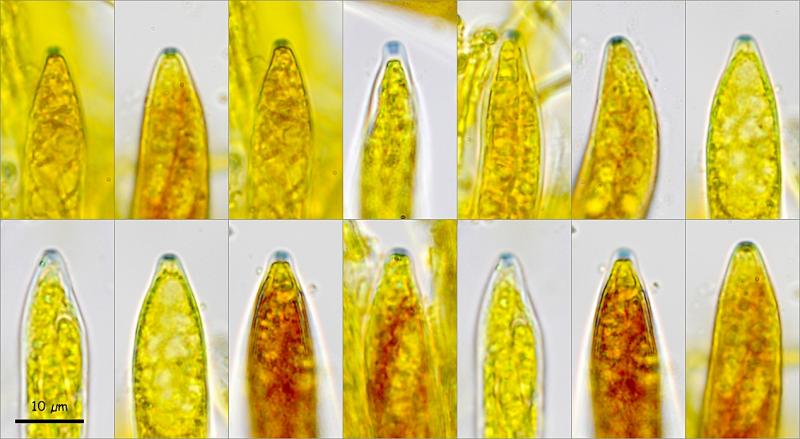
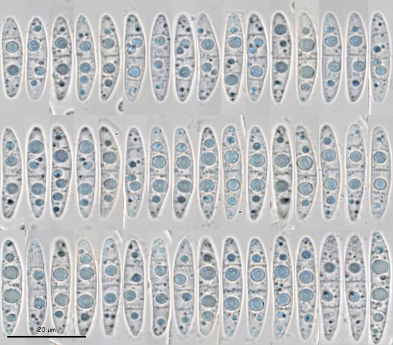
Asci octosporic, biseriate, with croziers, IKI+. Spores cylindrical-fusiform, usually asymmetric, with one side flatter and the other more convex, up to 3 septa, even inside the asci, with 1 large LB in each cell, at least in the 2 central cells. Paraphyses cylindrical, somewhat thickened at apex, with a large VB occupying the entire terminal cell, KOH+ intense yellow, with brown epithecium.
Asci: (76) 83.9 - 106.5 (111.9) Ã (10.3) 11.6 - 13.3 (13.9) Âĩm, Me = 95.5 Ã 12.3 Âĩm
Spores: (20.3) 21.8 - 24.9 (28.6) Ã (4.1) 4.6 - 5.2 (5.6) Âĩm, Q = (3.9) 4.4 - 5.3 (6) ; N = 66; Me = 23.6 Ã 4.9 Âĩm ; Qe = 4.8
I can find no Mollisia, Pyrenopeziza, Belonidium or similar with these spores. The closest spores I can find are those of M. ventosa. The spores of M. pilosa also have 3 septa, but they are larger and the asci are rr. I have also seen a picture of M. minutella with similar spores, but not the same.
Thanks for your help.
Miguel Ã. Ribes
Thanks

Very kind.Â
Best wishes.