

06-04-2026 21:36
 Viktorie Halasu
Hello, could anyone please send me the article wi
06-04-2026 19:40
 David Gibbs
Help with this one much appreciated, on rotting Fa
06-04-2026 11:07
Louis DENYBonjour forum, Trouvé sur bois de feuillu très d
06-04-2026 16:24
Juuso ÄikäsLast Tuesday I found some tiny white Helotiales gr
05-04-2026 20:40
 Robin Isaksson
Hi!Found i Japan on bark of Abies sp. Spores 35-4
06-04-2026 08:15
 Lothar Krieglsteiner
some days ago, on the lower surface of leaf of Que
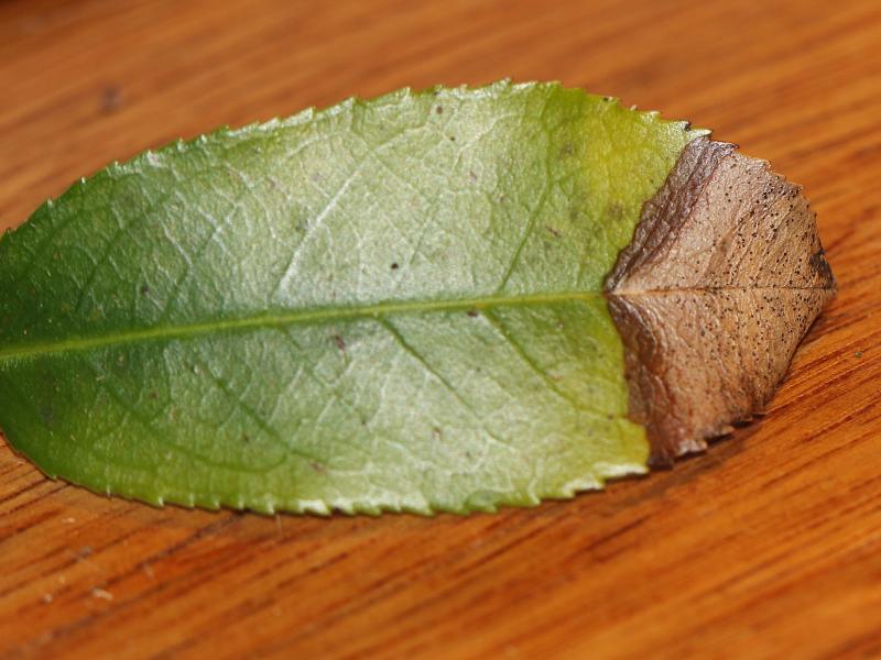
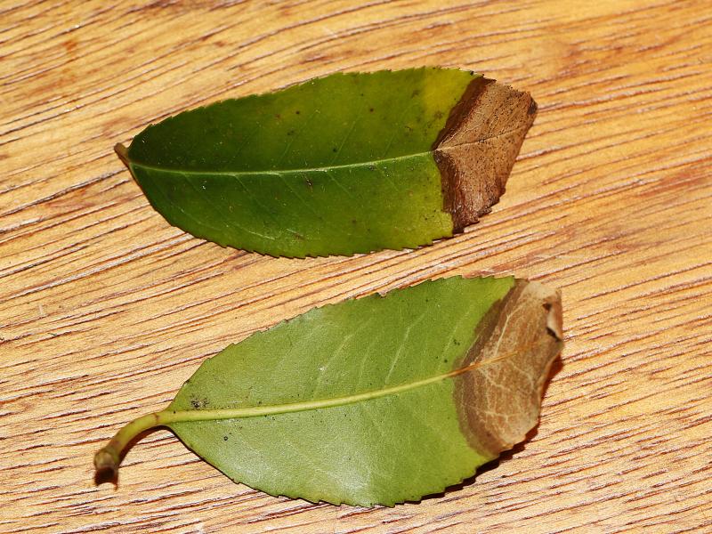
 On 6 february 2025 i stumbled upon a leafspot on Prunus lusitanica in Aerdenhout (The Netherlands). Can someone confirm that it is indeed Coleophoma prunicola? or maybe something completly different?
 For more photo's see: https://waarneming.nl/observation/338565267/
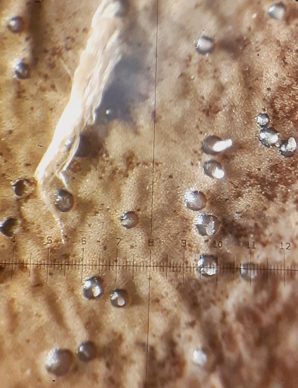
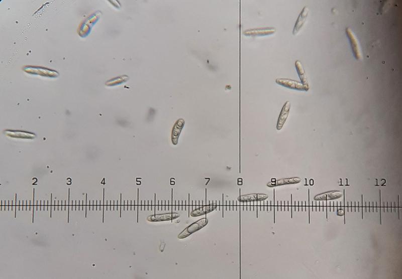
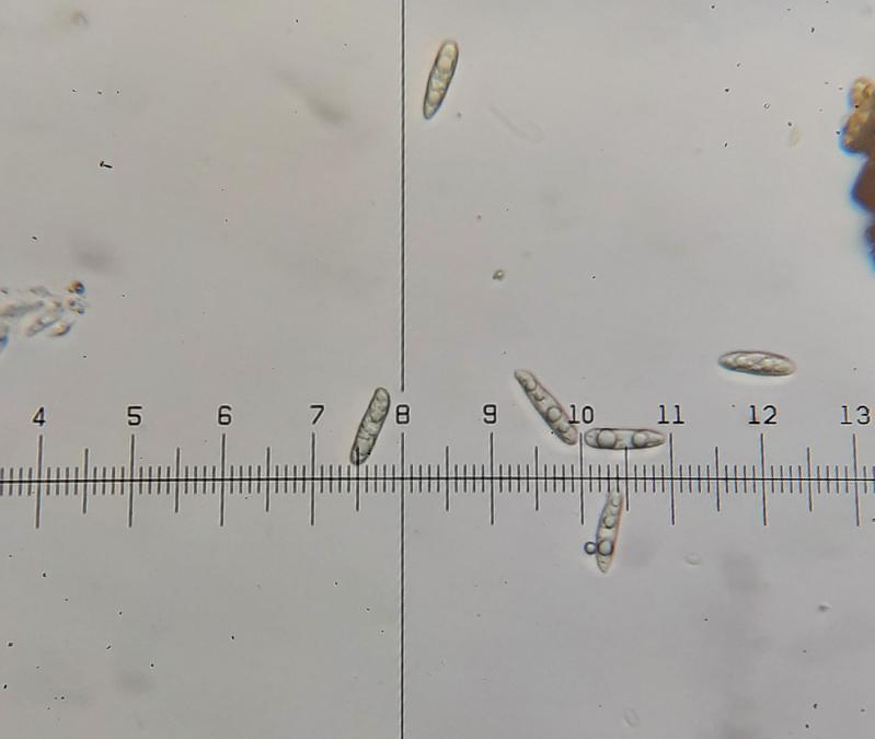
Conidiomate pycnidia, ca. 150- 200 µm diameter (N=10).
Conidia holoblastic, hyaline, aseptate, cylindrical, apex obtuse, base truncate, thin-walled, smooth, guttulate with several large guttules.
Conidia 20,8 - 23,4 - 26 ?m × 4-5,2 ?m (N=25)
400 X mafnification: 1 div. = 2,6 ?m
Following the Key for Coleophoma in the article: 'Reinstatement of Coleonaema for Coleophoma oleae and notes on Coleophoma' it should be Coleophoma prunicola
(Duan, J.X., Wu, W.P. and Liu, X.Z. (2007). Reinstatement of Coleonaema for Coleophoma oleae and notes on Coleophoma. Fungal Diversity 26: 187-204.) https://www.researchgate.net/publication/237440307_Reinstatement_of_Coleonaema_for_Coleophoma_oleae_and_notes_on_Coleophoma

To know this you should see the conidiogenous cells and compare them with those in the article.
Best wishes
Angel
Thanks for your comment. I can try another time to see the conidiogenous cells and compare them with those in the article. I already tried once but failed to see the conidiogenous cells sadly.
Kind regards,
Jorian