12-11-2019 10:32
 Miguel Ángel Ribes
Hi againExactly at the same place than my previous
25-12-2019 17:54
Valencia Lopez Francisco JavierHola a todos/asEstas supuestas pezizas estaban en
28-07-2011 18:31
 Alex Akulov
Dear FriendsToday I made the pdf file of Velenovsk
12-07-2015 00:05
 Nedim Jukic
This one from the same locality as the previous on


30-05-2026 21:12
Philippe PELLICIERSur branche de mélèze (Larix) près de la neige,
31-05-2026 10:35
Hulda Caroline HolteHello,I collected this species growing on a rather
25-05-2026 16:35
 Bernard CLESSE
Bonjour à toutes et tous,J'ai trouvé récemment,
29-05-2026 15:35
daniel FERREBonjour à tous,Je voudrais votre aide pour cette
Thanks in advance!
Bharati

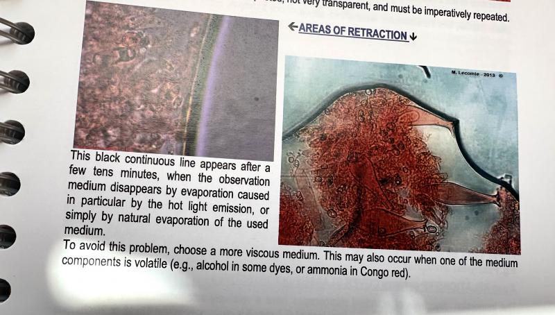
>"this is a very common error, apparently impossible to erase. Not less mature but alive!
>You have plenty of ejected living spores, which are fully mature, as I defined the term mature (1992).
>"It seems to me that the mature spores are only 1-septate and get 3-septate only when overmature. But >this you can find out when testing the numerous ejected spores with MLZ."
Will do.
>"I am not sure why your first micro image shows dead elements only, although being in water. I suspect pressure or drying and rewetting´as reason?
>Your ascus measurements sound like referring to dead asci, considering the low width. I see no living ascus in your pics."
Pressure is what I suspect. I will review my current collection of photos (I have more and of different mounts) and better yet make a new set of slides after I read the 1992 paper.
>"The spores are quite large, so L. lonicerae is impossible."
Noted. Based on the Raitviir key I get from 1-->2-->3-->9 which would rule out L. lonicerae – am I missing something again?
Bharati
Doi 10.5943/mycosphere/15/1/25) and copied an account by Peter Johnston which I found interesting:
I did not forget my homework assignment on this collection. :-) Life sometimes interferes with mycological pursuits.
I updated my inaturalist observation to include: an image of the dead spores showing septation (see photo 6; and one showing living asci with those guttulate spores (see photo 7); and relabeled photo 5 to reflect the fact that they are living spores.
Thanks again,
Bharati
All this is very illuminating for me – what structures within spores are what and what is visible in what media - , so some more questions in my ongoing tutorial: :-)
1) What do you make of the septation in the version with KOH?
2) In your Google Drive collection for L. subflavidum
a) How would you interpret the MLZ image embedded in 100x_spo_koh+rc_24038_apomad0420.jpg?
b) In the image titled Lasiobelonium subflavidum cf., 23.VI.94-2a.jpg, (JM Castro 23-3-94), the note below the spore drawing says that 1 septum is clear, 2 (others) are less clear. The next drawing from this series, labeled 2b shows a variation in septa.
Thanks
Bharati
I am attaching a photo of that paragraph from p. 77, in case others don't have a copy.
On pp. 41-42, he includes some options for viscous media including glycerinated water.