
05-04-2026 22:46
 Lothar Krieglsteiner
on wood of Ceratonia, Algarve, 3.4.2026.The color
05-04-2026 20:40
 Robin Isaksson
Hi!Found i Japan on bark of Abies sp. Spores 35-4
31-03-2026 21:18
 Miguel Ángel Ribes
Good evening. oes anyone have the original descrip
31-03-2026 20:57
Stefan BlaserHello everybody, I hope somebody can help me with
26-03-2026 15:31
 Åke Widgren
Hello,I found this one in October last year, on r
31-03-2026 16:20
 Mlcoch Patrik
Hello, Please about help with determination. On
31-03-2026 08:19
 Bernard CLESSE
Bonjour à toutes et tous,Pourriez-vous m'aider à
 • Chloroscypha was suggested to me, macro and habitat agree, presumably imported at some point with the non-native trees.
• C. seaveri is reported on this substrate and the micro seems to fit the current concept, but this sp. originally described in North America on Thuja with smaller spores.
• The host range seems broader than other species in the genus, including North American Cupressoideae and Asian Taxoidiaceae.
• Despite broad morphological similarity this may warrant further investigation.
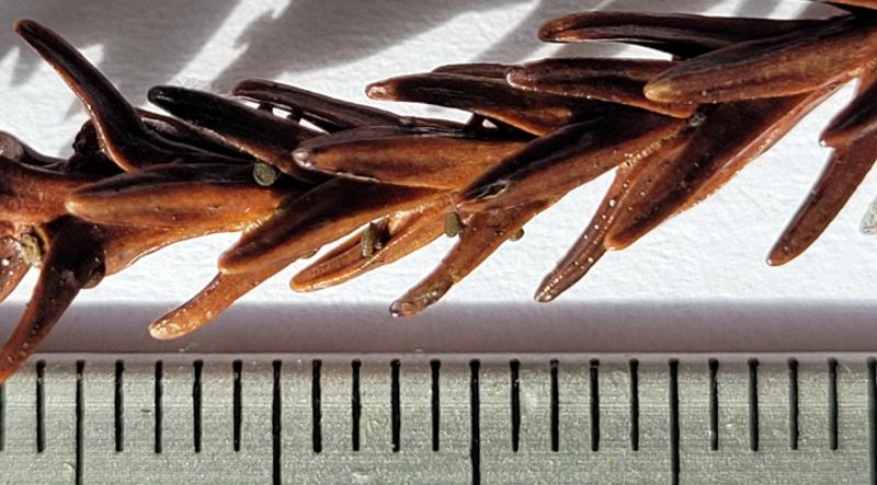
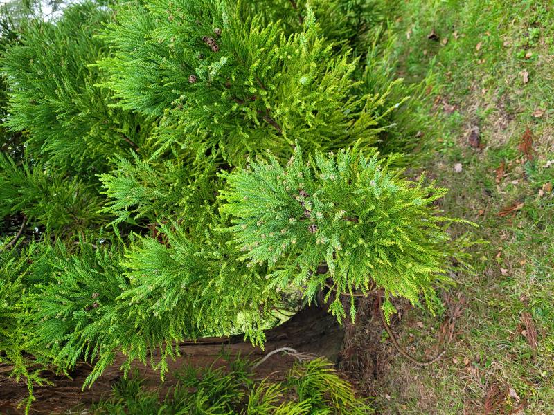
Habitat: On dead needle-like leaves of cultivated Cryptomeria japonica (monotypic), on the faces but not the edges, still attached to a fallen twig, lying on the floor, under the tree, presumably xeric, mature tree, no overt signs of disease, on a lawn, private garden, High Weald, southern England, mid-October, after rain.
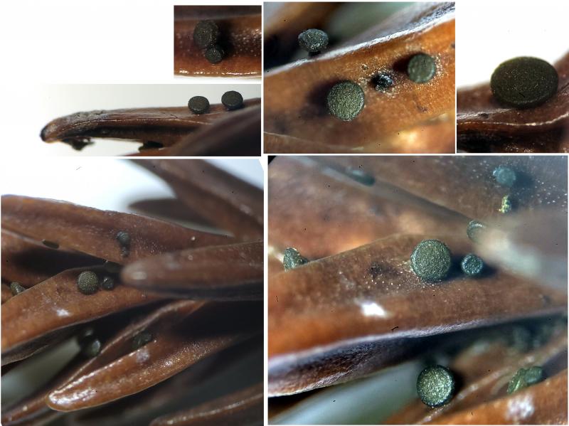
Apothecia: Localised to leaves on one part of the twig, several groups on some leaves, mostly < 0.5 mm diameter, one ~1 mm, dark greenish (olive) to black, apparently darkening and becoming more opaque with age, turbinate-discoid, sessile to short stipe, solitary to 2-3 caespitose, no other evidence on the surface of the needles.
Low magnification: Granular appearance, receptacle slightly to distinctly darker (when fresh), receptacle with whitish lumps at the flanks (druses), margin like receptacle, round, not exceeding the disc, indistinct meeting with disc, disc lighter, more greenish-yellowish, usually plano-concave, more uneven appearance compared to margin, again no further evidence on the needles.
Asci: Clavate, short and broad, with croziers, rings very(!) weakly bb or inamyloid, only seen on flaccid asci, apex rounded when turgid, more acute rounded and eventually hemispherical-subtruncate when flaccid, due to large thickening, 2-2.5-seriate when turgid, spores sometimes almost horizontally oriented but orientation still consistent, contents becoming yellowish after dying, mature pores opening easily with pressure on the asci, then spores discharging serially but ~4 spores remaining in ascus.
Spores: Broadly cylindrical-fusoid, slightly reniform or subcurved in profile view, ends abrupt and acute-rounded to acute, usually heteropolar with the basal end more elongated, filled with many small to medium LBs, looks like some small granular contents too, apparently uninucleate and aseptate, some spores showing dextrinoid contents.
Free living mature spores in water: (29.8) 32.3-35.6 (36.6) × (11.1) 12.0-13.2 µm, Q = (2.5) 2.6 - 2.8 (3.0), n = 12, mean = 33.8 × 12.5 µm, Q mean = 2.7.
Paraphyses: Narrow filiform, not to medium inflated at the apices, sometimes branching at the middle or near the apex, somewhat moniliform with inflation below the septa.
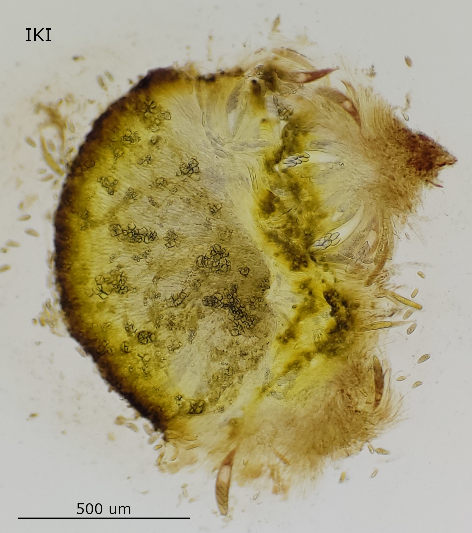
Ectal: Text. prismatica-porecta, long cells, gelatinised, with refractive contents (globose SCBs?), at the base cells wider, with a dextrinoid reaction, internally and at the margin cells narrower, looser, patchy amyloid reaction seen.
Medullary: Not seen clearly, possibly reduced and like inner part of ectal or subhymenium.
Exudate: Brownish to yellowish, browner in concentration, on the surface, receptacle +/- densely covered with smallish druses, individual crystals appear irregularly shaped, dense epithecium with smaller crystals, exudate extending into the hymenium.
 Section-0001.jpeg
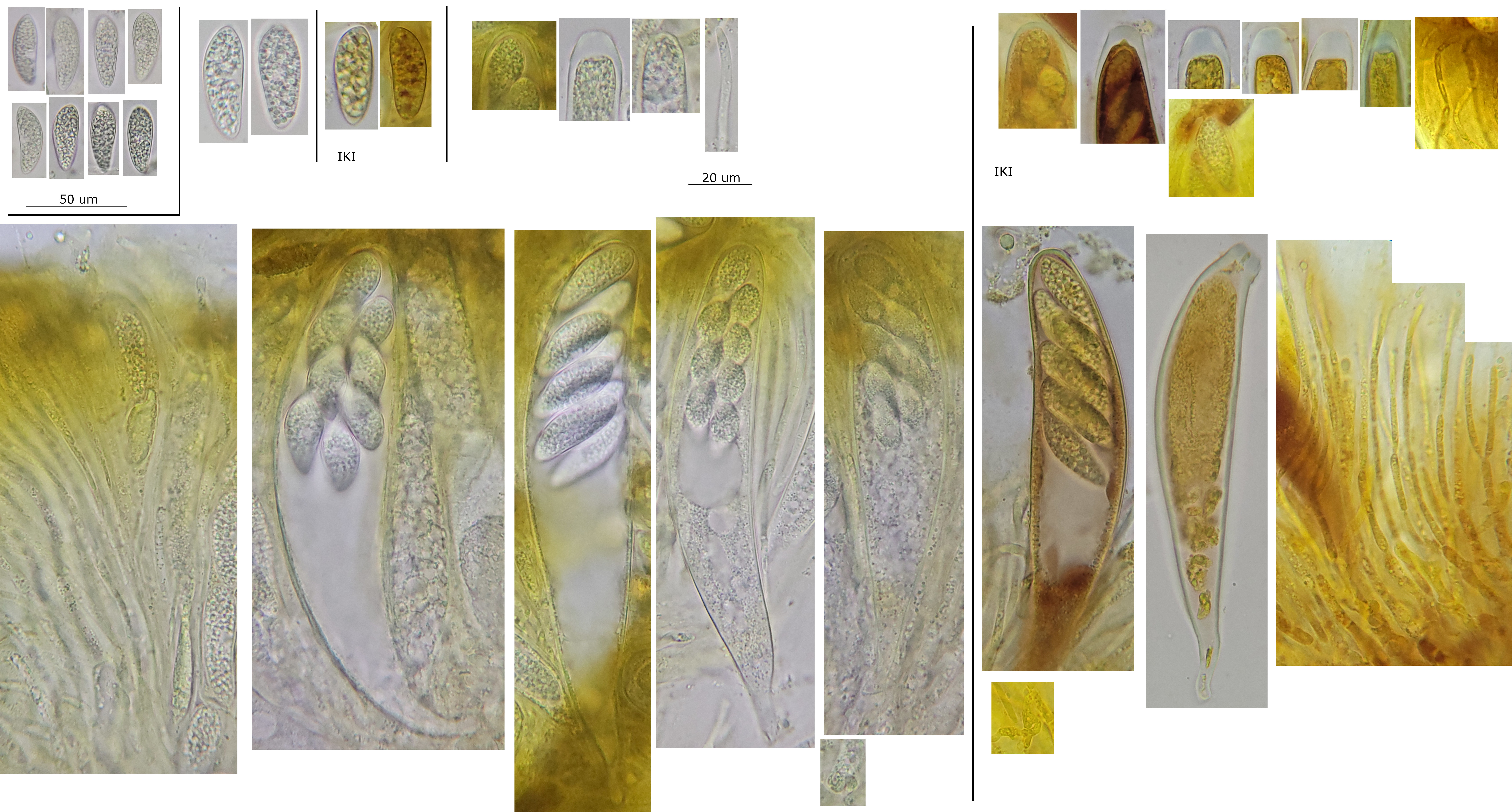 Hymenium-0020.jpeg
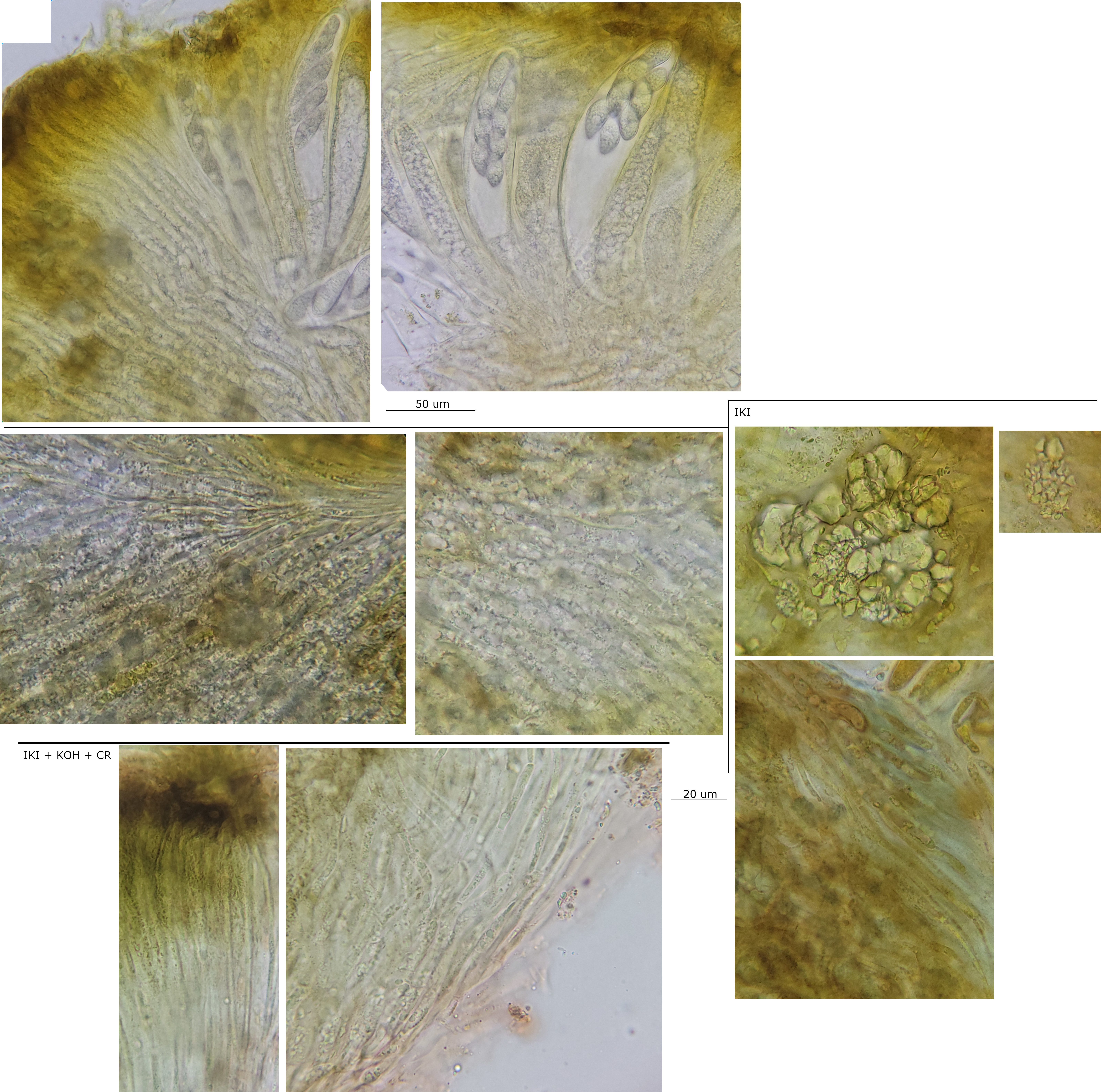 Excipulum-0013.jpeg
From what I read, mostly spore size and host was used by Seaver when he introduced Chloroscypha. He says that C. juniperina (= sabinae) is similar to C. seaveri but with smaller spores, and this does seem to be in agreement with your folder and https://www.centrodeestudiosmicologicosasturianos.org/?p=17124. The macro seems indistinguishable.
When I read the protolog of C. cryptomeriae, I did not feel that a different species was clearly distinguished morphologically (the measurements overlap C. seaveri). Kobayashi reports broad host ranges and his table of morphology and host data reported by various authors seems convincing.