
28-05-2026 16:15
James MitchellHello,Does anyone have the original publication of
28-05-2026 11:06
Thomas Læssøehttps://svampe.databasen.org/observations/10596750
23-05-2026 11:44
 Charles Grapinet
Hello, I am having trouble identifying this copro
25-05-2026 16:44
François BartholomeeusenHi forum members,During an excursion organised by
26-05-2026 21:25
Dirk GerstnerHello everyone, I'm completely stumped by this li
26-05-2026 22:44
Ethan CrensonHi all, I think I have Incrucipulum capitatum her
22-05-2026 14:44
 Lothar Krieglsteiner
in unripe condition citrine yellow, then soon fadi
25-05-2026 16:35
 Bernard CLESSE
Bonjour à toutes et tous,J'ai trouvé récemment,
22-05-2026 13:29
Gernot FriebesHi,I am curious to hear your opinion on this mater
23-05-2026 18:57
Sylvie Le GoffBonjour à tousRécolté sur une branchette de Sal
 Cudoniella tenuispora: Distinctive macro and habitat.
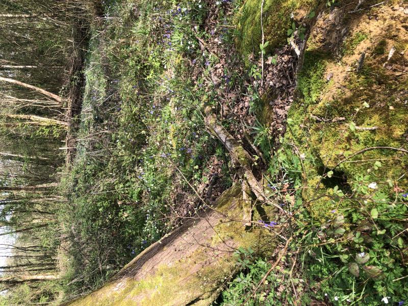
Habitat: On top of a bryophyte in sandy-looking soil, some small unidentified twigs and roots in the soil, at the base of a Quercus robur that was partially uprooted in the past, no bodies of water in the immediate vicinity, mixed deciduous woodland, mostly Betula pendula and Quercus robur, Low Weald, England (south), mid-April, after rain.
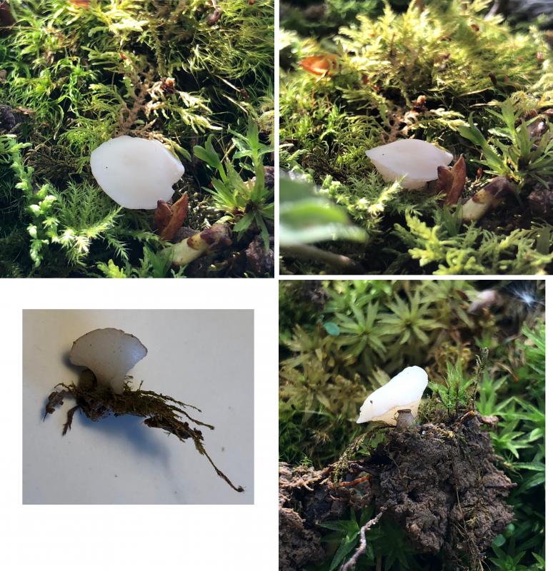
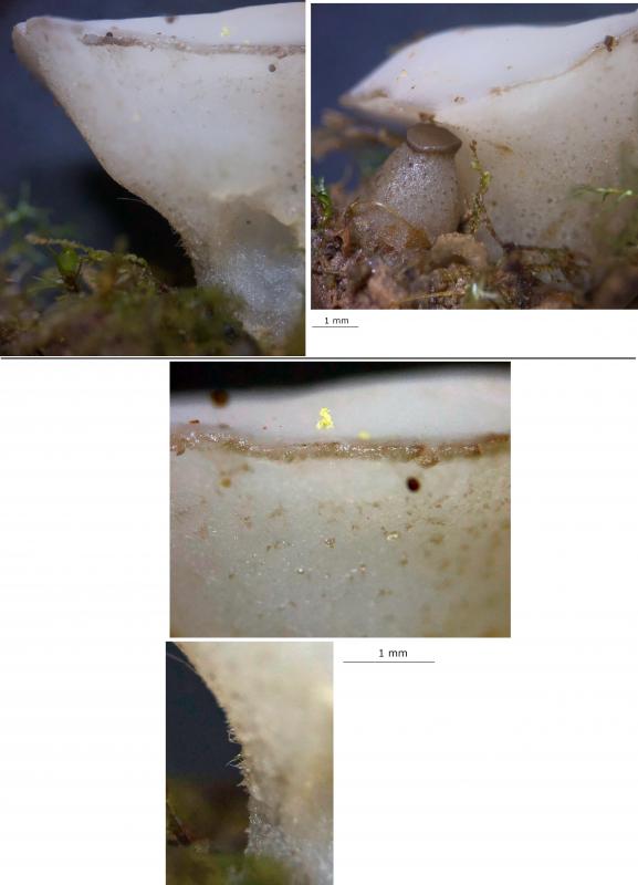
Apothecia: Two together, one mature and the other immature (pin), whitish-cream, opaque, initially ampulliform with a plane disc, then turbinate to funnel-shape; receptacle white, more translucent, greyish-brown spots, texture firm-gelatinous, golden-yellow lumps; stipe thick, initially bulbous and eventually slightly obconical, surface like receptacle, also with short felty hairs starting around the lower flanks, signs of grazing (presumably by a slug or similar); margin distinct, narrow, greyish-brownish, more out-rolled (from hymenium) and undulate in maturity; disc initially greyish-brown like margin then whitish, more opaque, with pruinose appearance, uneven and concave in maturity, many asci discharging at the same time twice in visible whitish clouds of spores when first disturbed.
Associates: Bryophytes on the soil around (possibly several species).
Storage and methods: Stored in a fridge for several days in a damp box, sliced in half and then a central section taken from one side, mounted in water, IKI added to water mount.
IKI: Rings very faintly bb, hymenoscyphus-type when distinct, easier to see in flaccid and juvenile asci, paraphyses dextrinoid (reddish) possibly VBs deforming and contents then yellow, standard reactions otherwise.
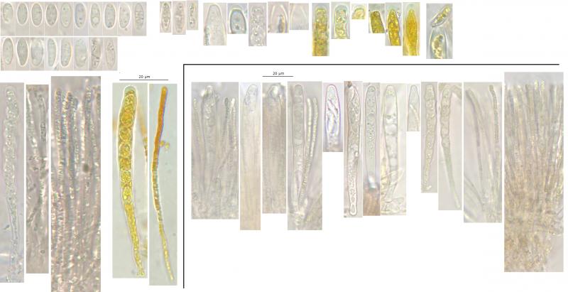
Asci: Hymenoscyphus-type rings, apex rounded to truncate and +/- conical, croziers, 1-2.5-seriate, poricidal opening, often apical thickening still extended, abruptly widening.
- Vital mature: ~82-93 x 8-9.5 µm, pars sporifera ~30-40%, apex rounded and often slightly conical, 1-2.5-seriate, one spore at apex, spores obtusely arranged.
- Dead: ~68-82 x 6-8.5 µm, pars sporifera ~60%, apex truncate and often acute-conical (apex narrower than spores), sometimes appears capitate when fully flaccid, distinct apical thickening < 2.5 um high, usually uniseriate, often irregularly shaped.
Spores: Shape seems to vary between more symmetric ellipsoid-ovoid and longer ellipsoid-fusoid, possibly due to maturity as more of the longer spores appeared over mature, usually not curved but often inequilateral in profile view, often heteropolar with base more attenuated and acute, apparently with (1-?) 2 nuclei, many small to medium and greenish LBs towards each pole, OCI 2-3; when over-mature, cytoplasm detaching from the wall, contents more yellowish.
Vital spores measured in water: (8) 9.3-12.6 (13.1) × (3.6) 4-4.7 (4.9) µm, Q = (1.8) 2.1-3 (3.3), n = 19, mean = 10.9 × 4.4 µm, Q mean = 2.5.
Paraphyses: Cylindrical, 2-2.5 (3) um wide, apex rounded, apparently with 3-4 septa, full of many medium-size greenish guttules – refracting yellowish and more brownish in concentration, central cells emptier, branching around the basal septum.
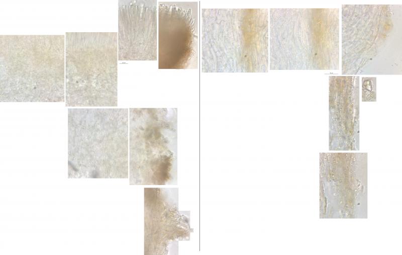
Exudate: Hyaline to greenish crystals on the exciple surface; yellowish to orangish-brown in concentration, clumping, thicker and gelatinising at the margin and parts of the stipe, apparently in the hymenium, patches throughout excipulum and on the surface; stipe surface appears gelatinised at least in places, hyaline.
Subhymenium: Light brown, apparently due to paraphysis bases.
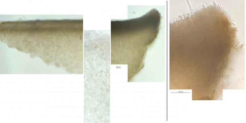
Medullary ex: Light brown, textura intracata-epidermoidea.
Marginal cells: Short and broad, at upper flanks not projecting, at the margin similar to paraphyses.
Ectal ex: Textura intricata, hyaline, greenish VBs in external cells (very active in water); hair-like hyphae projecting, sinuous to convoluted, often with exudate.
Basal attachment: Not viewed.

After your comments, I ran a blast on the sequence data of HB 6927, which I presume you have identified with morphology. The similarity with C. clavus is nice as the characters of this species that I saw in your folder look very similar. It would be good to know how the identification was achieved for the closer matches, especially those without species identification.
It seems interesting to me that species like H. imberbis are not there, you have this in the same folder, and my observations of these species have some similar characters. Although there are some sequences labelled as other H. sp. and some Cyathicula/Crocicreas sp. within 5-10%. I remember that there could be issues with using ITS alone and maybe there is not enough sequence data to resolve these relationships.
In the 2019 phylogeny paper, I read that Cudoniella is in the Tricladium subclade (Tricladiaceae), which appears basal to the Heliotaceae, and I realised Tricladium is traditionally an anamorphic genus. Within 5% there are two sequences labelled as the anamorph Dendrospora tenella, and Dendrospora is mentioned in the Seifert description of Tricladium. I guess D. tenella is too distant to be an anamorph of C. tenuispora, but seems to be an anamorph of a somewhat related species.
It would be nice to understand how these Heliotaceae s.l. species are related and how the genera split, but it seems there is a little work to do.