
11-06-2026 19:03
 Nicolas VAN VOOREN
Chers membres d'Ascofrance,Le site sera placé en
11-06-2026 19:01
 William Slosse
Hello all,In an attempt to make a culture of a sus

09-06-2026 18:32
Camille MertensSur morceau de roseau immergé 0,5 - 0,7 mm de dia
10-06-2026 12:54
Steve ClementsBonjour encore, Pouvez-vous m'aider, s'il vous pl
10-06-2026 21:16
 François Freléchoux
Bonsoir,Le dernier du jour, en attendant votre avi
10-06-2026 21:07
François Freléchoux
Toutes les tiges de gentianes jaunes de l'an pass�
10-06-2026 13:41
François Freléchoux
Bonjour à nouveau, Voici une trouvaille d'hier.
10-06-2026 11:53
Steve ClementsBonjour, This disco is abundant on dead stems of
Hymenoscyphus sp.
B Shelbourne,
08-01-2024 12:22
 I think this could be Hymenscyphus imberbis (Bull.) Dennis, 1964. Any feedback appreciated.
Macromorphology and VBs in paraphyses suggest Hymenoscyphus, and I have tried to use the unpublished key to Hymenoscyphus s.l. in Belgium by Declercq (2004) and the Hymenoscyphus folders of Baral.
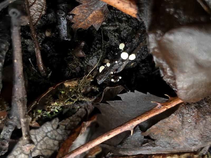
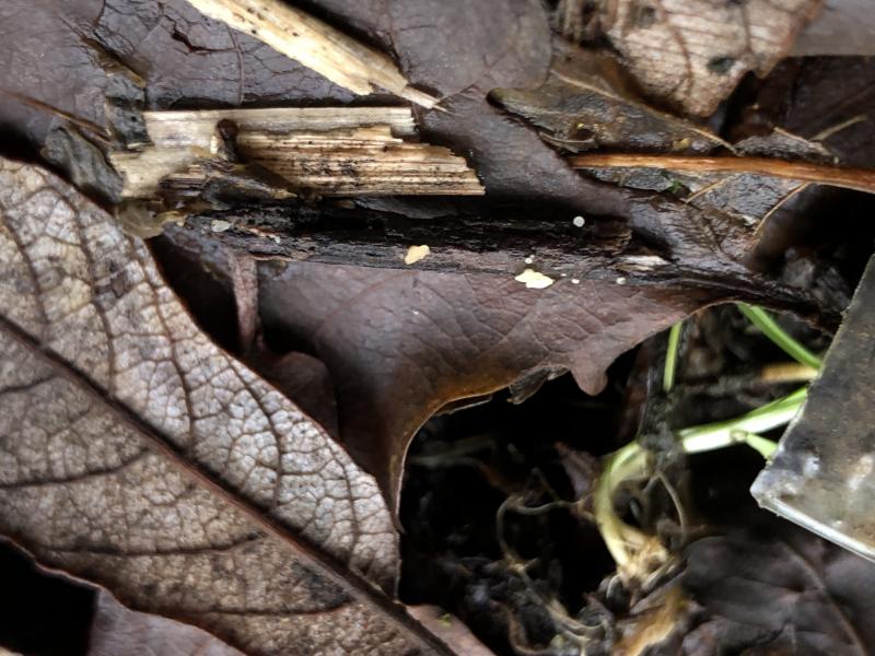
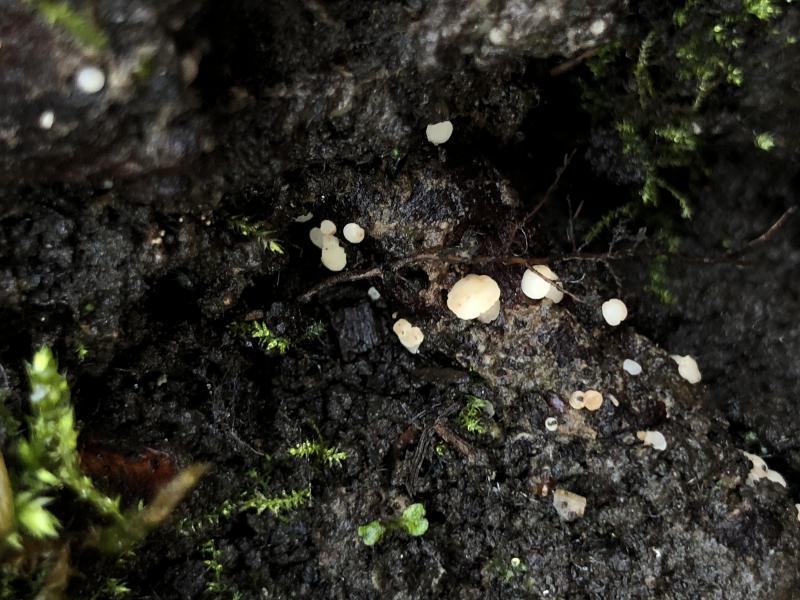
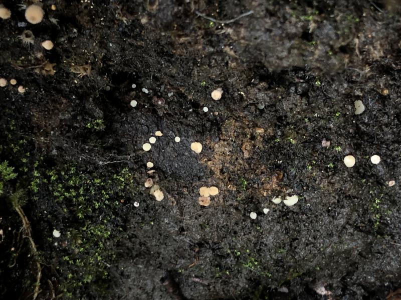
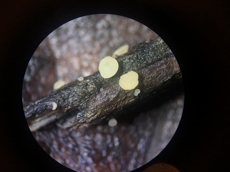
Habitat: Collected a few days ago (January), observed fruiting gregariously for several weeks prior, on the ground, appears saprotrophic on twigs and woody debris (and soil?), on a very damp and shady hill, close to drainage pipe and stream that feeds a pond, periodically disturbed by runoff, bryophytes close, Alnus and Salix nearby, in mixed deciduous woodland, southern England.
Preparation: Kept on wood in damp container to catch ejected ascopores, microscopically examined within 24 hours. One ascoma used, sections mounted in tap water, progressively squashed, and then IKI applied.
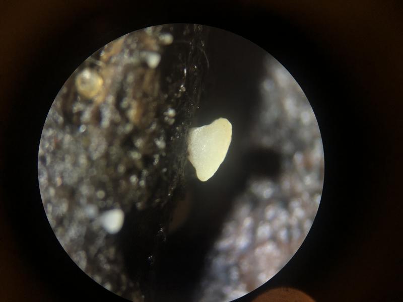
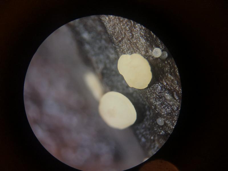
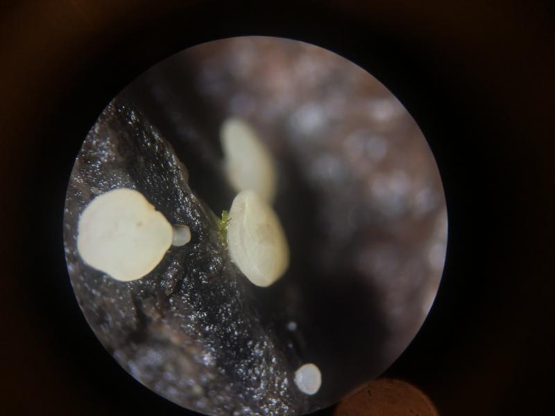
Ascomata: Apothecial, discoid, diameter <= ~2 mm, initially turbinate, sessile to short and broad stipe, disc widening and eventually going to convex, margin even or slightly wavy, initially whitish hyaline, going yellowish and finally ochraceous-apricot when aged, redding when bruised, hymenium convex in maturity, resistant to squashing.
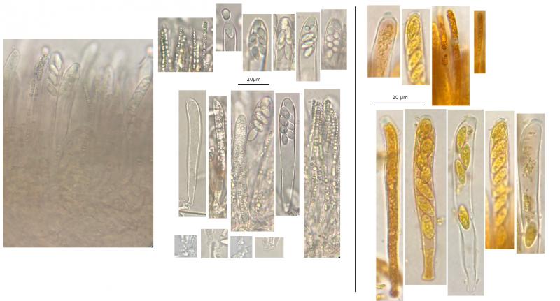
IKI: Apical ring bb, weak reaction, harder to see in dehisced or dead/opaque asci, faintly blue asci walls?, paraphysis VBs going red, some ascus cytoplasm red, spore cytoplasm yellow.
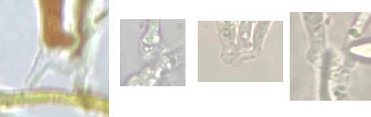
Asci: 8-spored, croziers +, apex more truncate and ring more visible in front view (resp. apex rounder in profile view), lots dehiscing in water mount.
• Vital – due to turgescence, clavate (wider), spores biseriate but one spores at apex, spores grouped towards apex, pars sporifera ~30% of length,
76.5 – 90.3 × 10.7 - 12.8 µm, N = 5.
• Dead – cylindrical-clavate, apical dome more visible, spores uniseriate, pars sporifera ~70% of length, often opaquely full of small and medium-size LBs,
66.6 – 82.5 × 5.3 - 8.7 µm, N = 7.
Paraphyses: Narrow filiform; apex filled by a row of small (squarish) and medium-size (rectangular), yellowish, strongly refractive VBs, globose lower down; not protruding beyond asci
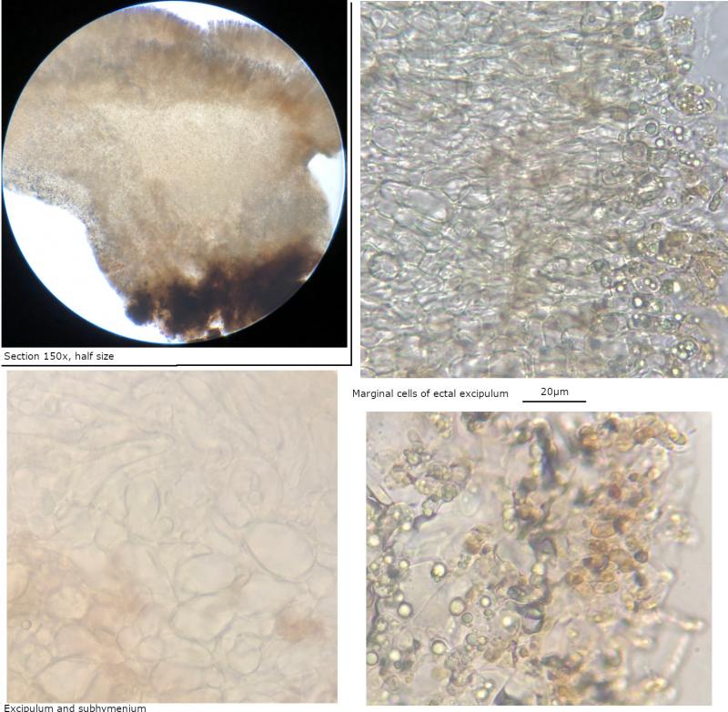
Ectal excipulum: A mixture of textura prismatica and angularis-globose, base brownish.
Marginal cells/hairs: Two or three terminal cells filled with several medium to large, strongly refractive VBs, these cells turning (microscopically) orange-reddish when traumatised, on stipe some terminal cells more like paraphyses.
Subhymenium: Densely woven hyphae (textura intrica?).
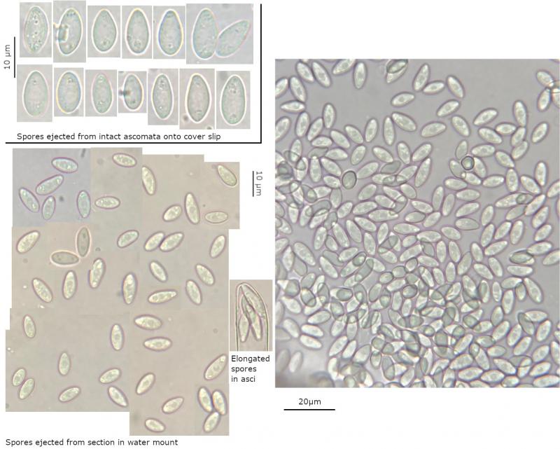
Spores: Mature and vital – narrowly-ellipsoid to ellipsoid-ovoid, aseptate, slightly inequilateral in profile view, large shadowy circle in the centre (nucleus with nucleolus sometimes visible?) and several LBs at the poles, often a distinct medium-size LB visible at one or both poles. Occasionally inside vital asci, some elongated, more fusiform.
• Ejected from intact ascomata in damp environment:
(8.4) 9.4 - 11.4 (11.7) × (4.3) 5 - 5.7 (6) µm,
Q = (1.7) 1.8 - 2.1, N = 17,
Me = 10.3 × 5.3 µm, Qe = 1.9.
• Ejected from section in water (no pressure):
(8.4) 8.9 - 11 (14.1) × (4.1) 4.2 - 5.1 (5.3) µm,
Q = (1.7) 1.9 - 2.5 (2.7), N = 40,
Me = 10.1 × 4.6 µm, Qe = 2.2.
• Together:
(8.4) 8.9 - 11.4 (14.1) × (4.1) 4.3 - 5.5 (6) µm
Q = (1.7) 1.8 - 2.4 (2.7) ; N = 57
Me = 10.1 × 4.8 µm ; Qe = 2.1
Subiculum: Maybe some anchoring hyphae identified in slide but not visible in macrophotos.
Anamorph: A crescent-shaped conidia found, but nothing related identified.
Hans-Otto Baral,
08-01-2024 12:30

Re : Hymenoscyphus sp.
Yes there is no doubt in my opinion. The resolution of the asci is too low to see the ascus base, but <i suppose you saw croziers.
B Shelbourne,
08-01-2024 12:50
Re : Hymenoscyphus sp.
Thank you for looking and your evaluation of the photos concerning croziers. It was hard to separate out the cells of the hymenium and there seemed to be fewer opportunities.
I have attached the best views I have but I understand the resolution is poor.
I have attached the best views I have but I understand the resolution is poor.
Hans-Otto Baral,
08-01-2024 17:19
Re : Hymenoscyphus sp.
Yes, clearly with croziers.