
10-06-2026 12:54
Steve ClementsBonjour encore, Pouvez-vous m'aider, s'il vous pl
09-06-2026 18:32
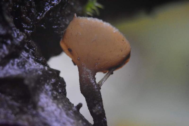
Camille MertensSur morceau de roseau immergé 0,5 - 0,7 mm de dia

10-06-2026 21:16
 François Freléchoux
Bonsoir,Le dernier du jour, en attendant votre avi
10-06-2026 21:07
François Freléchoux
Toutes les tiges de gentianes jaunes de l'an pass�
10-06-2026 13:41
François Freléchoux
Bonjour à nouveau, Voici une trouvaille d'hier.
10-06-2026 11:53
Steve ClementsBonjour, This disco is abundant on dead stems of
10-06-2026 10:45
François Freléchoux
Bonjour à nouveau, Encore une détermination qui
08-06-2026 10:16
 Spooren Marco
I don`t have a clou about this fungus,it is not in
10-06-2026 09:24
François Freléchoux
Bonjour, J'imagine que cette détermination ne do
Rutstroemia sp?
Nogueira Héctor,
28-10-2023 04:14
Hans-Otto Baral,
28-10-2023 08:06

Re : Rutstroemia sp?
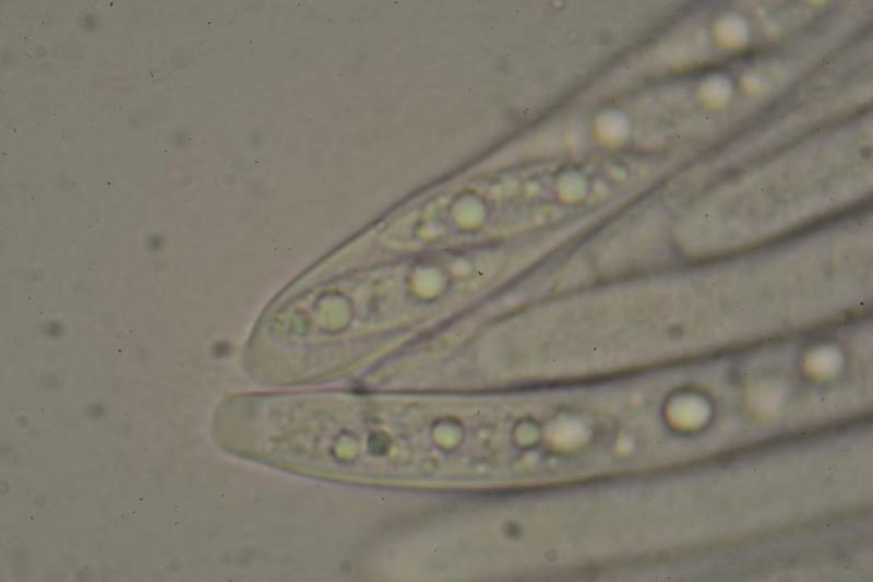
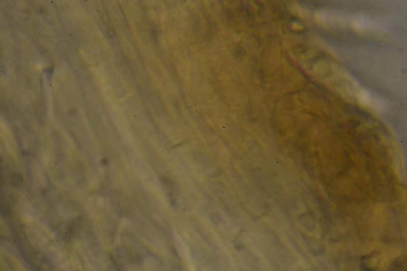
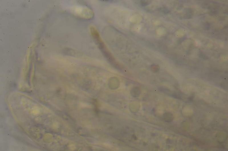
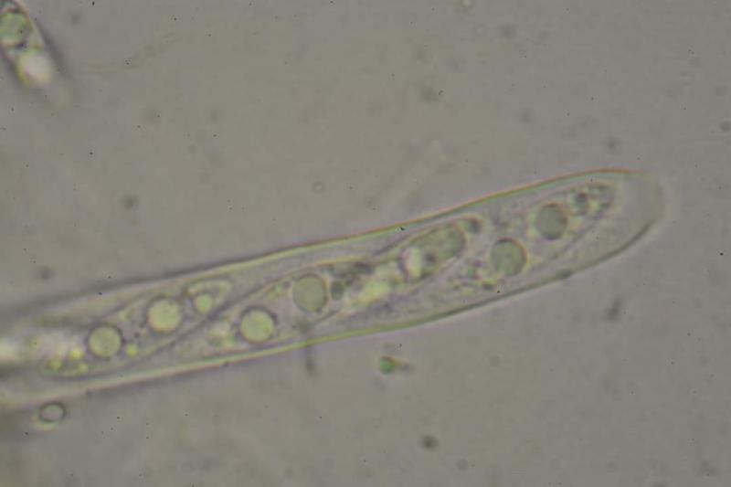
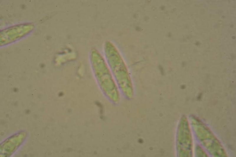
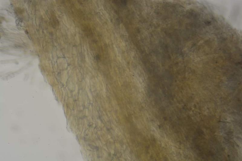
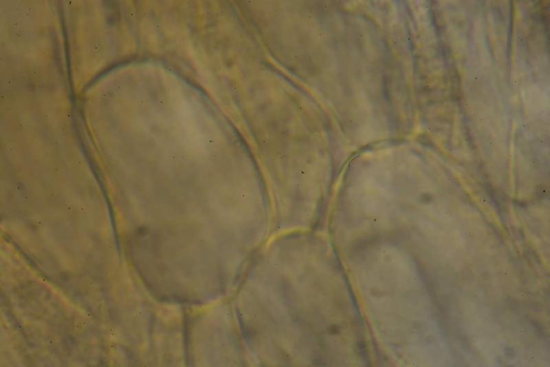
Hmm, I did not see a survey photo of the excipulum, I understand that the ectal is prismatic, non-gelatinised? Only those spores are alive in your pics which have two distint globose drops, the others have an obscure content because of turgor loss (perhaps because of pressing on the slide? Or did you study the dried apo?). I miss the contents of the living paraphyses.
R. alnobetulae, named after the host Alnus alnobetula, has refractive contents in the living paraphyses which stain blue in IKI. And it has a gelatinised excipulum if I remember right.
Nogueira Héctor,
28-10-2023 09:42
Re : Rutstroemia sp?
I don't know why the spores look like that (I saw the live-looking spores during the study of the sample, but I thought it was significant to add them since the size and content seemed notable to me). The specimen was fresh, I left it spore the same day and studied it the next day (I use a fixative for samples with spores, maybe that could have influenced it?) I'm not good at cutting, I'll try to see if I can take a photo of the excipulum of the small piece I have left.
Thank you
Thank you
Hans-Otto Baral,
28-10-2023 09:54
Re : Rutstroemia sp?
A fixative sounds indeed like toxic for the spores. Tap water is the medium to study living cells.
Nogueira Héctor,
28-10-2023 10:03
Re : Rutstroemia sp?
I use the fixative because it makes it easier for me to photograph the spores, sometimes they move a lot and it is complicated, I have never had problems (the fixative was given to me by a veterinarian friend of mine), I will avoid using it from now on. Thank you
Hans-Otto Baral,
28-10-2023 10:13
Re : Rutstroemia sp?
The movement of the spores is no problem when you use a strong lamp. It is usually no problem to achieve a shutter speed of 1/60 which is enough for sharp photos.
I once tried to use protein solutions for this purpose because they were said to avoid osmotic effects, but then I realised that this is not necessary.
Nogueira Héctor,
28-10-2023 10:22
Re : Rutstroemia sp?
I'll keep that in mind from now on, thanks. If I get any progress I will continue commenting. Thanks and regards
Nogueira Héctor,
29-10-2023 11:15
Re : Rutstroemia sp?
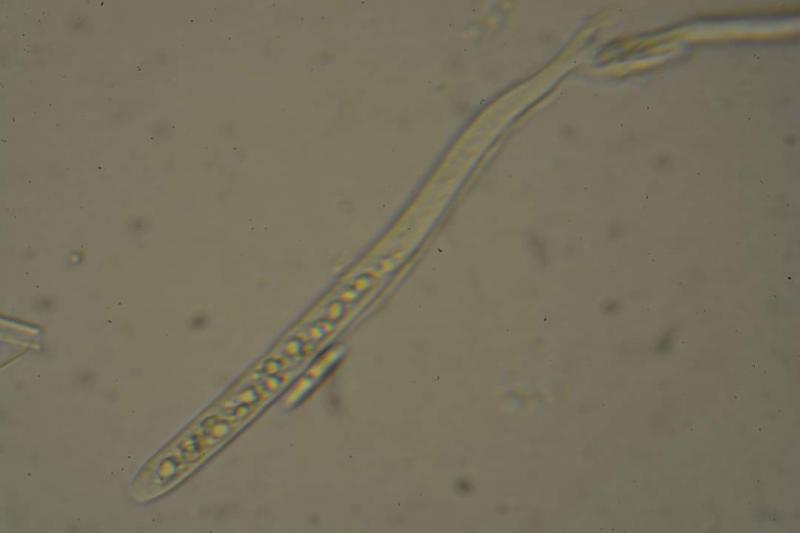
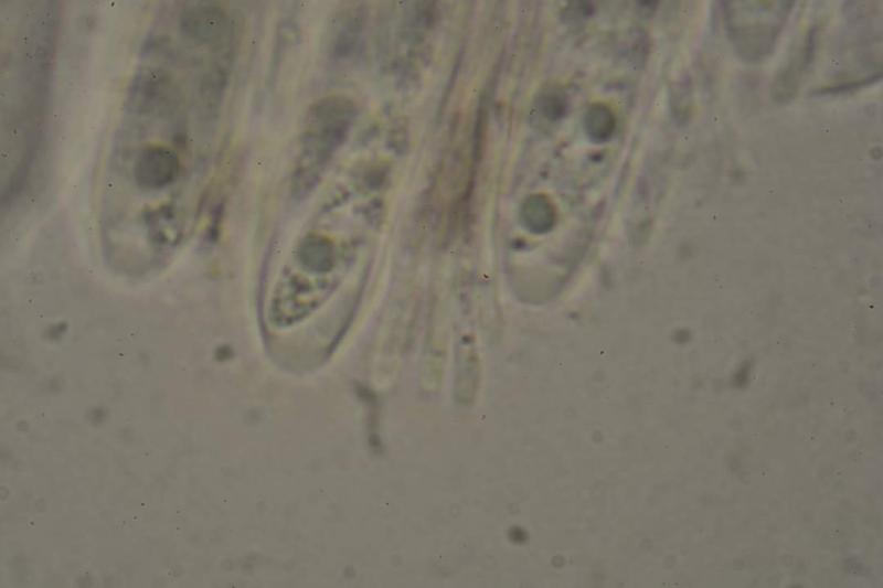
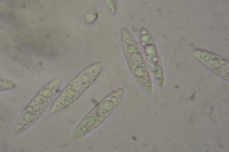
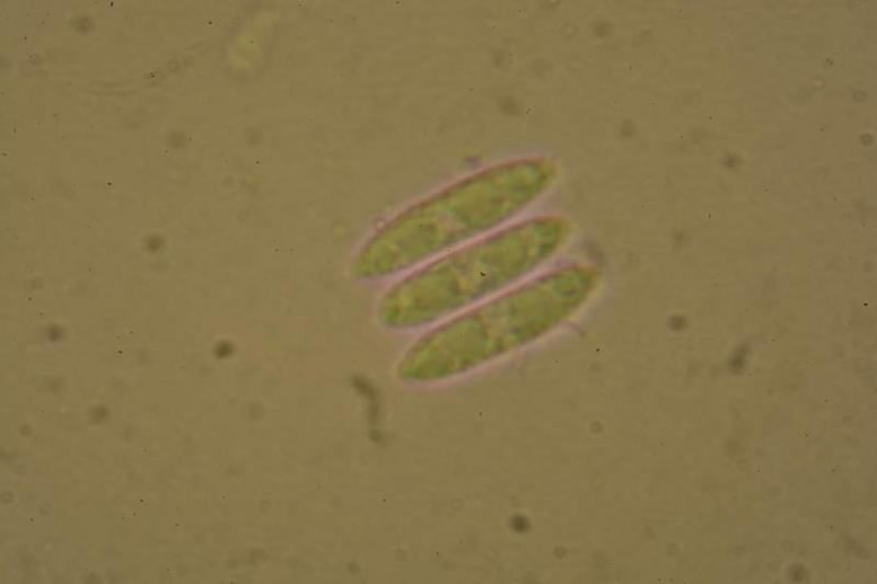
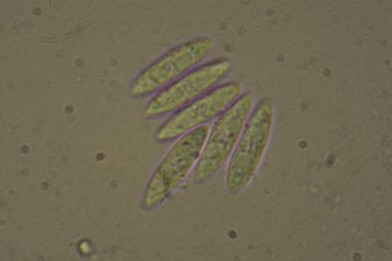
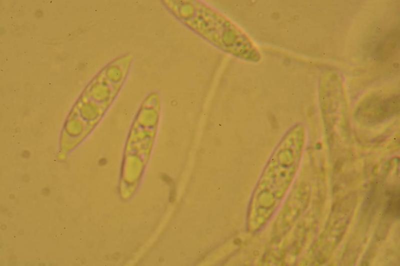
I have not been able to get free sporade. Spores measured in water during sample observation:
(21.4) 22.8 - 25.3 (25.9) × (4.9) 5.3 - 6.3 (6.5) µm
Q = (3.6) 3.7 - 4.4 (5.2) ; N = 23
Me = 24.1 × 5.9 µm; Qe = 4.1
(21.4) 22.8 - 25.3 (25.9) × (4.9) 5.3 - 6.3 (6.5) µm
Q = (3.6) 3.7 - 4.4 (5.2) ; N = 23
Me = 24.1 × 5.9 µm; Qe = 4.1

Hans-Otto Baral,
29-10-2023 12:03
Re : Rutstroemia sp?
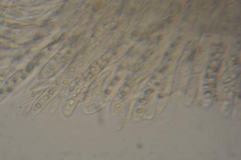
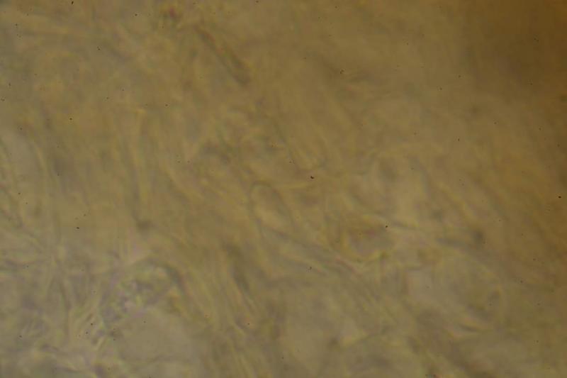
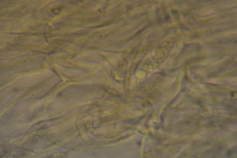
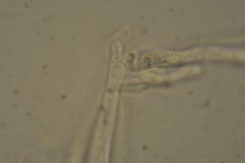
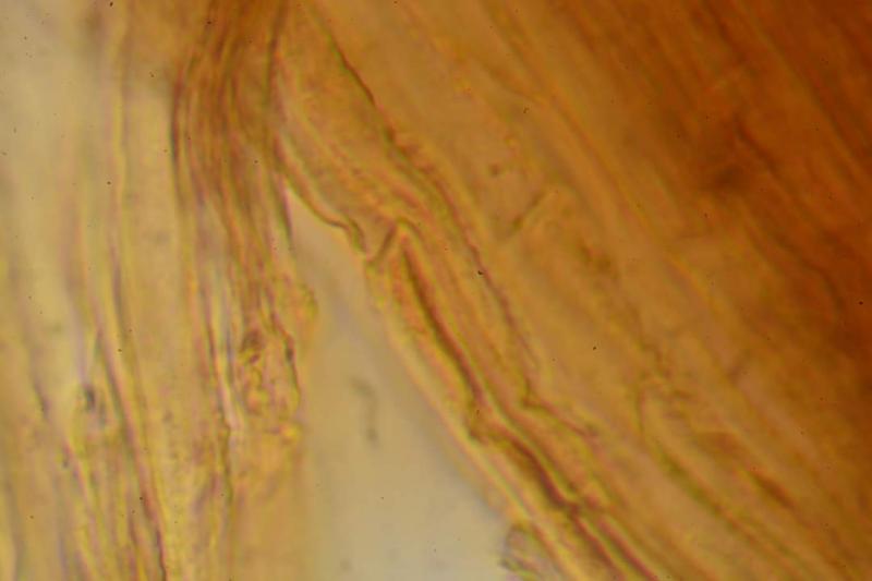
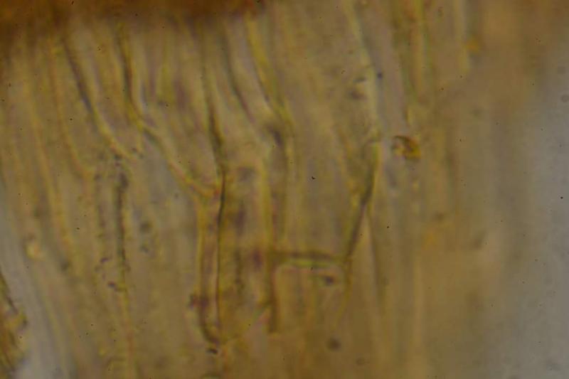
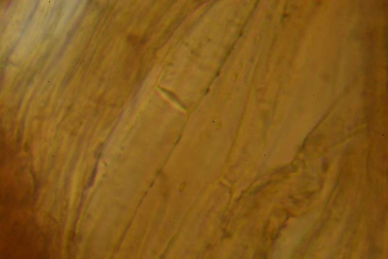
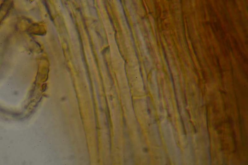
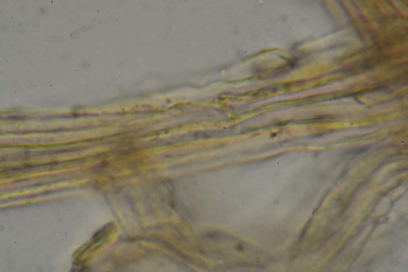
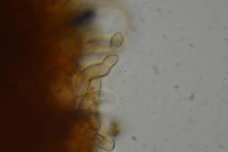
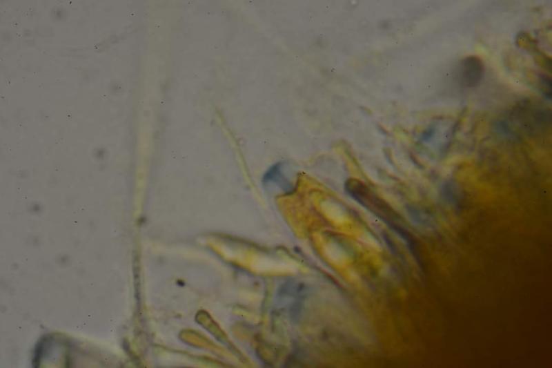
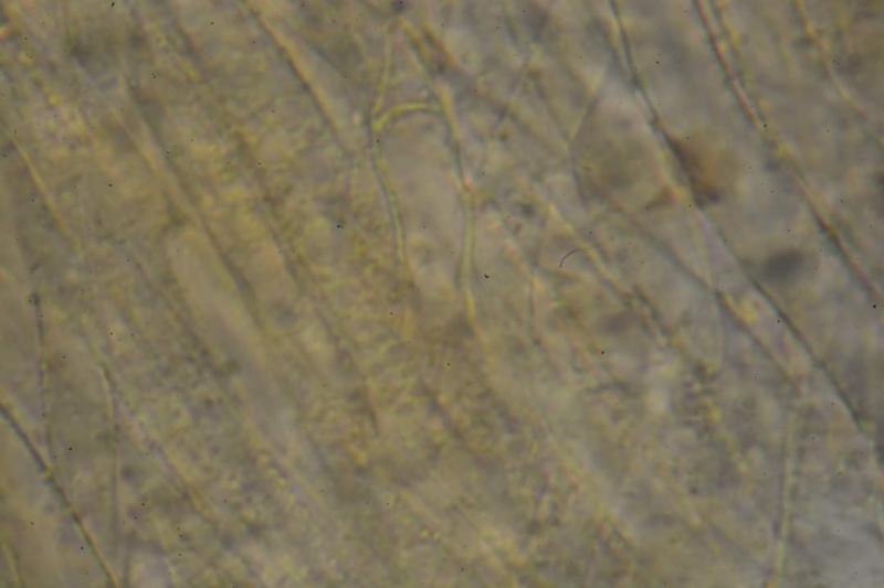
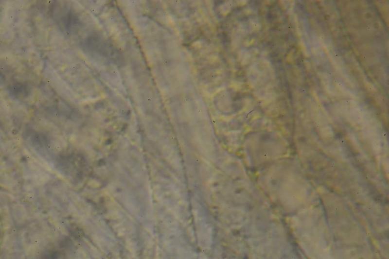
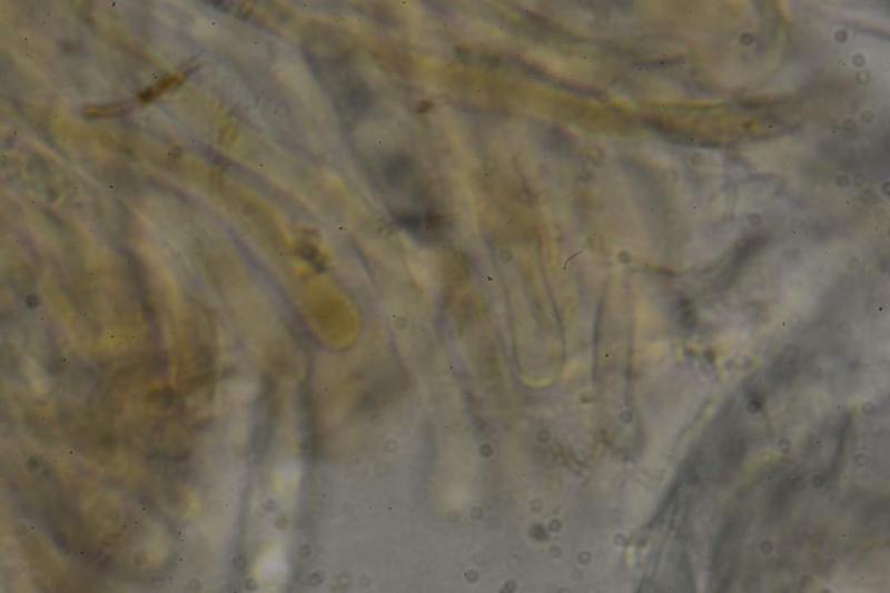
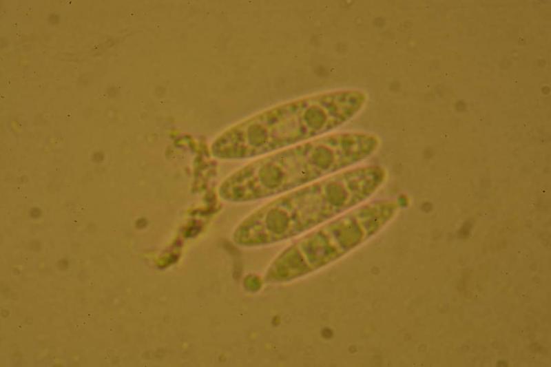
Now it is very clear: Thin-walled broad prismatic cells of ectal and narrow warted hyphae of medullary excipulum. The lack of gel appears to exclude R. alnobetulae. The paraphyses contain yellowish-brownish vacules - typical of many Rutstroemia. The four spores of the beforelast pic look splendid, mature and alive. Probably the overmature spores would get septate and produce conidia. Not sure if there is a name for it.
Nogueira Héctor,
29-10-2023 13:13
Re : Rutstroemia sp?
Thank you very much for the help and teachings! I will try to find another copy in case anyone is interested in studying it in depth.