12-06-2026 14:50
 François Freléchoux
Bonjour, Voici la brève description d'une Mollis
10-06-2026 21:16
François Freléchoux
Bonsoir,Le dernier du jour, en attendant votre avi
11-06-2026 19:01
 William Slosse
Hello all,In an attempt to make a culture of a sus
11-06-2026 19:03
 Nicolas VAN VOOREN
Chers membres d'Ascofrance,Le site sera placé en

09-06-2026 18:32
Camille MertensSur morceau de roseau immergé 0,5 - 0,7 mm de dia
10-06-2026 12:54
Steve ClementsBonjour encore, Pouvez-vous m'aider, s'il vous pl
10-06-2026 21:07
François Freléchoux
Toutes les tiges de gentianes jaunes de l'an pass�
10-06-2026 13:41
François Freléchoux
Bonjour à nouveau, Voici une trouvaille d'hier.
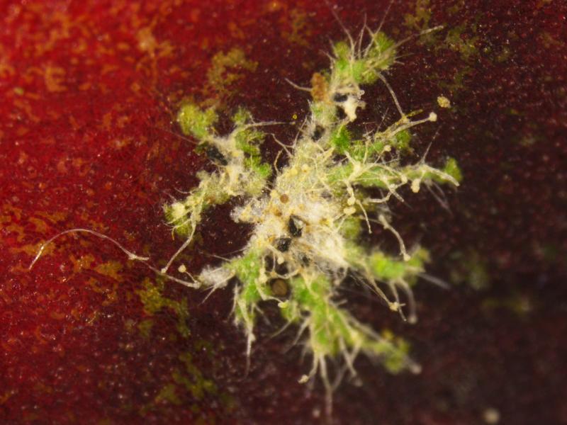
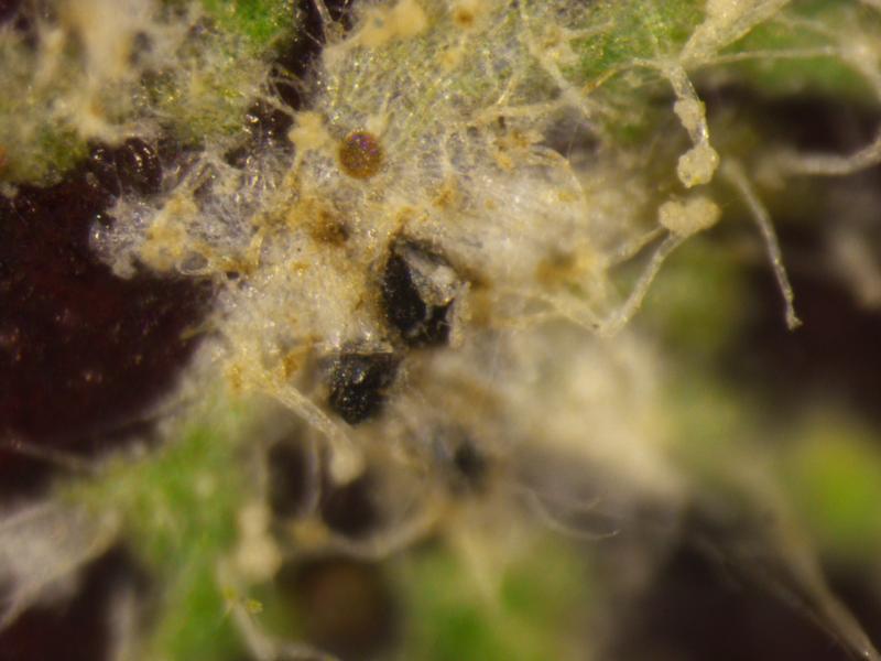
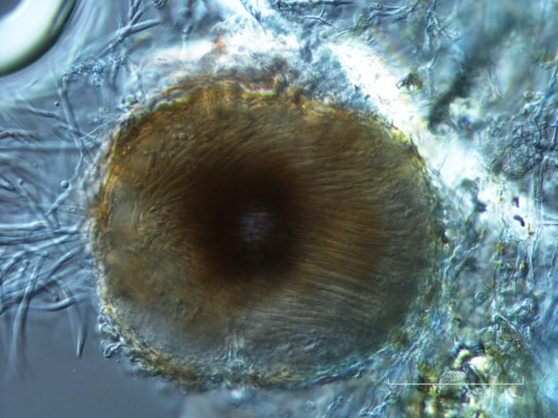
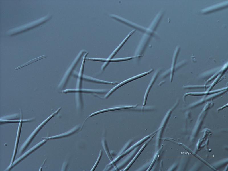
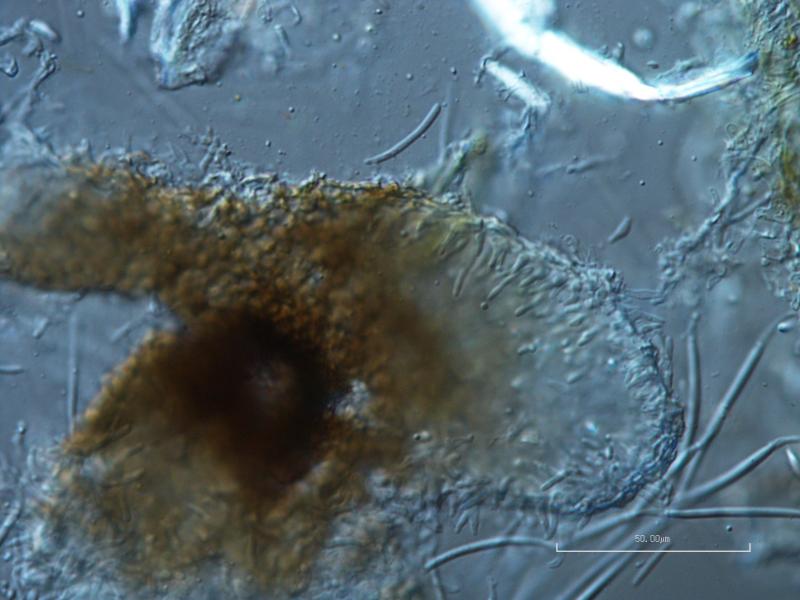
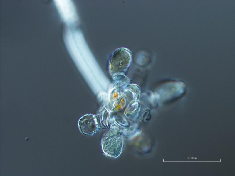
 Hello! I wanted to share a fascinating thing I saw this week. This is the coelomycete anamorph state of pyrenolichen Phyllocharis orbicularis (=Strigula orbicularis), which is apparently not illustrated anywhere. I was lucky enough to be able to ask Robert Lücking for help, and he ID'd it right away, having seen it before; when I asked where I could find an illustration, he admitted that he didn't think one existed, only the ascomata and the macromorphology of the thallus. The thallus of this particular example is somewhat poorly lichenized, and looks more like the photobiont (Cephaleuros virescens) than the typical thallus, but the conidia are the really fun part anyway.
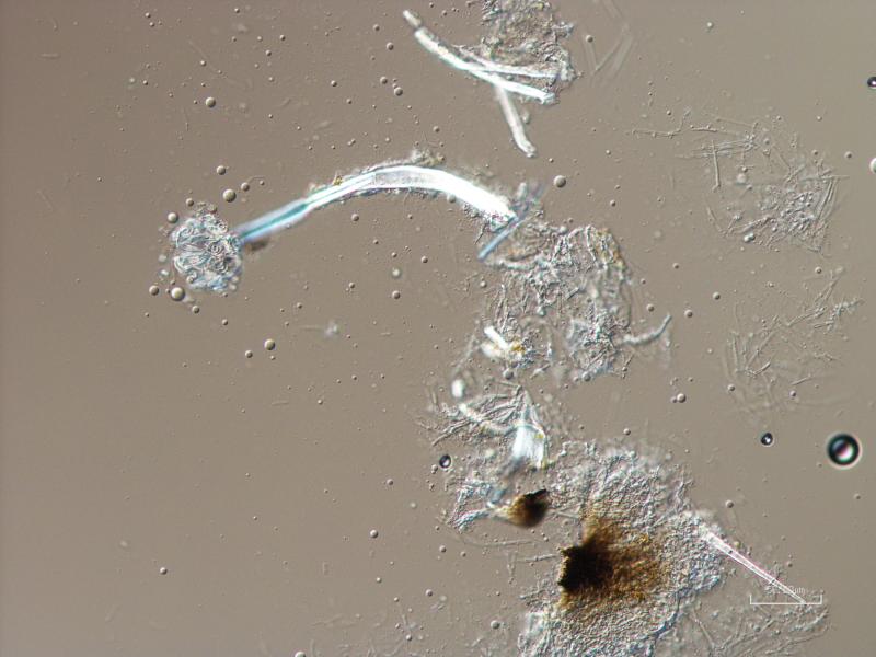
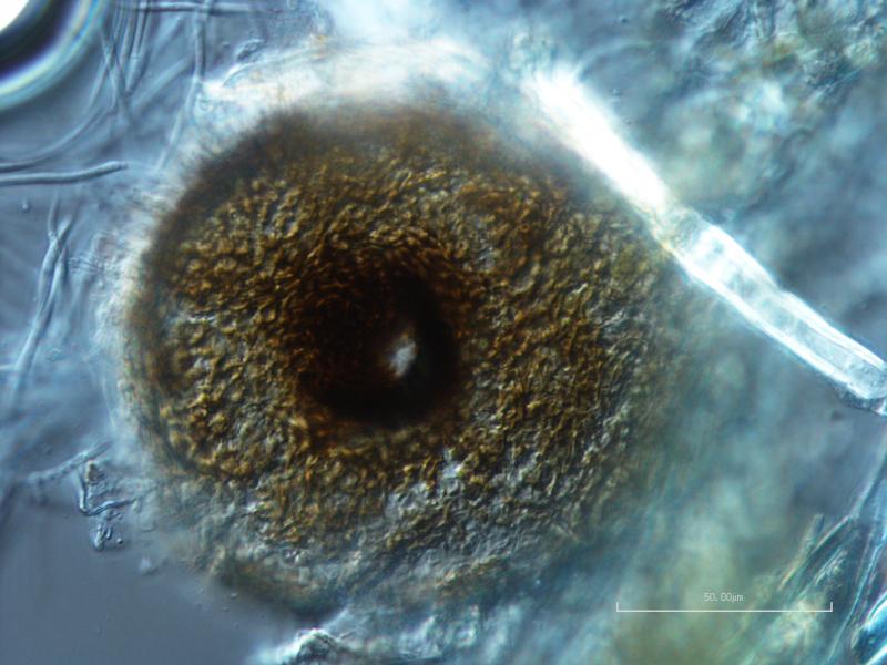
The conidia are hyaline, 4- to 6-septate, 40-45 x 2-3.5 ?m excluding the appendages, with a non-cellular, mucoid appendage at each end, which are quite variable in length, but generally less than 10 ?m, and often curving into a hook. Conidiophores are small, lageniform, reduced to conidiogenous cells, and integrated into the inner wall of the pycnidium.
I really wanted to put these photos out there, so that if anyone else is struggling to identify this beautiful and distinctive anamorph they'll be able to find some reference images! I can't thank Dr. Lücking enough for his kind help in the identification of this fungus.