10-06-2026 12:54
Steve ClementsBonjour encore, Pouvez-vous m'aider, s'il vous pl
09-06-2026 18:32
Camille MertensSur morceau de roseau immergé 0,5 - 0,7 mm de dia
10-06-2026 21:16
 François Freléchoux
Bonsoir,Le dernier du jour, en attendant votre avi
10-06-2026 21:07
François Freléchoux
Toutes les tiges de gentianes jaunes de l'an pass�
10-06-2026 13:41
François Freléchoux
Bonjour à nouveau, Voici une trouvaille d'hier.
10-06-2026 11:53
Steve ClementsBonjour, This disco is abundant on dead stems of
10-06-2026 10:45
François Freléchoux
Bonjour à nouveau, Encore une détermination qui
08-06-2026 10:16
 Spooren Marco
I don`t have a clou about this fungus,it is not in
10-06-2026 09:24
François Freléchoux
Bonjour, J'imagine que cette détermination ne do
08-06-2026 17:00
François BartholomeeusenGood day everyone, On June 5 2026, I collected de
White microfungus (airborne contaminant ?) with swellings along its hyphae
Zetti Mario,
16-07-2022 17:01
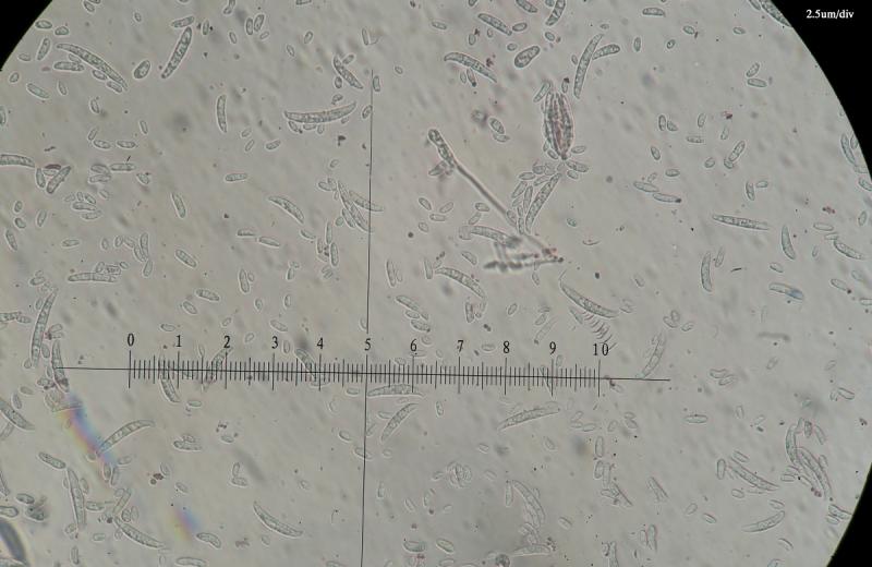
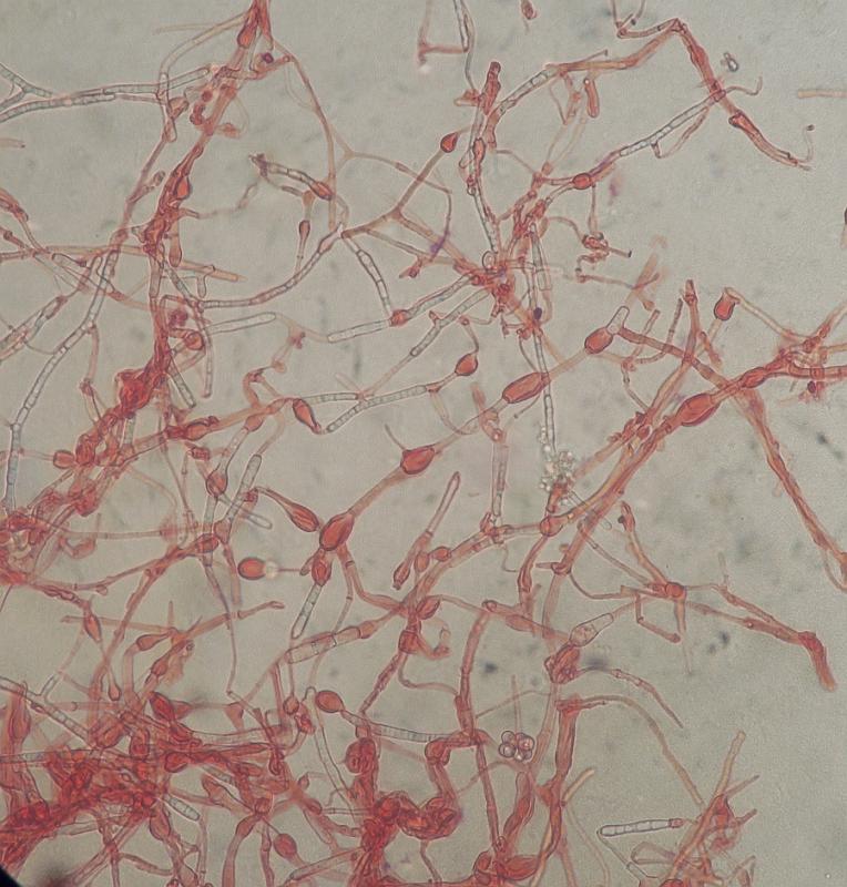
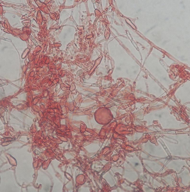
Under the microscope, I cannot observe free conidiospores but the hyphae were regularly swollen into thick-walled vesicles (zoospores?). Any suggestions are welcomed. Reminds me in the distance to Phythphtora...
David Malloch,
16-07-2022 20:35

Re : White microfungus (airborne contaminant ?) with swellings along its hyphae
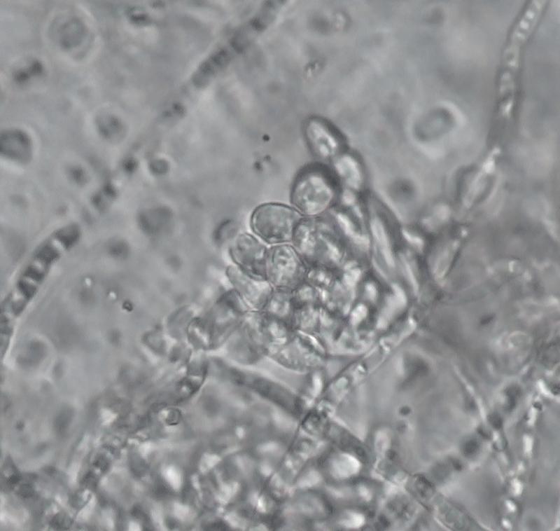
That will be difficult to identify. The hyphae are producing thick-walled intercalary cells and chlamydospores, but nothing else. It could be a Fusarium, but many other things as well. When I encounter fungi like that, I try a different medium. Maybe V8 or something with a lot of cellulose but not much sugar. Good luck!
Zetti Mario,
17-07-2022 08:07
Re : White microfungus (airborne contaminant ?) with swellings along its hyphae
Thank you, interesting. I was working with a Fusarium oxysporum recently. I have available Sabouroud agar, CZapeK DoX, PDA, Oat Agar and MEA. Which medium do you suggest (Oat perhaps?) - TNX
David Malloch,
17-07-2022 13:45
Re : White microfungus (airborne contaminant ?) with swellings along its hyphae
Those media mostly have a high C/N ratio that may encourage vegetative growth at the expense of sporulation. Perhaps the oat agar would be the best choice.
Zetti Mario,
18-07-2022 08:47
Re : White microfungus (airborne contaminant ?) with swellings along its hyphae
Thank you, Oat Meal and PDA innoculated and I will report back the results.
Zetti Mario,
08-08-2022 18:32
Re : White microfungus (airborne contaminant ?) with swellings along its hyphae
Hello David and everyone.
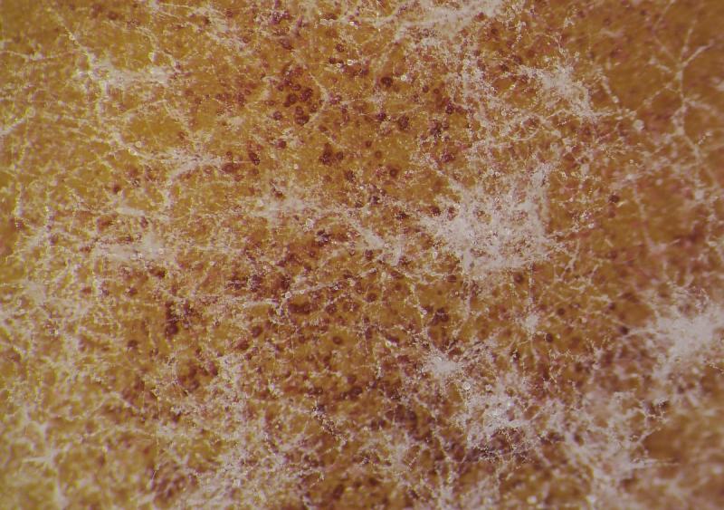
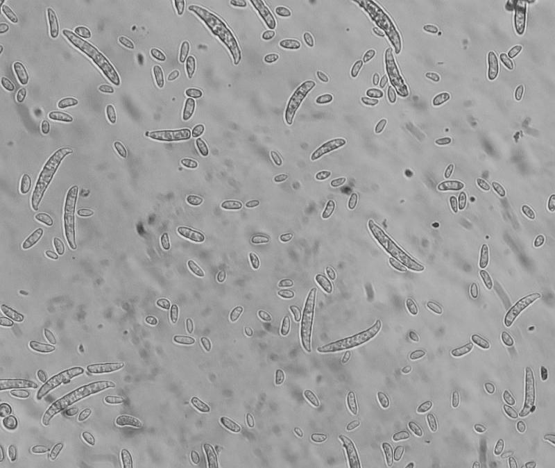
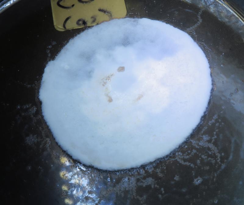
I managed to find time to reply you back. I managed to get a pure colony on PDA and Oat indeed it was a Fusarium, probably F. oxysporum s.l. (maybe lycopersici) I followed a simple key so dont take this as final determination of course!
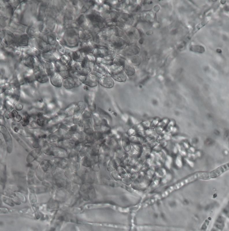
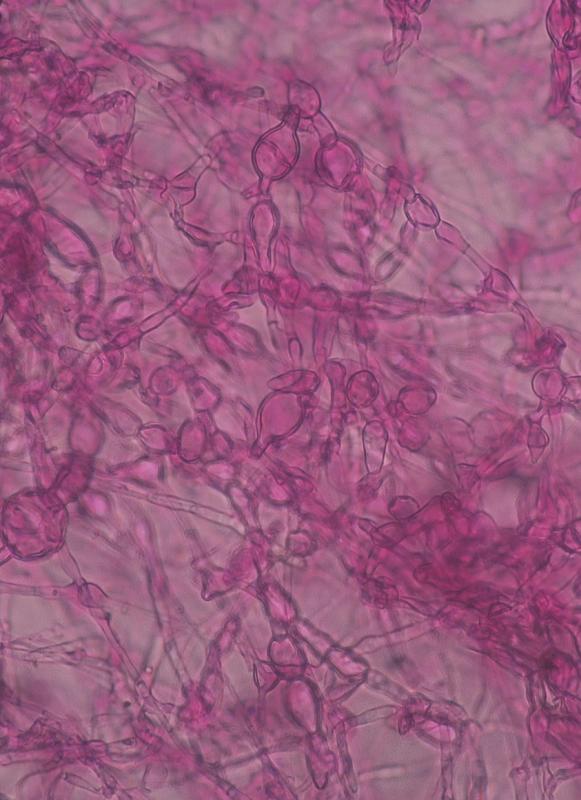
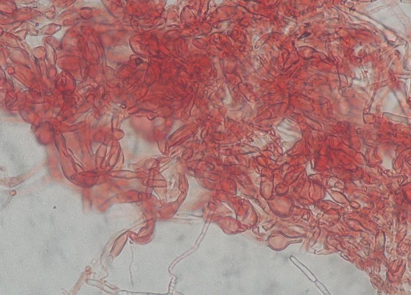
I was intrigued by pinkish-peach coloration (images below has enhanced colour) in the 12 day old colony. They were like soft non encapsulated tissue. Under the microscope they looked like bunch of elongated grapes. Is this characteristic of the species ? Are they the sporodochia ?
I saw some papers showing this pinkish-peach coloration by F. oxysporum s.l. - e.g. https://www.frontiersin.org/files/Articles/335707/fmicb-09-00420-HTML/image_m/fmicb-09-00420-g001.jpg
from https://www.frontiersin.org/articles/10.3389/fmicb.2018.00420/full
and this is a good reference paper too
https://repository.naturalis.nl/document/668094
I managed to find time to reply you back. I managed to get a pure colony on PDA and Oat indeed it was a Fusarium, probably F. oxysporum s.l. (maybe lycopersici) I followed a simple key so dont take this as final determination of course!
I was intrigued by pinkish-peach coloration (images below has enhanced colour) in the 12 day old colony. They were like soft non encapsulated tissue. Under the microscope they looked like bunch of elongated grapes. Is this characteristic of the species ? Are they the sporodochia ?
I saw some papers showing this pinkish-peach coloration by F. oxysporum s.l. - e.g. https://www.frontiersin.org/files/Articles/335707/fmicb-09-00420-HTML/image_m/fmicb-09-00420-g001.jpg
from https://www.frontiersin.org/articles/10.3389/fmicb.2018.00420/full
and this is a good reference paper too
https://repository.naturalis.nl/document/668094