12-04-2026 17:56
 Hardware Tony
Found on dead stems in February earlier this year
17-04-2026 19:16
 Enrique Rubio
Hi to everybodyI would appreciate any assistance r
14-04-2026 05:32
Ethan CrensonHi all, A few weeks back a friend pointed out som
17-04-2026 15:14
 Bruno Coué
Bonjour.Récoltes du 16/04/2026, sur feuilles mort
12-04-2026 15:52
Gernot FriebesHi,I'm looking for help with this anamorph collect
14-04-2026 21:52
Gernot FriebesHi,found on dead leaves of Carex elata. Conidia: 4
16-04-2026 22:09
Buckwheat PeteHello, I'd like to ask about this older specimen:
15-04-2026 19:33
Fátima Durán ManzanequeHi!! I need help, I found this Ascomycete but I d
14-04-2026 20:31
Gernot FriebesHi,can this be Psilachnum lateritioalbum on Phragm
12-04-2026 12:22
 William Slosse
In a dune grassland in Oostduinkerke (Belgium), on
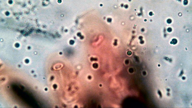
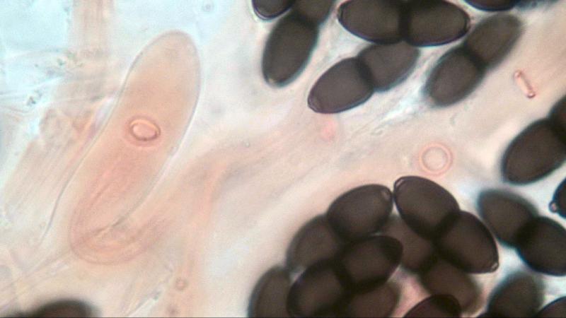
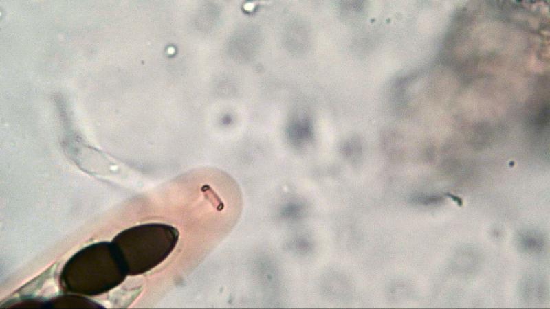
 When we are investigating characters of species under a light through microscope we do observe that in a 2D picture.
So we have to think in 3D but that is not always possible because our mindset cannot cope with the optical illusion we are looking at.
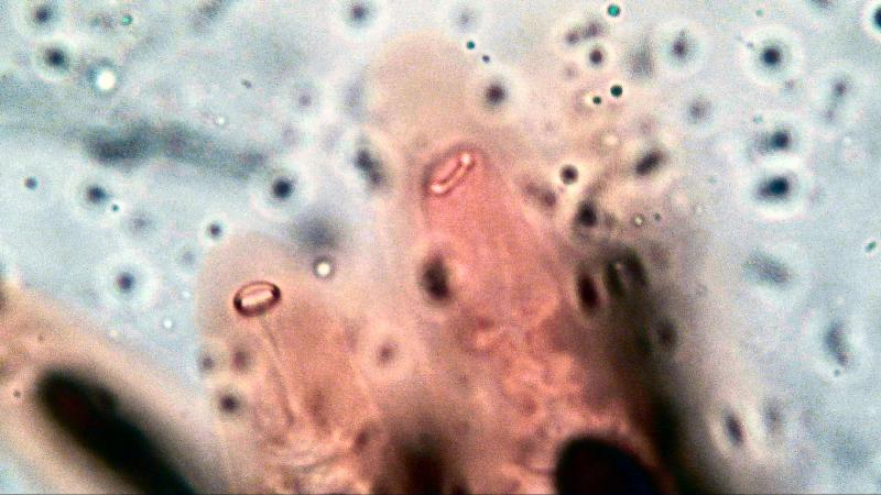
Accidentally I did find out that we can create a 3D picture by changing the focal distance from the lens to the object using a Plane Objective 100x/1.25 (photo 1 & 2). Probably by stacking photo's you will create the same effect.
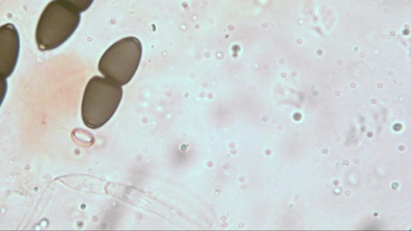
The ring is elastic and the distance when the apical system is not fully developed is as follows: Diameter of the outer circular ring is 0,9 um; total diameter is 4,6 um and inner diameter is 2.8 um. Photo-3 is a ring clearly visible with a spore ready to enter.
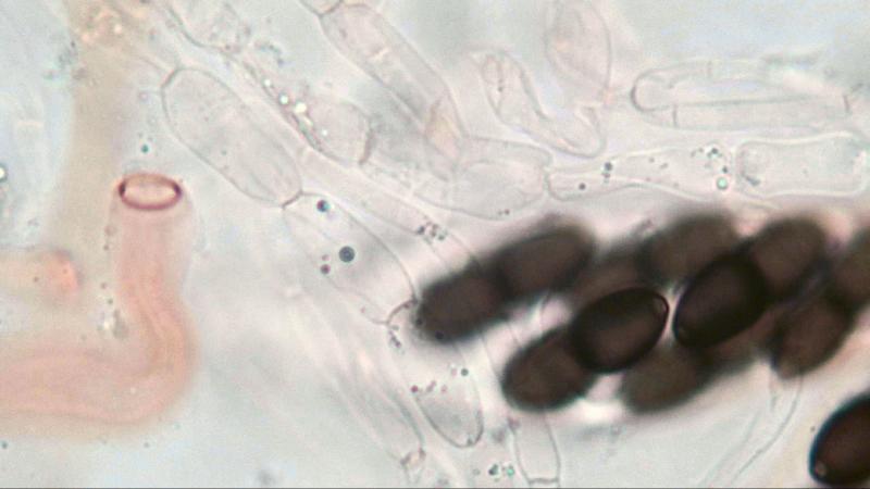
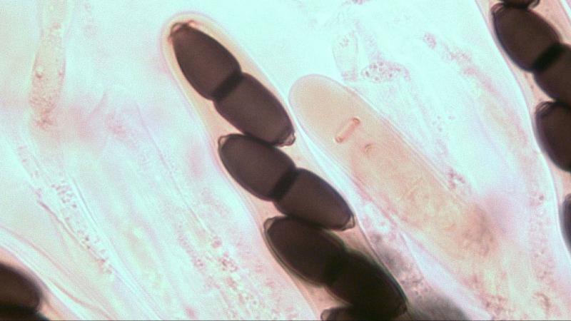
Photo 4 the ring is connected to an ampty inner wall, photo 5 is the same situation but inside a still present outer wall.
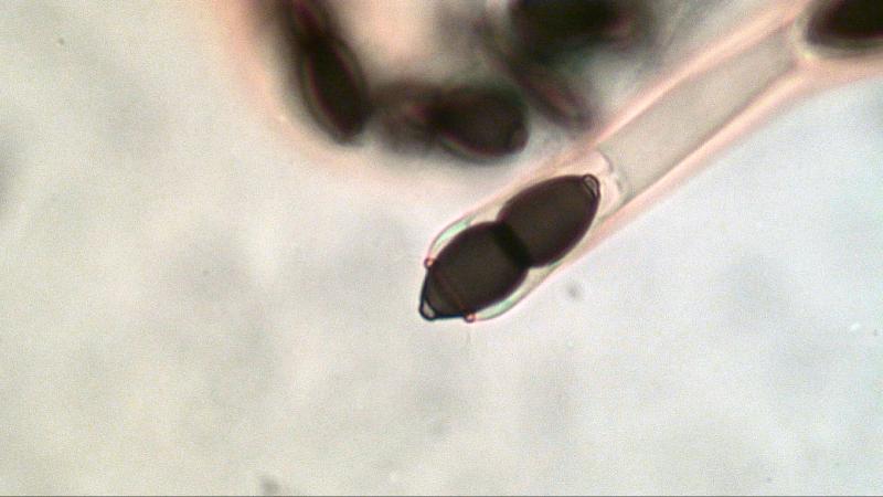
Photo 6 shows the apical ring in the end phase with spore clicked inside and the outer wall still present.
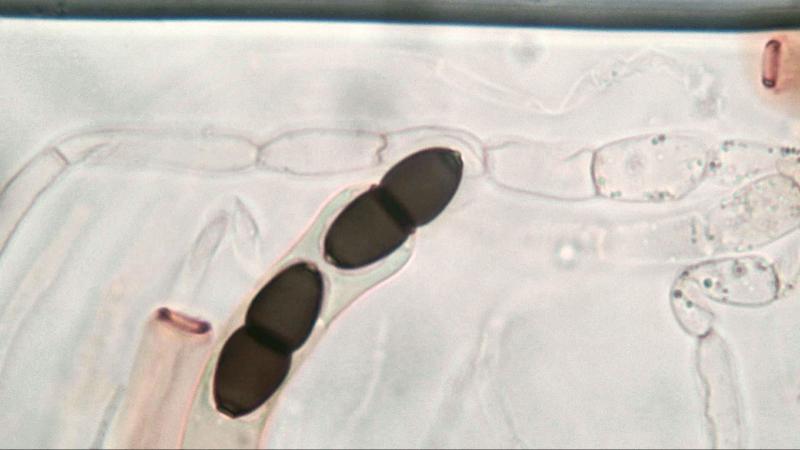
Photo 7 & 8 show spores inside the ring and outer wall gone.
The ring itself is more oval than it is circular. (photo 9)
Kind regards,
Joop