
12-04-2026 17:56
 Hardware Tony
Found on dead stems in February earlier this year
17-04-2026 19:16
 Enrique Rubio
Hi to everybodyI would appreciate any assistance r
14-04-2026 05:32
Ethan CrensonHi all, A few weeks back a friend pointed out som
17-04-2026 15:14
 Bruno Coué
Bonjour.Récoltes du 16/04/2026, sur feuilles mort
12-04-2026 15:52
Gernot FriebesHi,I'm looking for help with this anamorph collect
14-04-2026 21:52
Gernot FriebesHi,found on dead leaves of Carex elata. Conidia: 4
16-04-2026 22:09
Buckwheat PeteHello, I'd like to ask about this older specimen:
15-04-2026 19:33
Fátima Durán ManzanequeHi!! I need help, I found this Ascomycete but I d
14-04-2026 20:31
Gernot FriebesHi,can this be Psilachnum lateritioalbum on Phragm
12-04-2026 12:22
 William Slosse
In a dune grassland in Oostduinkerke (Belgium), on
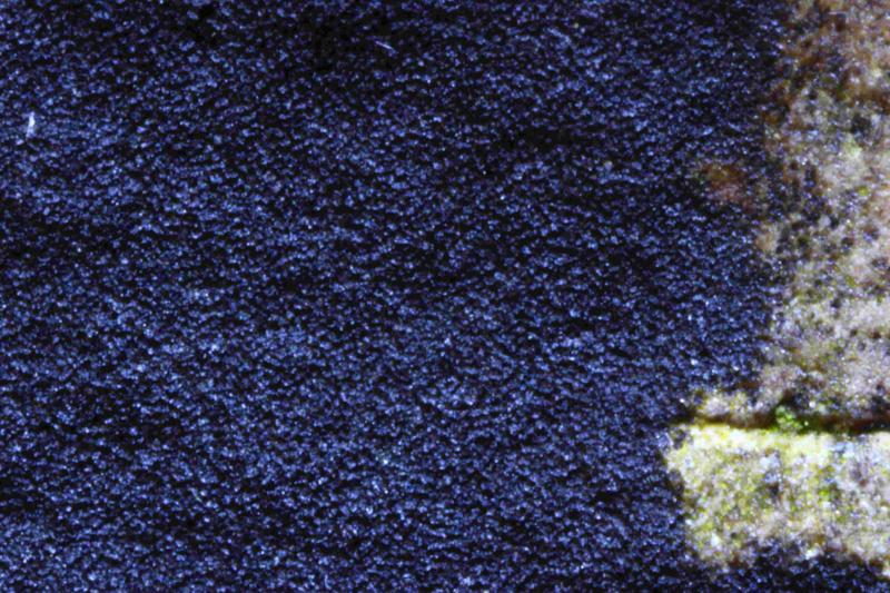
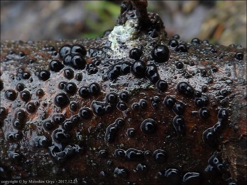
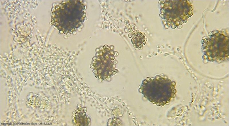
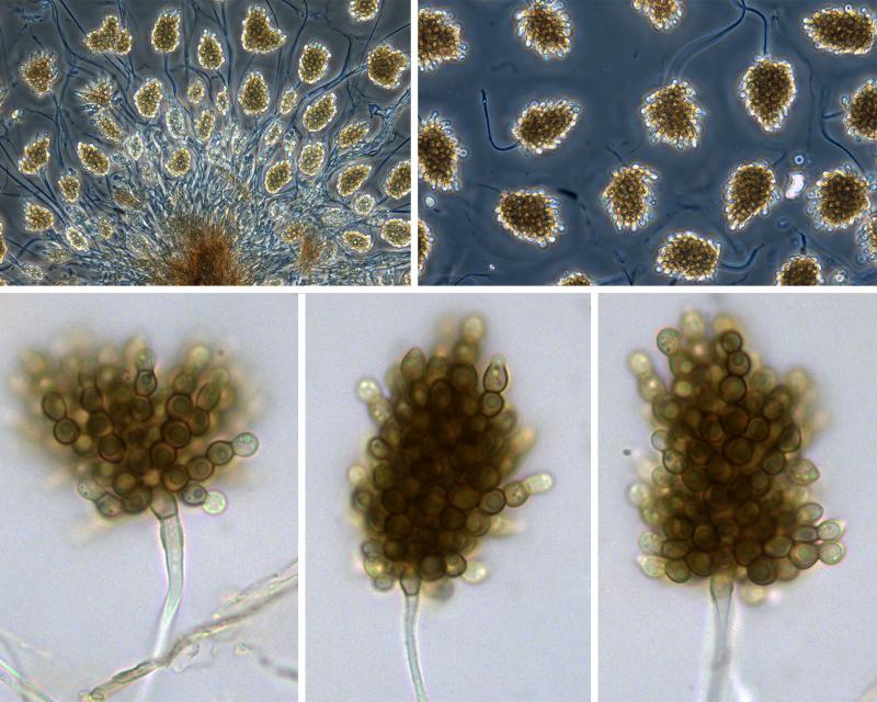
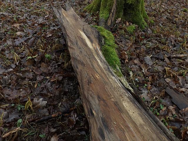
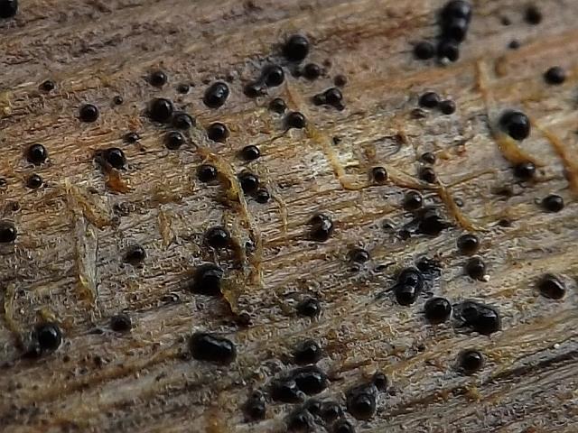
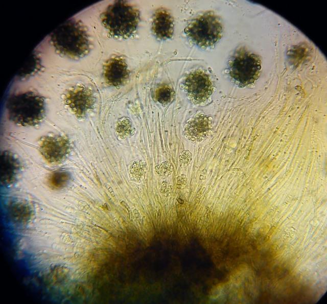
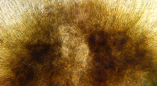
I have found what appears to be an effuse layer of fruit bodies expanding across the bark of a dead trunk of Corylus avellana about 50 cm above ground level. Macroscopically, they look like tiny spheres tightly clustered together.
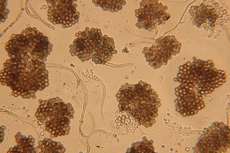
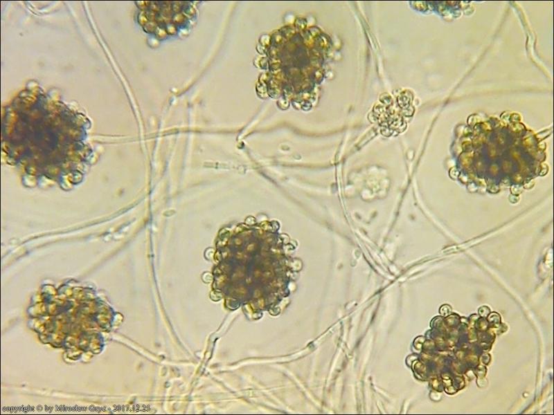
Microscopy shows what would appear to be septate hyphae / conidiophores with branches, on which initially hyaline spheres form. As they mature, these clusters of spheres become brown and most seem to contain a refractive guttule. Each tiny sphere is smooth and about 5 um in diameter and most do not separate when prepared for microscopy.
I wonder if this effuse layer could be a hyphomycete, but I can not find any species with clusters of smooth conidia resembling these.
I have attached photos of the effuse layer and the available microscopic chararacteristics.
I wonder if anyone has any ideas about what this might be?
Thank You,
With Best Wishes,
Peter.
Thank you for your interesting reply.
I am quite sure that my fungus matches very well with your Ascofrance post dated 26th November 2017.
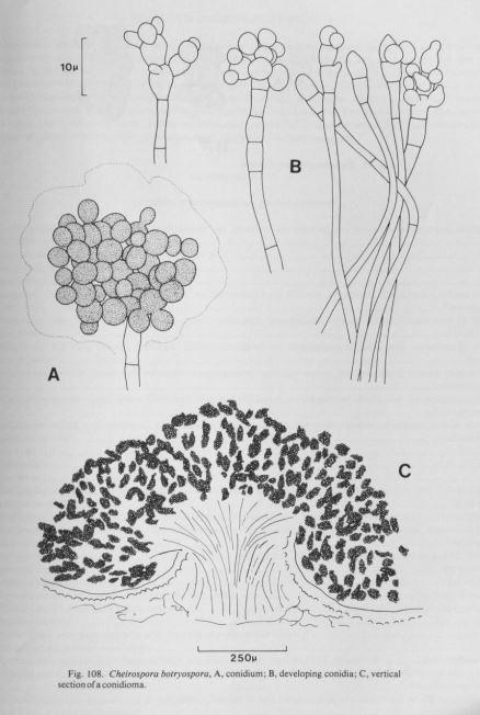
I had not considered the possibility of it being a coelomycete. Looking in Ellis & Ellis, the description of Cheirospora botryospora being tremelloid, with clusters of conidia contained in gelatinous sheaths both look wrong, so perhaps Ellis & Ellis have misinterpreted the taxon.
Interestingly, Zotto Baral has mentioned DNA sequences clustering around Mollisia albogrisea. I collected a pale mollisioid fungus which was growing nearby, which may or may not be M. albogrisea.
I will need to look into these samples further, if it is possible that they may be related.
With Best Wishes,
Peter.


Your image panel certainly looks different from what I have found, both macroscopically and microscopically.
Your image (and some others on the Internet) shows spheres resting on the host, whereas mine are effuse. There had been a lot of rain before I collected my sample, so I wouldn't have thought that it could have dried out from spheres to become effuse. Perhaps I can test this, by adding water.
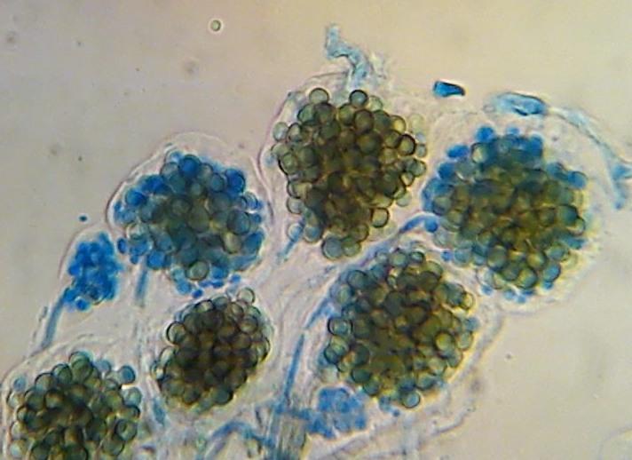
Your micro photo differs from mine as you rightly say that yours look like the fingers of a hand. Mine are irregularly rounded clusters of conidia, varying in size, but seemingly intact and not resembling fingers.
Two different species in the same genus, perhaps? If so, I wonder which is Cheirospora botryospora and which might be something else.
With Best Wishes,
Peter.
Yes, the conidiogenous cells of my sample match well those shown in the very recent Ascofrance post which you refer to.
Also mine seem to match another Ascofrance post from 28th February 2018, where the host substrate was also Corylus avellana.
There seems to be a lot of confusion regarding the hyphomycete Periconia cambrensis and the coelomycete Cheirospora botryospora. Nick Aplin mentioned this later in the same 28th February 2018 forum post.
To me, the microscopy looks more like that of a hyphomycete than a coelomycete.
Perhaps I should investigate for the first time the DNA testing process, which I think is still quite primitive in Britain.
With Best Wishes,
Peter.
Fruit bodies grey to blue grey, seated on a subiculum of brown hyphae, 4 - 5 um in diameter. The subiculum also extends across adjacent decorticated wood. KOH reaction shows bright yellow leaching out of the fruit body.
Paraphyses are strongly refractive in their upper half. Ascus tips are clearly blued by lugol. Spores contain a few small drops, mainly towards the poles. They typically measure 11 - 14 x 2.25 - 2.5 um.
End cells are almost spherical and strongly refractive.
It would seem that the presence of the mollisioid fruit bodies is coincidental rather than being related to the nearby effuse conidial state fungus.
With Best Wishes,
Peter.
Yes, this a tough problem.
There's a definite conflict of interpretations between
Cheirospora=Like a hand
Botryospora=Like a bunch of grapes
I disregarded the possibility of Periconia being the correct name because of the hyaline conidiophores.
There are so many names synonymised with Cheirospora botryospora it's difficult to pick apart the taxonomy. Illustrations in the literature are often schematic but there are some good ones (Like Sutton) which indicate to me that the anamorph of P. atrovinosa (what Peter has here) has surely been included in the concept of C. botryospora in the past.
Cheers,
Nick
Oh yes, I see some light brown conidiophores in your folder of P. atrovinosa. Perhaps there's some natural variation between hyaline->brown.....
I also thought the formation of spores along the length of this Periconia (rather than a cluster at the tip in P. atrovinosa) was a separating feature.
Cheers,
Nick
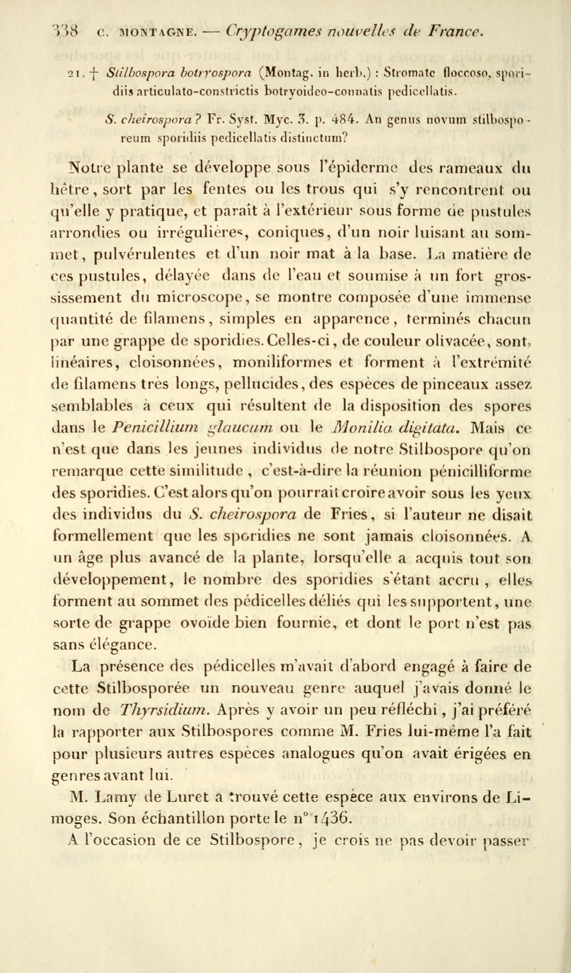
My current theory is that Montagne's Stilbospora botryospora was different from Fries' Stilbospora cheirospora, for reasons that Montagne explains in his description (also the epithets are quite descriptive!).
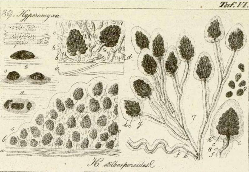
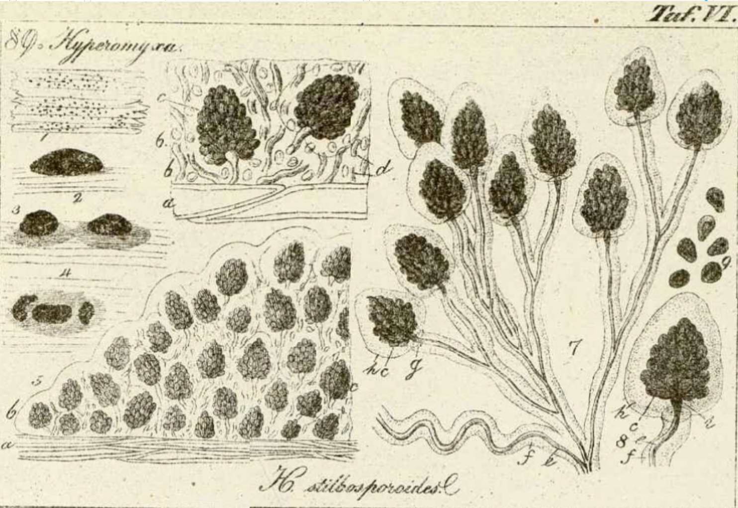
For example, here's an illustration of Corda's Hyperomyxa stilbosporoides that Montagne assumed was the same as his Stilbospora (later Cheirospora) botryospora in 1843 - Note the lack of chain-like digitate spores. In my opinion this drawing is probably also the same as Peter's fungus (and the anamorph of Patellariopsis). Sorry for the poor quality of the file - Perhaps someone has something better?
I have trouble trying to interpret what Fries described....
Regards,
Nick
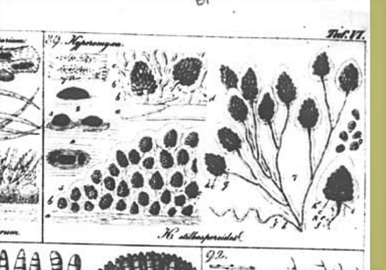

Corda's work is online here: https://bibdigital.rjb.csic.es/viewer/11086/?offset=#page=69&viewer=picture&o=bookmark&n=0&q=
Viktorie
 Clip-9-0001.jpg
Nick
Thanks for your latest interesting contributions and images.
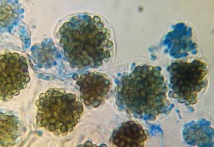
I have had another look at the sample under the microscope and am happy to confirm the following:
a) The hyphae / conidiophores are hyaline or very pale, but certainly not brown
b) The conidiogenous cells are sometimes attached to short branches, but usually to much longer branches.
c) The bunches of conidia, the largest of which have 80 to 100, are not surrounded by gelatinous sheaths
Also, I have rehydrated part of the sample and this has caused a swelling of the thin, effuse layer into low domes. Although adding a few drops of water has not resulted in enough swelling to produce perfect spheres resting on the surface, I think that it is possible that saturating the sample could potentially have this effect over time.
From the available information, it does not seem that either Periconia cambrensis or Cheirospora botryospora are perfect matches for this sample.
With Best Wishes,
Peter.
Initially, I marked him as Cheirospora botryospora.
But it seems that this is not it.
I add to the discussion my photos and some archival works.
Best regards
Mirek
 annalesdesscience26pari-0342-0001.jpg
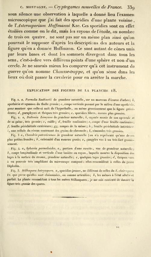 annalesdesscience26pari-0343-0001.jpg
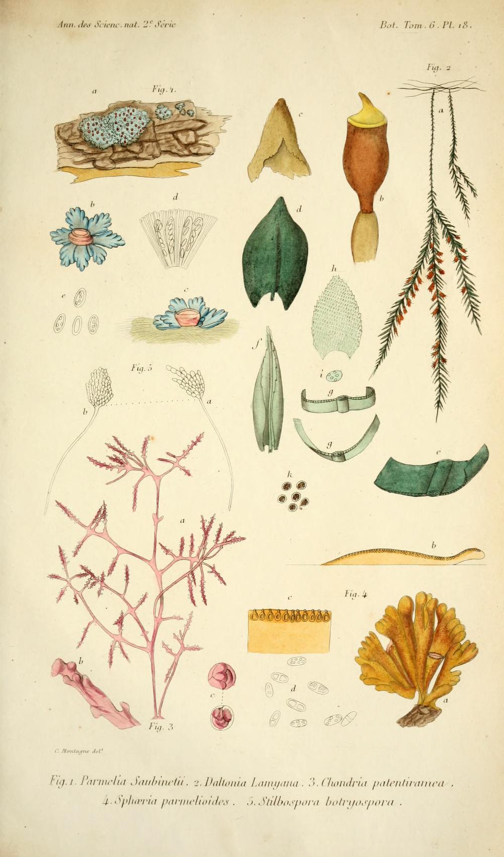 annalesdesscience26pari-0423-0001.jpg
In my collections, the conidia fit the description and illustration of Montagne and were characterised by gelatinous sheathes (easy to miss when mounted in water if you aren't looking for them), long pedicels, etc. although the branches were looser than Montagne illustrated them and sort of resembled a palm tree or top of a pineapple in outline. They fit Sutton's (1980) illustration but Fig. 108 A is either immature or something different, because the chains of cells are not parallel and appear more "botryose", while the conidia (E & F) in Crous' figure seem somewhat more hand-like and more strongly organized into parallel rows than my collections and what Montagne and others illustrated.
It would be nice to compare with specimens of Endobotryella oblonga and especially Endobotrya elegans, which was described from F. grandifolia and could be C. botryospora. I will resurrect some of my C. botryospora cultures and try to sequence them in the near future to see if they correspond with Crous' sequences.
Mirek, it looks like the Cheirospora-like conidia in your third image might be enveloped in a thick gelatinous sheath based on the way the surrounding conidia are crowded away from them?
This topic seems to be becoming more complex. It looks to me as if the species which I have found is the same as those found by Eduard and Mirek.
Zotto's illustration, resembling the fingers of a hand, is different and the species shown by Joey, with a very regular shape to all clusters of conidia looks like a third taxon.
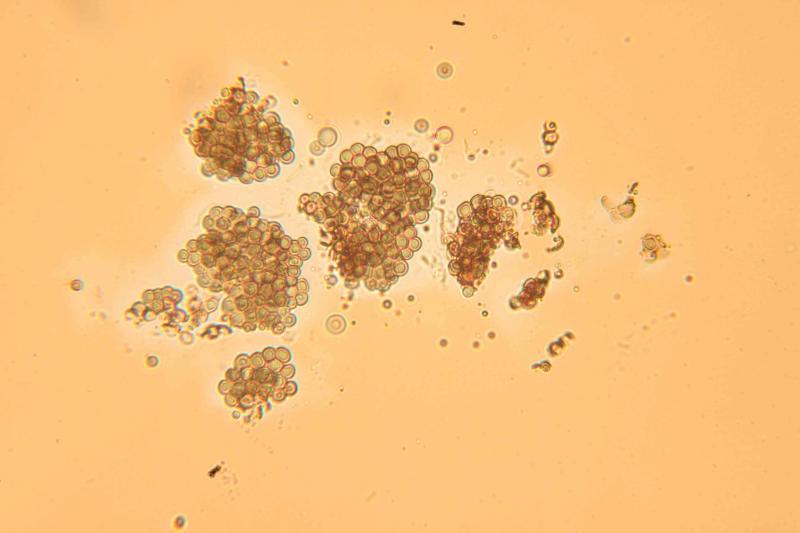
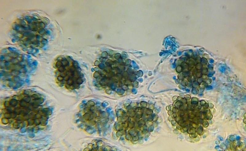
I have looked again at my collection and have now concluded that there is a gelatinous sheath surrounding the clusters of conidia. A photo is attached, mounted in Congo Red, with a very clear, broad, pale zone surrounding the conidia.
With Best Wishes,
Peter.
As you can see mine doesn't have chain-shaped conidia.
So maybe there are actually several species?
This time the substrate was Carpinus betulus wood.
Mirek

I am pleased to be able to say that Nick Aplin has kindly DNA tested this Cheirospora sample and Zotto Baral has cleaned up the resulting sequence.
The conclusion reached is that my sample is 100% identical with Guy Marson's sequences in the ITS region. My sample lacks the S1506 intron which was present in all of Guy's samples.
The sequence data will, I believe, be filed in GenBank so that it is available to anyone who is interested. I can send it by email, with the key data highlighted, if anyone would like to receive it that way.
With Best Wishes,
Peter.
 Cheirospora-botryospora-Karunarathna-et-al.-2020-0001.jpg
 Patellariopsis-atrovinosa-anamorph-Karunarathna-et-al.-2020-0001.jpg
Thanks for drawing our attention to these plates, they illustrate the subtle differences nicely. I think it draws the discussion on this thread to a nice conclusion!
Cheers,
Nick