
17-09-2025 19:43
Philippe PELLICIERSur branche morte de Mélèze. Les ascospores sph�
17-09-2025 10:50
Heather MerryleesHi there!I am hoping for any advice on the identif
11-09-2025 16:57
 Jason Karakehian
Our revision of Marthamycetales (Leotiomycetes) is
16-09-2025 12:53
Philippe PELLICIERPézizes de 1-4 mm, brun grisâtres, sur les capsu
03-09-2025 12:44
 Enrique Rubio
Hi to somebody.I would like to know your opinion o
15-09-2025 14:40
 Nicolas VAN VOOREN
Hello.I'm searching for a digital copy of the seco
14-09-2025 22:16
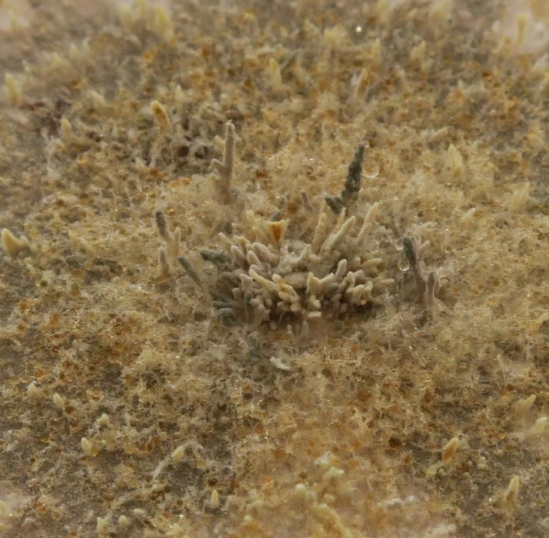
Philippe PELLICIERApothécies petites jusquà 3 mm, oranges, avec de
13-09-2025 14:01
Thomas Flammerdark brown apothecia, splitIKI-Spores biguttulate
10-09-2025 17:18
 Blasco Rafael
Hola, encontre este estiercol de vaca estos apotec
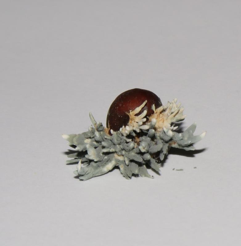
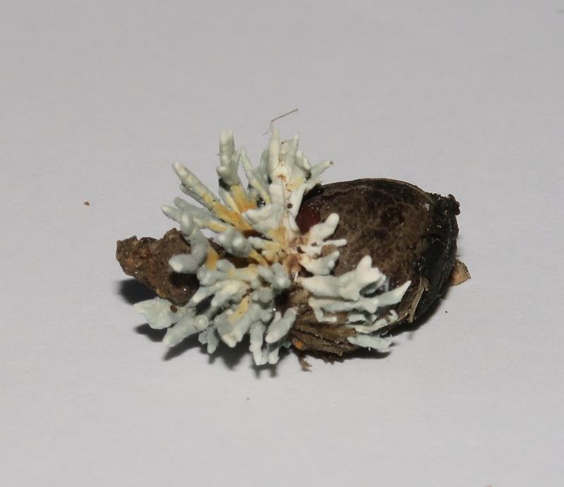
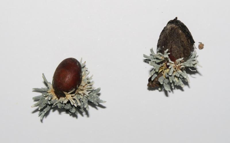
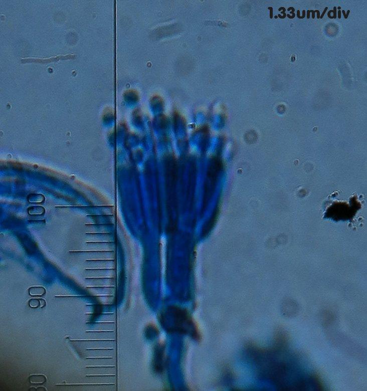
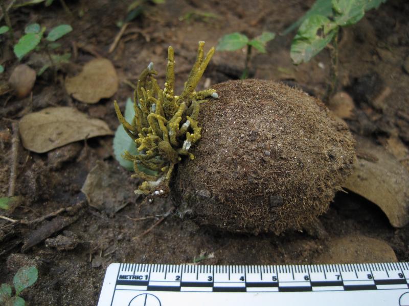
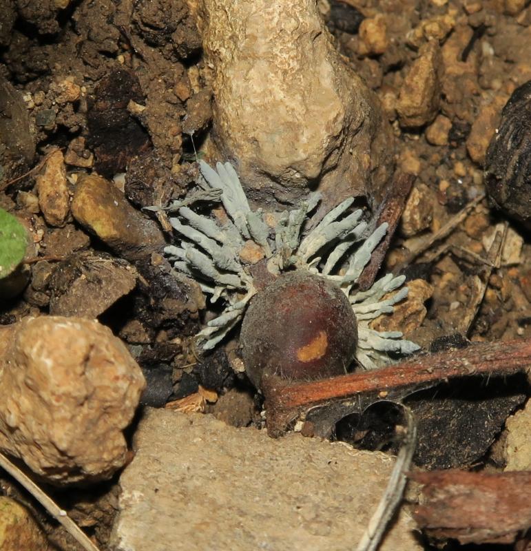
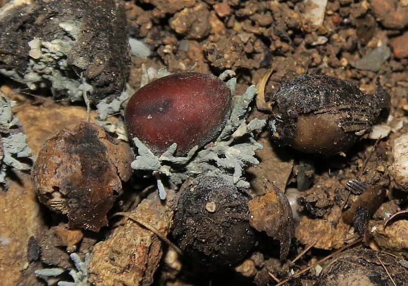
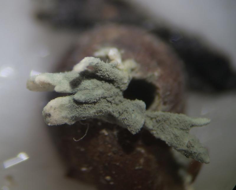
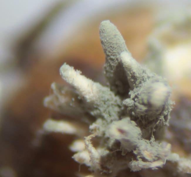
 Hi, I found numerous seeds of Washingtonia robusta (a species of plam tree) on soil infected with a clustered fungus having a club-like shape (Synnemata?) initially white then covered by blue-green spores (reminds me the colour of Penicillium and blue cheese).
Microscopy later tonight, but do have any suggestions (Family / genus) ?
This was picked up from the Mal tese islands

good to see that Ascofrance forum is back. Thanks Christian!
this looks like conidial stromata of a Xylaria. You should fing conidia at the white tips.
Make sure old mature stromata are not present around, they are black and less conspicuous but they would me more informative. Not so many species occur on palm seeds.
Cheers,
Jacques
you are right, definitely not a Xylaria!
Jacques
Thanks.
Lookin forward for you microscopy to compare. I have measurmenst and other photos, but quite useless withour refernces to work through the ID.
When it comes to cultivate fungi on media at home, what advise you can give me to keep as much as possible a sterile environment. I would like to add some chemical to act as an antibacterial agent (what please?) while apart from bacterial contamination, I tend to have Penicillium sp. and Mucor sp. colonies. Just one or two but still it spoils the plate :-( Is there any specific chemical to restrict these unwanted contaminants?
Also, in regards to our fungus, I found this online, have you seen this? http://mushroomobserver.org/observer/index_observation?q=j9Z. We can try to hunt down this literature if you have not seen this?
I have more advise on culturing but too much to write all here. I also have an article that was in Fungi magazine about a home sterile hood from a clear plastic bin but I can't find the reference!! Here's one I found but maybe this is too much for our purposes:
http://www.freshcapmushrooms.com/learn/keeping-it-clean-how-to-design-and-build-a-laminar-flow-hood
September 2001
Pages 509–510.
Let me know if you can't get access to this and I will get it for you. I have a photocopy and no PDF.
Best - Jason
I am indebted for your help, time and useful comments. I can't afford a laminar flow at home (space restrictions) but i try to keep sterile conditions as much as possible. From experience I adopted the following practices.
- When preparing media, I only make a small batch that will be used with a week. Storing media in fridge for a long period will unavoidably lead to contaminations.
- I place my prepared media in a top-sealed plastic bag and then this in a plastic container. This had reduced drasticallly contamination of stored media
- I do not have the facility of an autoclave but I don't think it is essential when preparing small batches. I pour the boiled media in plates when still very hot and cover with aluminium foil while cooling
- Problems of contamination start after inocculation and standing at room temperature. I try to achieve sterility by keeping my tweezers and inoculation needles in alcohol and occassionally put them in a flame of a spirit burner. Still I get 2/5 plates contaminated with Penicilium sp. and Staphylococcus or Pseudomonas. I am confident taht adding a broad range antibiotic like streptomycin will cut down the bacterial growth, but I can't find to buy very easily.
- I found that placing the innoculated dishes in a closed contained while standing helps a bit, but I think that contamination occurs on the process of inocculation, when the plate is open and some spores or bacteria fall on the surface of the media. I never experiened mite contamination.
So that is my home setup, simple and not ideal but soon I shall be working in a mycological Lab with a Laminar flow and hopefully I would get better results.
I did some further research on this synnematous fungus. I have found some depictions of Penicilliopsis palmicola (Sarophorum palmicola) and I have more doubt that our fungus is this species because, if the source I found is reliable, the metulae and branches of this species are distinctly swollen.
http://www.bcrc.firdi.org.tw/fungi/fungal_detail.jsp?id=FU200802290000
Moreover, google images of P. palmicola brings a lot of yellow, long branched synnemata which is different from my specimen?!?!
I could not open the link of mushroom observer that you provided (it opens a non-rlated page) but my samples are definitely the same as these:
http://mushroomobserver.org/58980?q=nB7
http://mushroomobserver.org/221873?q=nB7
or actually all of these:
http://mushroomobserver.org/species_list/show_species_list/611
Interestingly all collections are from South America:
Regarding literature:
The following paper gives a good introduction on the genus but I could not find the full paper:
https://www.jstor.org/stable/3761788?seq=1#page_scan_tab_contents
Another very important paper with keys is this paper:
http://link.springer.com/chapter/10.1007%2F978-1-4757-1856-0_31
If you can find it would be very useful
Another thing I did was to check what species are ther in the genus Sarophorum and Penicilliopsis. In the former I only found two species (S. palmicola and S. ledermannii) while for the latter there were a dozen:
Penicilliopsis Penicilliopsis Solms, Annales du Jardin Botanique de Buitenzorg 6: 53 (1887) [MB#3806]
Penicilliopsis africana Penicilliopsis africana Samson & Seifert, Advances in Penicillium and Aspergillus Systematics: 408 (1985) [MB#114759]
Penicilliopsis bambusae Penicilliopsis bambusae Nag Raj & Govindu (1970) [MB#319256]
Penicilliopsis brasiliensis Penicilliopsis brasiliensis Möller, Botanische Mittheilungen aus den Tropen 9: 293 (1901) [MB#141876]
Penicilliopsis clavariaeformis Penicilliopsis clavariaeformis Solms (1887) [MB#274252]
Penicilliopsis clavariiformis Penicilliopsis clavariiformis Solms, Annales du Jardin Botanique de Buitenzorg 6: 53 (1886) [MB#120178]
Penicilliopsis dichotoma Penicilliopsis dichotoma Hauman, Bulletin de la Société Botanique Belgique 69: 113 (1936) [MB#141964]
Penicilliopsis dybowskii Penicilliopsis dybowskii Pat., Bull. Soc. Mycol. France: 54 (1891) [MB#142485]
Penicilliopsis dybowskii var. dybowskii Penicilliopsis dybowskii var. dybowskii [MB#421814]
Penicilliopsis dybowskii var. macrospora Penicilliopsis dybowskii var. macrospora Beeli, Bulletin de la Société Royale de Botanique de Belgique 59 (2): 160 (1927) [MB#141715]
Penicilliopsis flavidus Penicilliopsis flavidus (Berk. & Broome) A.H.S. Br. [MB#124185]
Penicilliopsis juruensis Penicilliopsis juruensis Henn., Hedwigia 44: 59 (1905) [MB#142319]
Penicilliopsis ledermannii Penicilliopsis ledermannii (Syd. & P. Syd.) Hauman, Bulletin de la Société Botanique Belgique 69: 111 (1936) [MB#269153]
Penicilliopsis longissima Penicilliopsis longissima Hauman, Bulletin de la Société Botanique Belgique 69: 110 (1936) [MB#142089]
Penicilliopsis microsequoia Penicilliopsis microsequoia Hauman, Bulletin de la Société Botanique Belgique 69: 112 (1936) [MB#142431]
Penicilliopsis palmicola Penicilliopsis palmicola Henn., Hedwigia 43: 352 (1904) [MB#247191]
Penicilliopsis pseudocordyceps Penicilliopsis pseudocordyceps H.M. Hsieh & Y.M. Ju, Mycologia 94 (3): 541 (2002) [MB#484663]
Penicilliopsis togoensis Penicilliopsis togoensis Henn., Botanische Jahrbücher für Systematik Pflanzengeschichte und Pflanzengeographie 30: 40 (1901) [MB#246624]
I think I've written a lot today but we keep in touch and hope to find a name for this mysterious fungus
The only thing I can think of here from what you describe is that you might try flaming your needles and tools for every transfer. I would definitely try acidifying the media if you can. Also, did you mention whether or not you wrapped plates in parafilm? I think that this cuts down on some contamination and dehydration. It also prevents me from carelessly picking up a plate by the lid and leaving the bottom on the countertop exposed to the open air! I would always be on the lookout for mites - keep checking those contaminated plates. The mites are so pernicious, they get into everything, including plates wrapped tightly in parafilm - they are, as a colleague once said, the closest thing to evidence for spontaneous generation! I myself have recently lost many plates of hard-gained cultures due to mite infestations. I am glad that you will soon have access to a laminar flow hood. Kind regards - Jason
Those Mushroom observer links that you provided I haven't seen, but definitely on the same track. I'm sorry those links didn't work. I will retry.
I will write to you now using the email address you sent. Best - Jason
A fungus such as this should be fairly easy to culture assuming your media is sterile. Less is often more, so simply touching the conidiophores with a heated-and-cooled pin and streaking it on the plate or doing a three-point inoculation should be more successful than using a lot of material. Avoid opening the plate too much, just lift one side 1-2 cm to allow you to place your pin/needle inside.
Very nice fungus! I hope you can get clean cultures, especailly if it is an unsequenced species.

that paper from Mycologia is online on cyberliber: http://www.cybertruffle.org.uk/cyberliber/59350/0094/003/0539.htm
Viktorie

I am excited to see such interest in these palm seed parasite fungi. They have been a favorite of mine for many years. I am the creator of the species list linked in a previous comment (http://mushroomobserver.org/species_list/show_species_list/611).
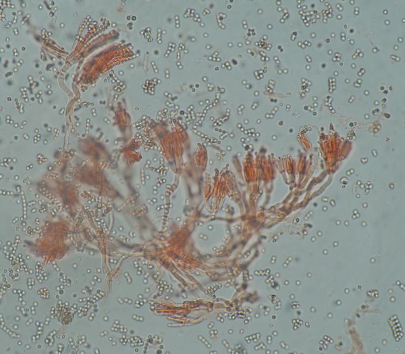
It has been very interesting to see that this also occurs in the old world. Jason's Mozambique material, this collection, and another from Hawaii currently going by the name Penicillium palmae (http://mushroomobserver.org/230574) are the only three I know of from outside the Neotropics with this characteristic abundant blue conidia. How many species are at work here remains a mystery; perhaps one cosmopolitan one, perhaps several specialized ones.
Below is a link to what for me has been one of the best (and only) resources on this group of fungi:
https://drive.google.com/open?id=0B6XMTmQ88ozaM28xRlRWN09xVTQ
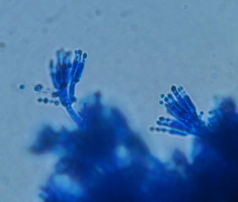
This is the first time I have seen a collection accompanied by microscopy, so it should hopefully be possible to move closer to an identification.
-Danny
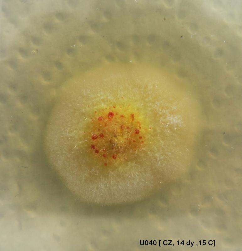
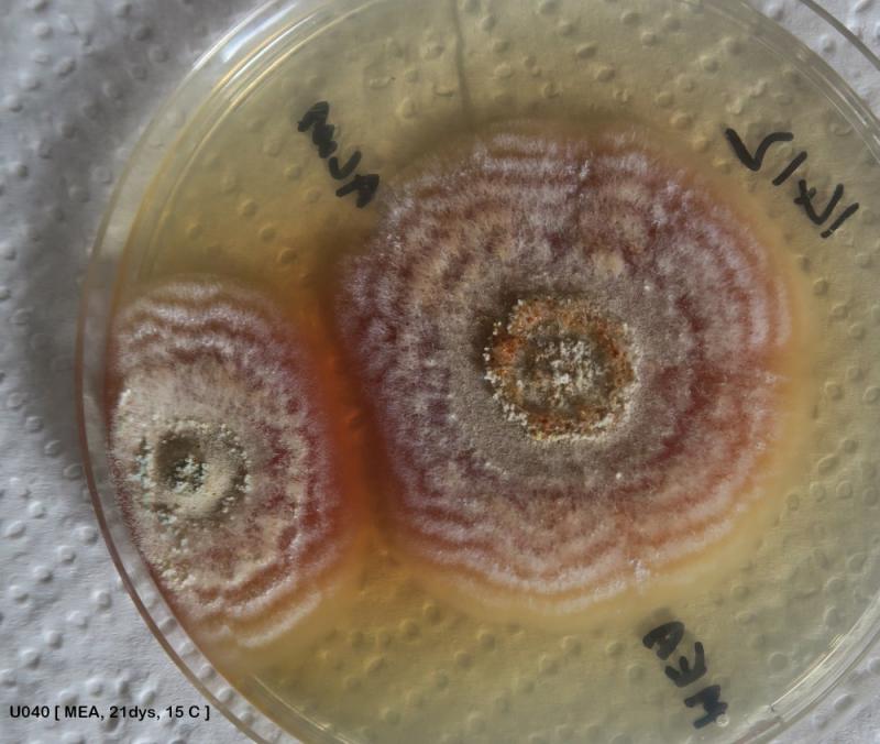
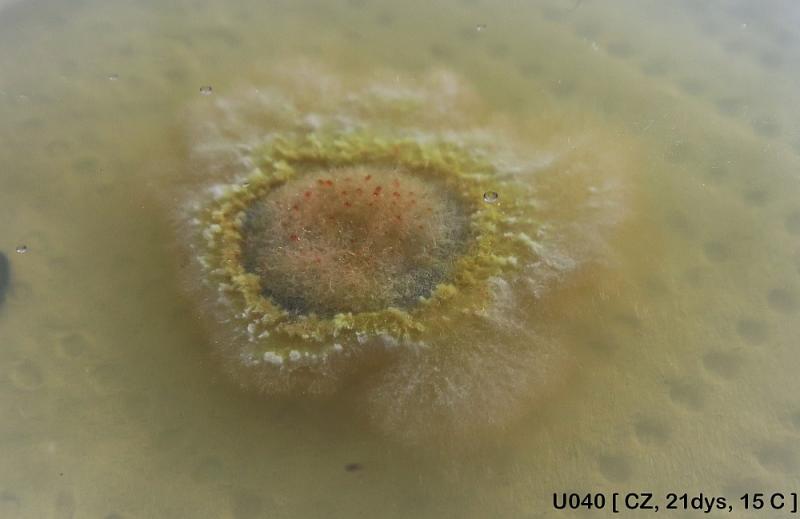
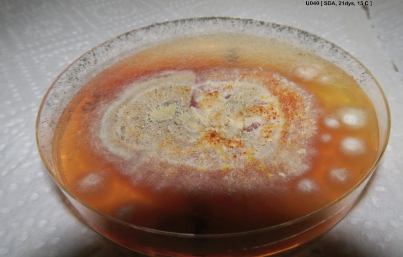
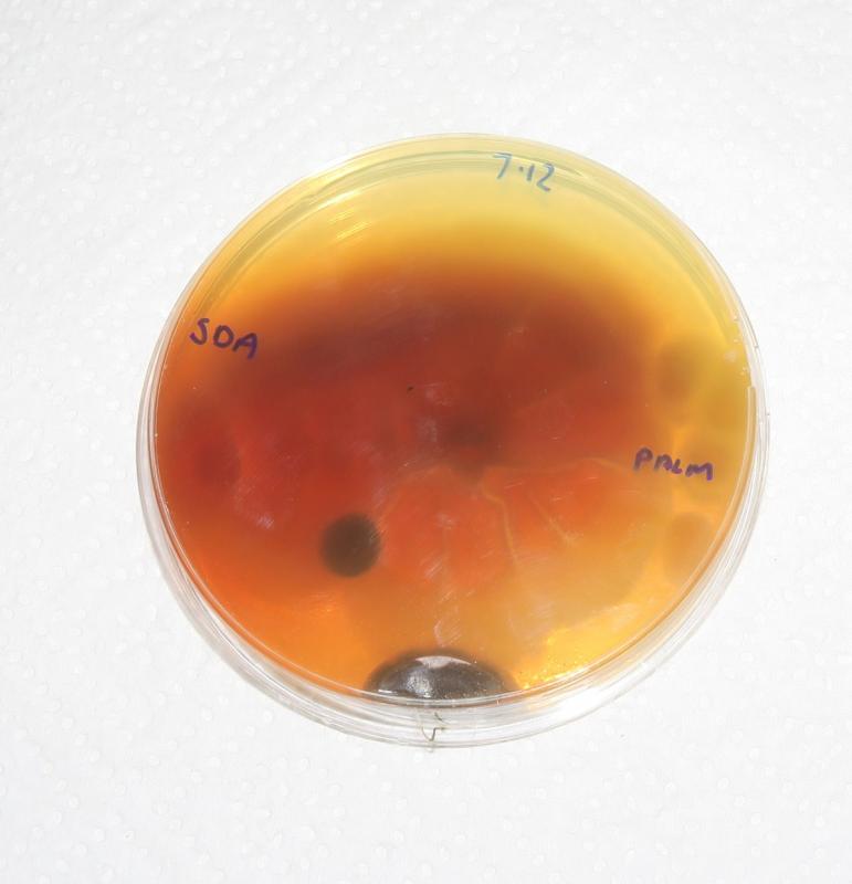
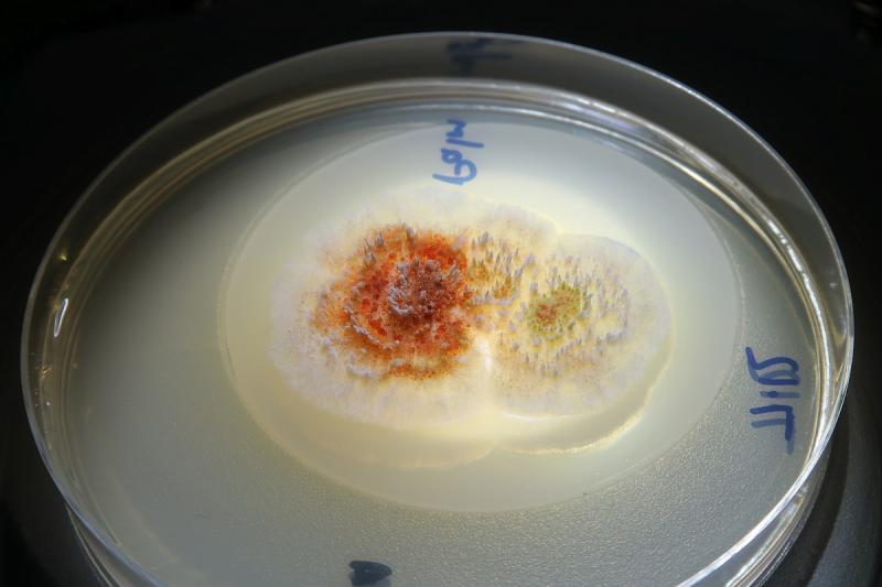
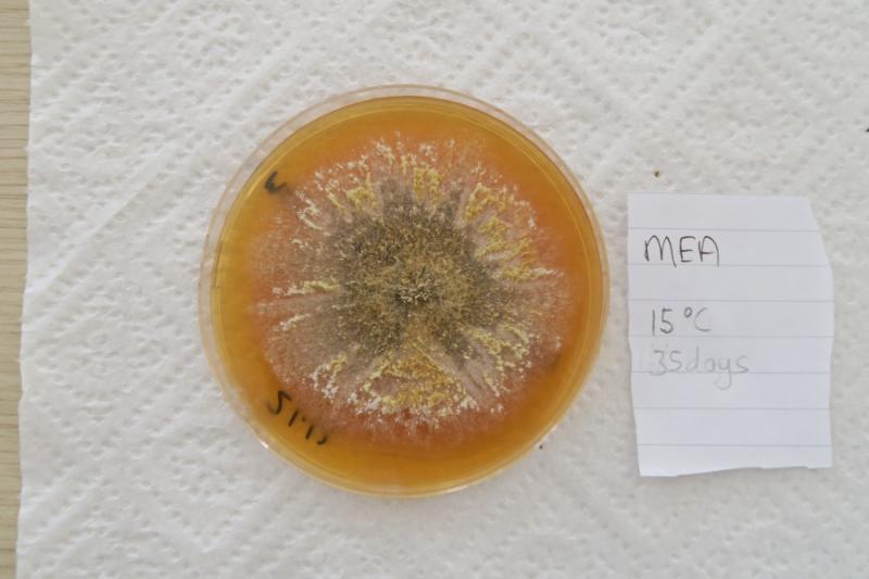
I am also glad that someone out there is also studying this fungus. This year I have seen it again in one of the two locations I know it from here in Gozo. and tried to follow it but I had no time to do the microscopy. However, I had successful isolates on culture media producing outstanding colonies with the production of synemata especially on MEA. As I said earlier, it is always growing on Washingtonia palms. The production of red dye is interesting too. At 15C colony growth is very very slow. I do not have an incubator. I think it is still in my LAB and I can send you some more data and if in a good state, even samples. I have not been in the Lab for a month now :-(
For a moment I understood you described Penicillium palmae, so I ask if you have the protologue of this species, please: Penicillium palmae Samson, Stolk & Frisvad, Studies in Mycology 31: 135 (1989)
Like me, you are not attributing this Washingtonia seed fungus with any species covered by Samson and Seifort? The closest I got was Sarophorum palmicola, but I know it is not that species and so you are suggesting to name this as Penicillium palmae, right?
The conversation on this and other, possibly-related palm seed parasites continues at https://mushroomobserver.org/262509. I hope you will join us there. MO is in great need of more ascomycetologists.