
23-05-2026 11:44
 Charles Grapinet
Hello, I am having trouble identifying this copro
25-05-2026 16:44
François BartholomeeusenHi forum members,During an excursion organised by
26-05-2026 21:25
Dirk GerstnerHello everyone, I'm completely stumped by this li
26-05-2026 22:44
Ethan CrensonHi all, I think I have Incrucipulum capitatum her
22-05-2026 14:44
 Lothar Krieglsteiner
in unripe condition citrine yellow, then soon fadi
25-05-2026 16:35
 Bernard CLESSE
Bonjour à toutes et tous,J'ai trouvé récemment,
22-05-2026 13:29
Gernot FriebesHi,I am curious to hear your opinion on this mater
23-05-2026 18:57
Sylvie Le GoffBonjour à tousRécolté sur une branchette de Sal
22-05-2026 21:35
Steve ClementsBonjour, I expected this find on old wood on our
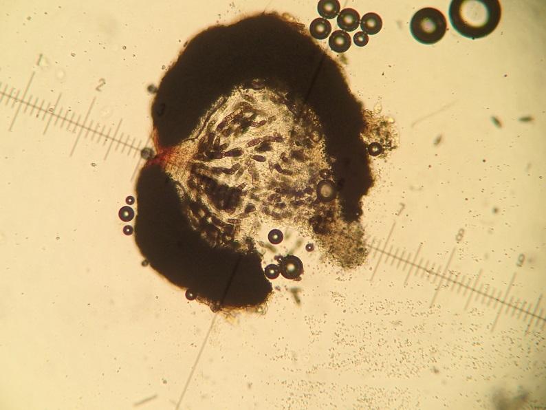
 Hello,
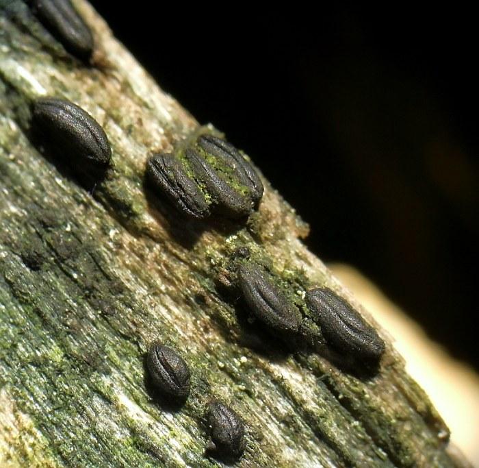
I found that in an herbarium sheet, probably on Fagus sylvatica, in France, Puy-de-Dôme (63). Do any one can confirm / infirm the ID ? I'm not conviced by the general shape...
Many thanks for the help!
Rémy
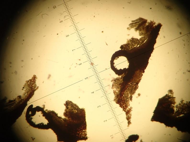
Hi Remy,
although the macrofoto is not very distinctive and does not show the form of the coffee beans I think there can be no doubt because of the very unique spores.
Regards from Lothar
Many thanks for your very quick answer !
I agree that I make very poor quality picture - not sure yet if it is because of the photographer, or the material ;)
In fact, I was not sure for this ID because many herbarium sheets I am currently reviewing (for epiphytic lichens) contains this species, and most of them are more "sphaerical" that coffee bean shaped... All are from the same locality.
Cheers,
Rémy
Hi Remy,
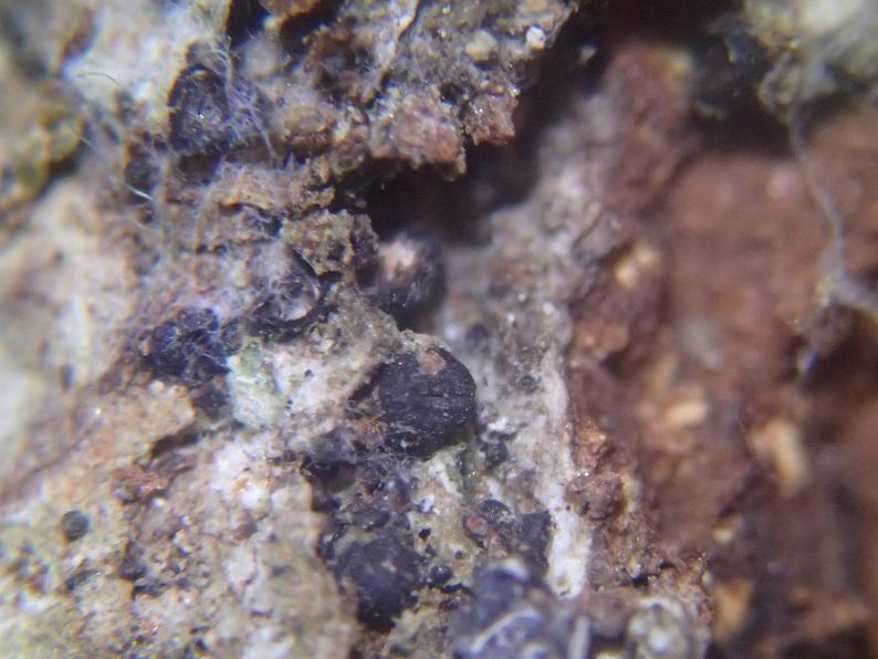
the growth on bark of living deciduous trees is very typical for H. pulicare. But - if the ascomata are not hysterothecia, it must be something different, anyway ...
Best regards from Lothar
Thank you for the precision ; I will try to ID other specimen in order to be sure !
Bests,
Rémy

so Hysterium pulicare would have to look.
Greetings Peter.
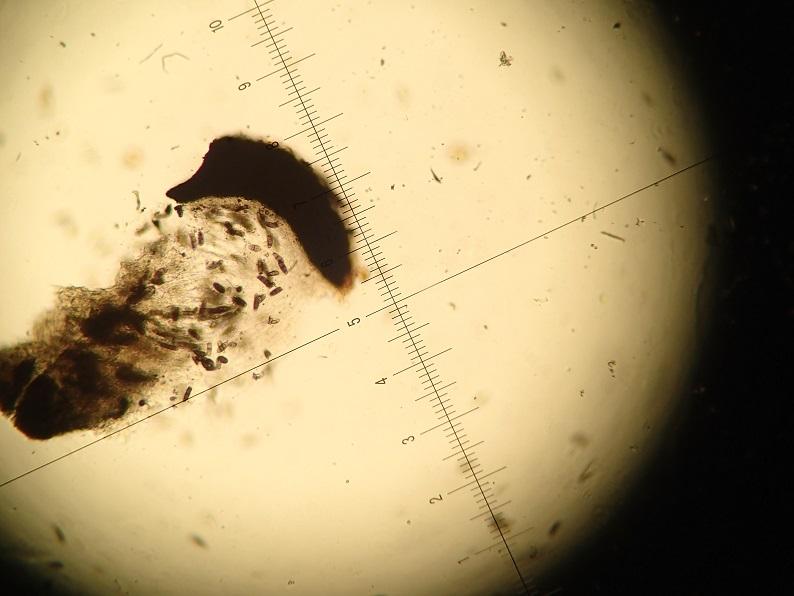
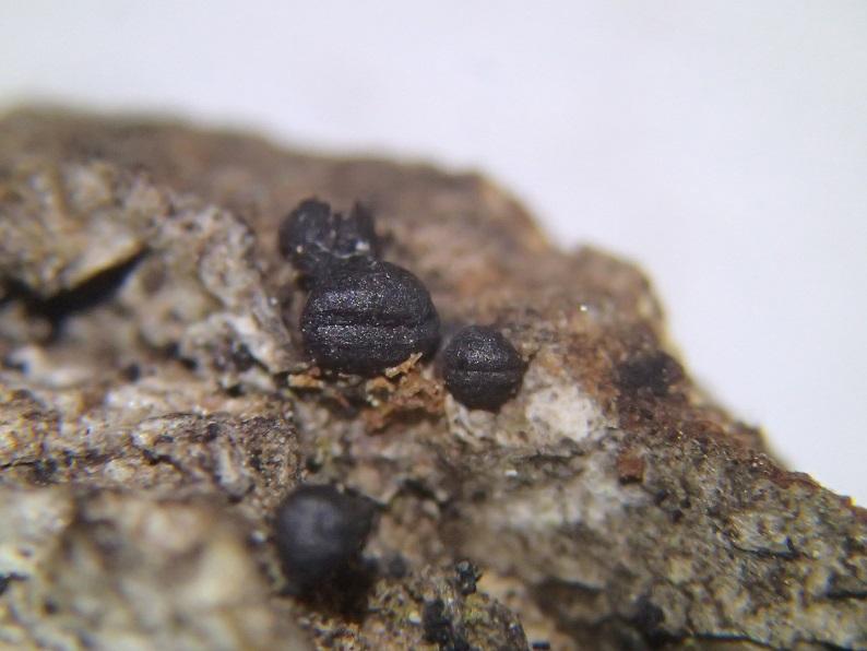
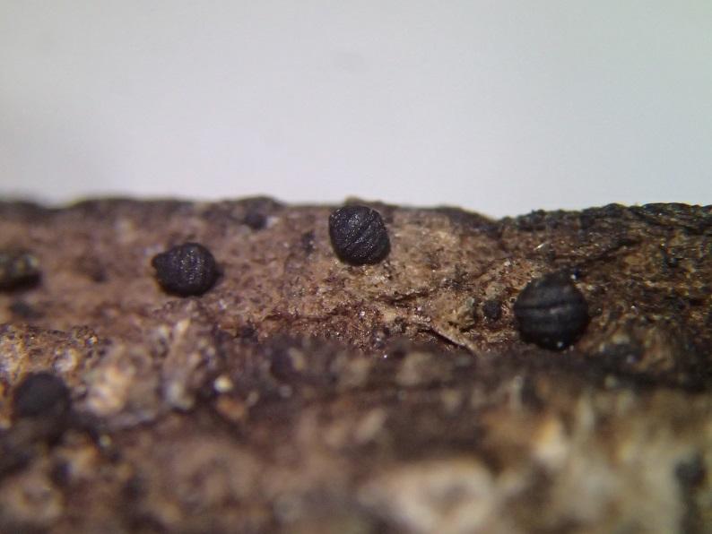
I found a new one in another herbarium sheet. The two pictures are taken on the same piece of bark... On has a coffe bean shape, and the second one not much. Is it beacause of dessication ? Or another species ?
Many thanks,
Rémy
Hi Remy,
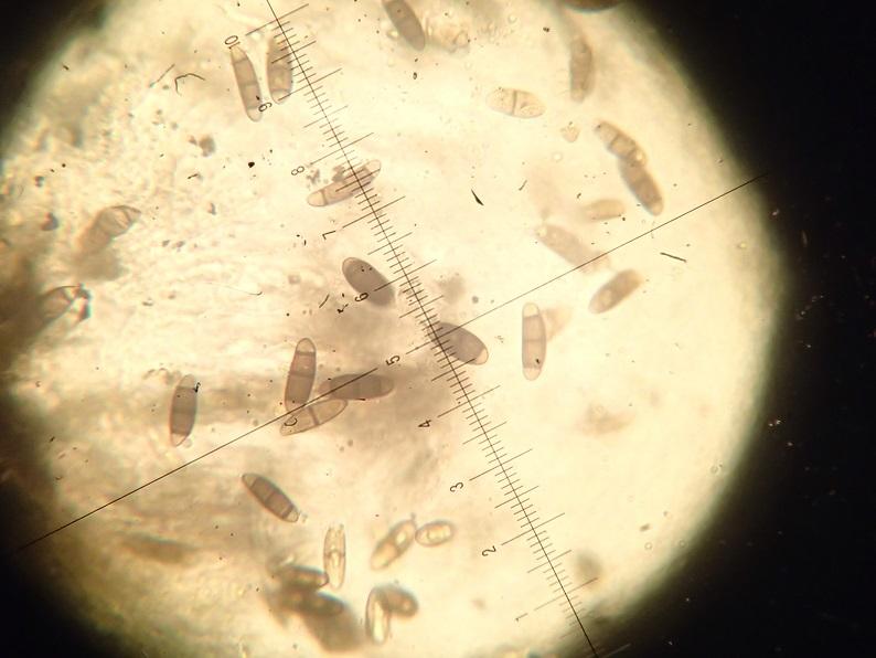
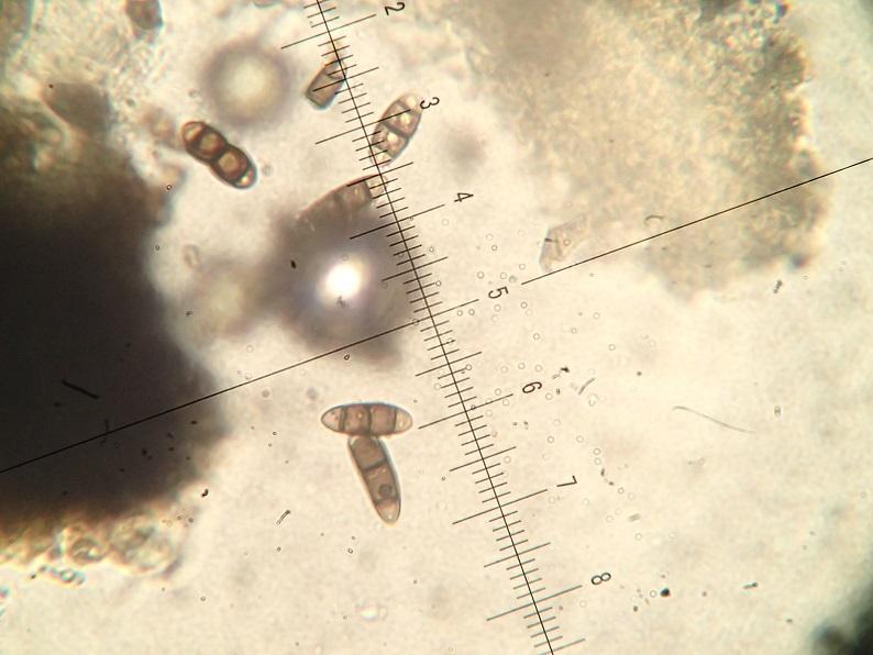
the pictures show clear hysterothecia in my opinion. Do they have the same spores (4-celled with terminal hyaline cells)? Then it ist H. pulicare.
Maybe unripe or badly developed specimens do not form the typical slit very distinctly. Peters fotos are very typical.
Regards from Lothar
this goes not without microscopic examination. If the scales of the photos 1:1 with the micro, it cannot be H. pulicare, the spores would be too small there.
Greetings Peter.
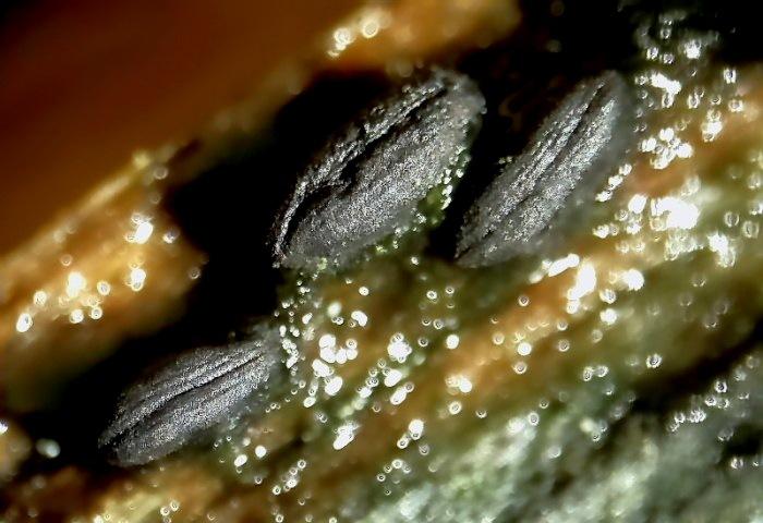
some new picture from the inside. Spores are about ~20-28µm.
Bests,
Rémy
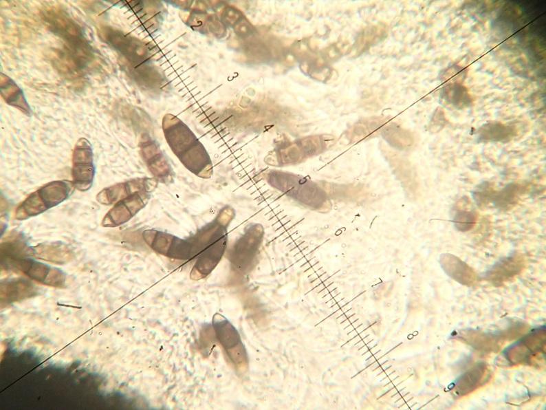
now everything is clear.
Greetings Peter.