
16-03-2011 14:31
roman vargas albertoHi. I would like some opinion about this Peziza
14-05-2026 05:36
Ethan CrensonHi all, I haven't paid much attention to Lachnu
10-05-2026 23:17
 Andreas Gminder
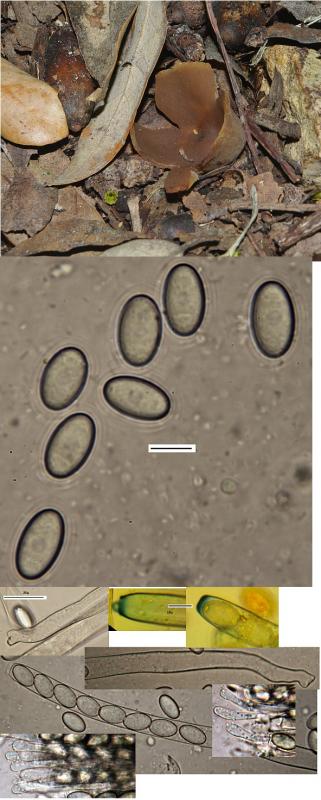
Hello,today we found in a moist steep decidous for
11-05-2026 12:32
 Bernard CLESSE
Pourriez-vous m'aider à identifier cette héloti
13-05-2026 15:26
 François Freléchoux
Bonjour,Voici une récolte faite il y a quelques j
12-05-2026 15:41
 Nicolas VAN VOOREN
Dear Ascolovers, especially interested in Pezizale
13-05-2026 12:05
 Thierry Blondelle
Bonjour à tous,J'aimerais avoir confirmation de c
28-04-2026 20:07
 Lothar Krieglsteiner
... on twig in the air at standing Ceratonia siliq
27-04-2026 20:52
Lothar Krieglsteiner
Found on hanging tiwg of Olea europaea in dried-ou
11-05-2026 20:22
Lothar Krieglsteiner
on attached twig of standing Ficus caricaquite uns
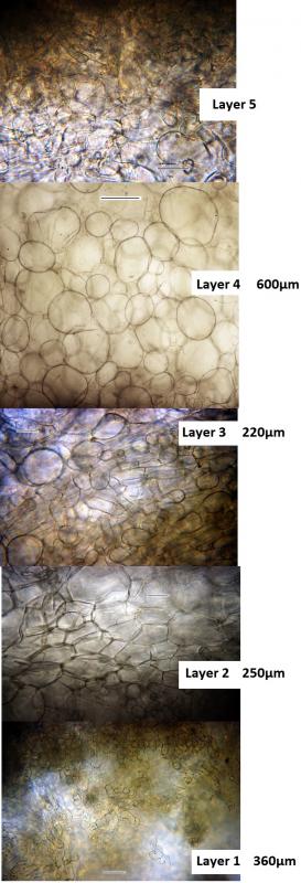
The subhymenium (layer 5 in Donadini) consists of a narrow layer composed of a mix of textura intricata with elements of t. globosa-angularis. Medullary excipulum is made up of 3 distinct layers. The upper layer (layer 4) is wide (600µm) and made up of textura globosa with some cells reaching 175µm in diameter. The middle med. excipulum (layer 3) consists of a narrow layer (220µm) of t. intricata with some globular elements reaching 50µm diameter. The lower med. excipulum (layer 2) consists of a wider (250µm) layer of t. angularis (some elements up to 70µm wide). The ectal excipulum (layer 1) is a dark coloured layer (360µm thick) composed of t. intricata.
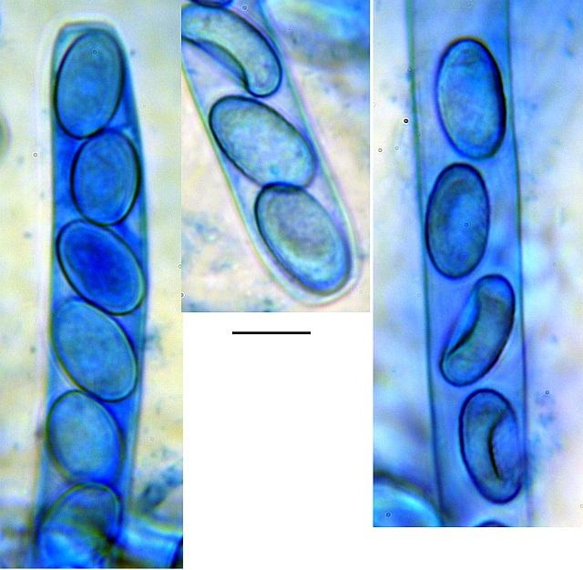
Asci 280 x 13µm, pleurorhynchus base. Paraphyses slightly inflated at the tips (6 - 9µm), septate, hyaline. Ascospores Mean 17.2 x 10.2µm; Qe = 1.69, ellipsoid, hyaline, eguttulate, rough (but no specific ornamentation seen with CB not with oil immersion unfortunately; when examining images, at high contrast, one can see a central oil drop which is not visible normally - perhaps an artifact of my setup.
The species appears somewhere between arvernensis and pseudosylvestris (particularly the wide upper med.excipulum) but P. pseudosylvestris is not a European species. Donadini separates the 2 species by the very wide 2 & 4 layers which this collection appears to have, but the spores do not show particular ornamentation. Has anyone collected P.arvernensis which such wide layers?
P. arvernensis has clearly verrucose ascospores, so it's important to check this character.
I had not considered P. varia as the middle t. intricata should be visible as a distinct layer.
I forgot to post a TS section before. In the attached image there is no distinct middle layer, but I have never seen P. varia, so I may be wrong.
Hello,
in contrast to Nicolas I do believe that the spores are verrucose. In my opinion the smooth spores of Peziza varia agg. are withour any content and are completely smooth. the shown spores do have something inside or on the spores. I believe it is an ornamentation.
best regards,
Andreas
The spores do not appear to be smooth. Varying the focus there seems to be some roughness on the surface but maybe because I did not use oil immersion it is difficult to be certain. Even if the spores are verrucose the medullary excipulum width is rather large for P. arvernensis. This was the reason for my post, as this is the first time I found this species I wanted to have opinions on what to look for to get a more confident identification.