08-06-2026 10:16
 Spooren Marco
I don`t have a clou about this fungus,it is not in
08-06-2026 17:00
François BartholomeeusenGood day everyone, On June 5 2026, I collected de
07-06-2026 15:10
 William Slosse
Hello everyone,On 05-06-26, I found following asco
05-06-2026 11:02
Thomas Læssøehttps://svampe.databasen.org/observations/10596691
07-06-2026 12:09
 François Freléchoux
Bonjour, Voici une brève description de ce qui m
07-06-2026 12:43
Steve ClementsBojour. This was a strange find on a stick on my
12-07-2015 00:05
 Nedim Jukic
This one from the same locality as the previous on
06-06-2026 17:44
Steve ClementsBonjour, This disco was on planed wood 3 x 1.5 cm
14-08-2016 23:15
 Alex Akulov
Dear friendsCan you help me to find the descriptio
 Hi to all
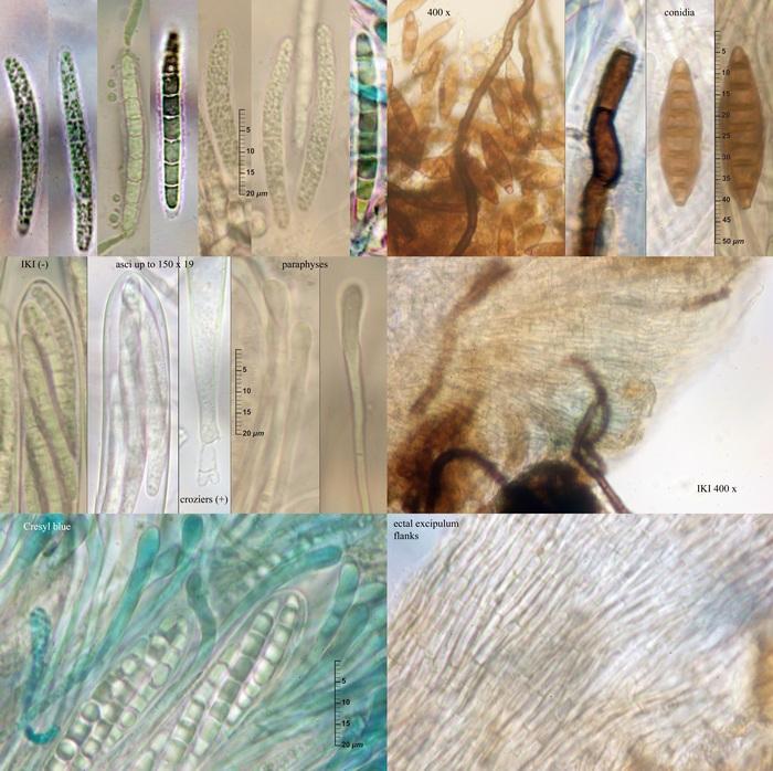
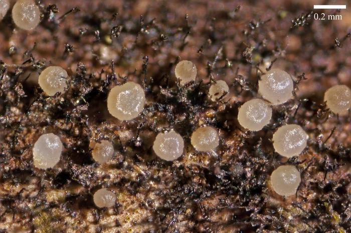
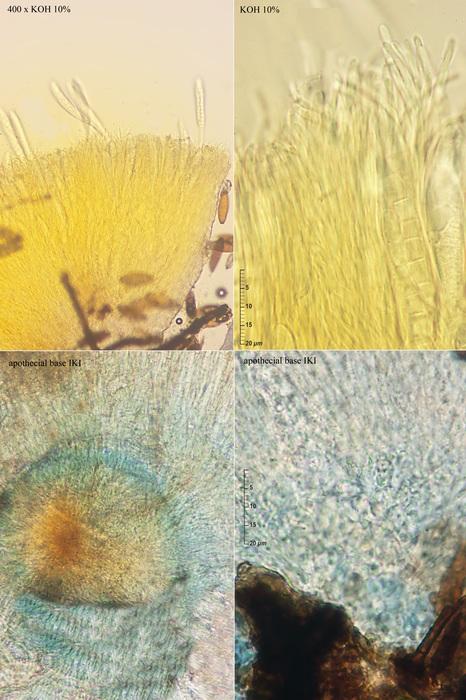
Few days ago I have found these small fruitbodys growing on old wet stems of Rubus grex fruticosus at the sea level. Here is my description of this fungus.
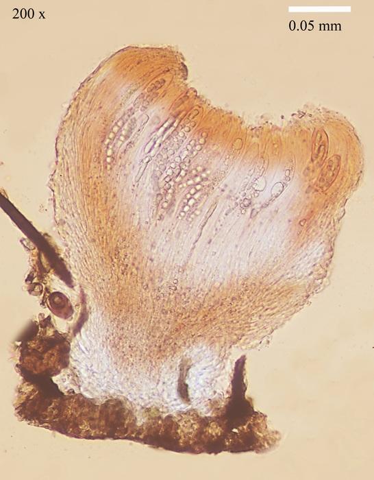
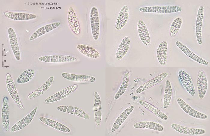
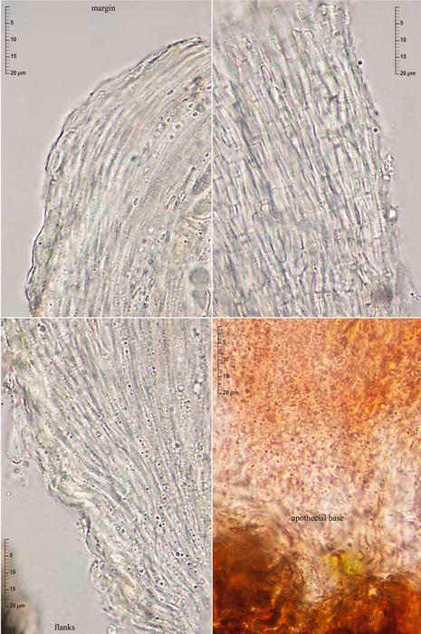
Apothecia superficial, gregarious, scattered among the blackish brown setae of the conidiophores of a dematiaceous mould (Pseudospiropes?), narrowly turbinate, more or less gelified, whitish to amber color, up to 0.30 x 0.25 mm, substipitate on a short and stout pseudostipe 0.12 high and 0.15 broad. Hymenium flattened, smooth or only slightly pruinose. Margin involute, glabrous, that exceeds the hymenium level. Excipulum glabrous and whitish.
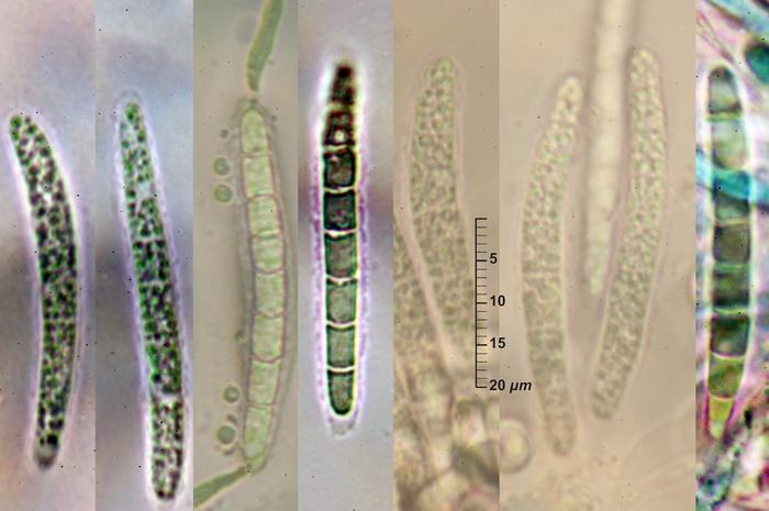
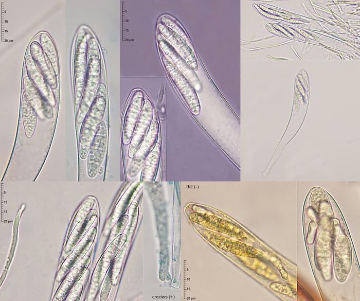
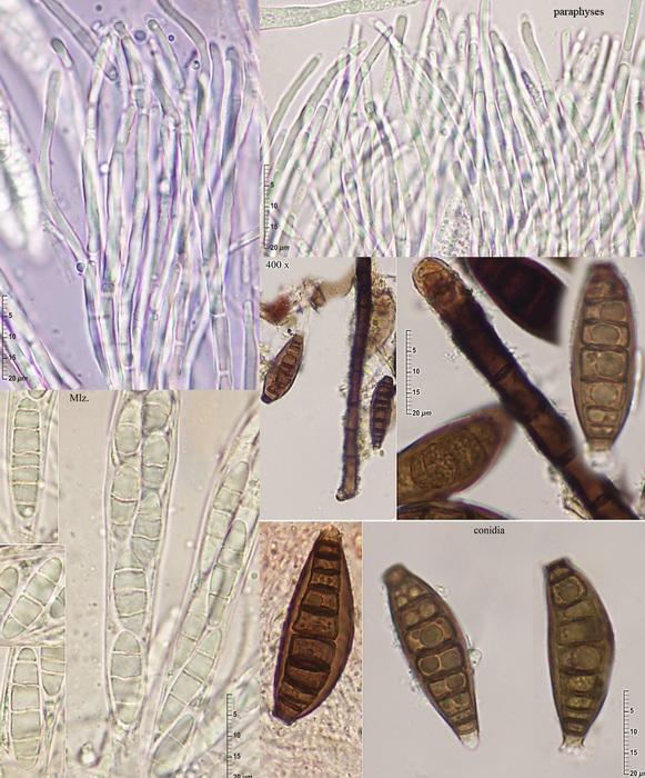
Asci narrowly clavate, inoperculate, unitunicate, 137-182 x 19-20 µm, 8-spored, IKI negative, arising from croziers. Ascospores biseriate, hyaline, smooth, with many small LBs, very polymorphic, ellipsoidal, cylindrical, subfusoid, obovoid, clavate, (19-)30(-38) x (5.2-)6.9(-9.8) µm; Q = (1.9-)4.6(-6.9), with 3(-5-6) cross septa only well visible in Melzer's reactive or congo red. A more or less smooth and complete gel sheath surrounds the fresh ejected ascospores. Paraphyses filiform, septate, not enlarged at their tips or only up to 2-2.5 µm, with cylindrical Vbs. Ectal excipulum textura oblita with cylindrical, elongate, septate, hyaline cells 2-2.5 µm, with a thin gel layer between them, wider at the apothecial base. Medullary excipulum is indistinguishable
The basal region of the ascomata stained blue in IKI. All the apothecial tissues extrude in fresh condition a bright yellow fluid in KOH 10%.
After reading Iturriaga & Korf's paper I don't know a species that fits well with this fungus.
Could you help me?
Thanks again


I have also my problems with Iturriaga's paper, I never came clear with it. But I have also problems to recognize more than one species in this genus. Maybe ther exist tropical species, but in our region I think it is always S. basitricha. Perhaps until someone finds genetical differences.
Did you see anything that is unusual in your fungus compared to S. basitricha on wood?
I remember an amyloidity of the spores in Iturriaga's paper, am I right? Perhaps to obtain only with overmature dead spores, similar as in D. connivens.
Hi Zotto
Yes. I see some differences with the 'common' basitricha on wood with more septate cylindrical ascospores with a narrower verrucose gel sheath and broader paraphyses tips. I have not seen the amyloid reaction on my fresh spores but I pressume that Iturriaga & Korf's observations were made allways on dried material.
This picture was take from an old collection from Robinia pseudoacacia
Thanks again