
23-05-2026 11:44
 Charles Grapinet
Hello, I am having trouble identifying this copro
25-05-2026 16:44
François BartholomeeusenHi forum members,During an excursion organised by
26-05-2026 21:25
Dirk GerstnerHello everyone, I'm completely stumped by this li
26-05-2026 22:44
Ethan CrensonHi all, I think I have Incrucipulum capitatum her
22-05-2026 14:44
 Lothar Krieglsteiner
in unripe condition citrine yellow, then soon fadi
25-05-2026 16:35
 Bernard CLESSE
Bonjour à toutes et tous,J'ai trouvé récemment,
22-05-2026 13:29
Gernot FriebesHi,I am curious to hear your opinion on this mater
23-05-2026 18:57
Sylvie Le GoffBonjour à tousRécolté sur une branchette de Sal
22-05-2026 21:35
Steve ClementsBonjour, I expected this find on old wood on our
Unguiculariopsis-like on Eutypa
Chris Yeates,
01-03-2015 16:25
 Bonjour tous
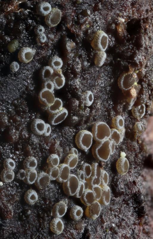
I collected this intriguing discomycete a couple of days ago on a small decorticated branch well covered with Eutypa (I suspect the tree was Acer pseudoplatanus).
When I saw the globose spores and iodine negative (both IKI & MLZ) asci, these combined made me think of Unguiculariopsis, a genus with few British records. I have collected U. ilicincola some years ago (on Ilex), and that is the total of my familiarity with the genus. I find Zhuang's monograph useful, but not completely so.
I would have assumed this to be U. ilicincola but for the fact that the excipular margin is completely different from what I expected. There is no evidence of the uncinate terminal cells one would expect, very well shown in Enrique's http://www.ascofrance.fr/search_recolte/3899
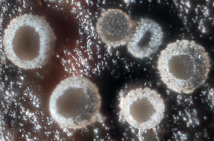
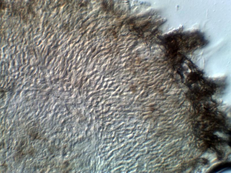
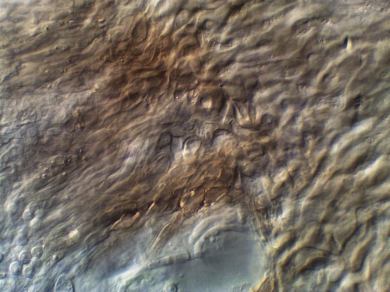
The excipulum consists of thick walled glassy cells which elongate towards the margin where they end as pigmented and lying more or less parallel. In addition a considerable number of these terminal cells bear an ornamentation of small globose bodies, which seem to be a part of the cell wall rather than a superficial coating (for example they were unaffected by 5% KOH). Perhaps the images will explain this better. I shall post images in more than one tranche as there are quite a few.
Details of the fungus are:
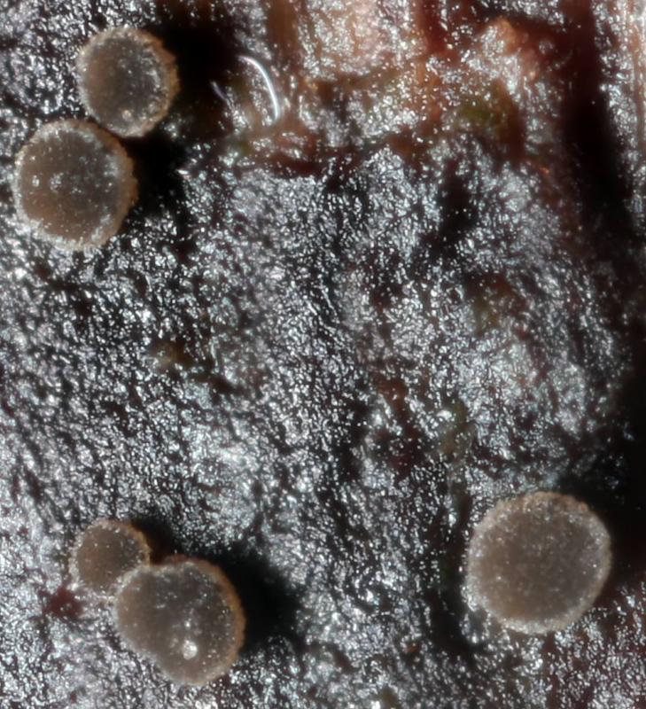
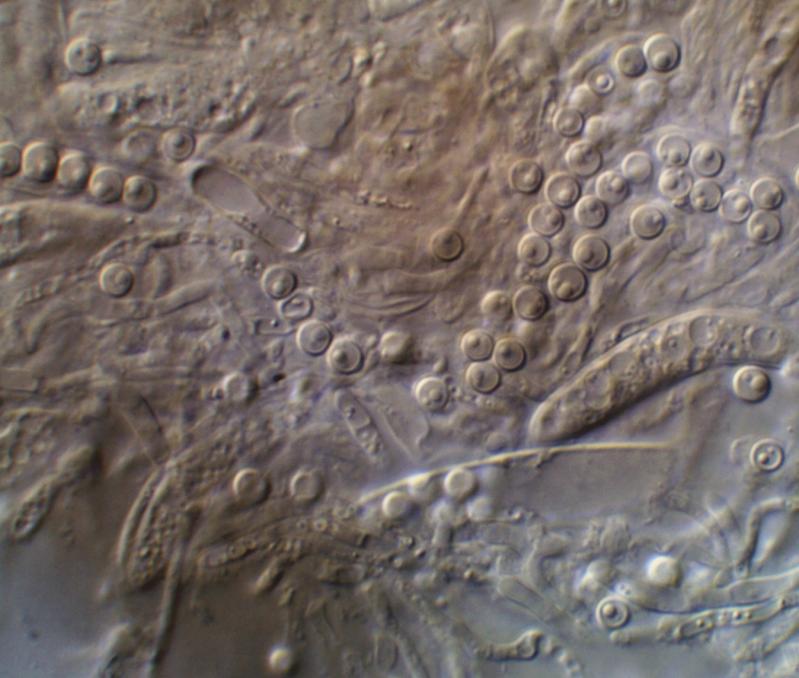
apothecia mostly in the region of 300-400µm when mature on ? Acer pseudoplatanus branch covered by a Eutypa species, many apo's on areas covered with damp algal growth
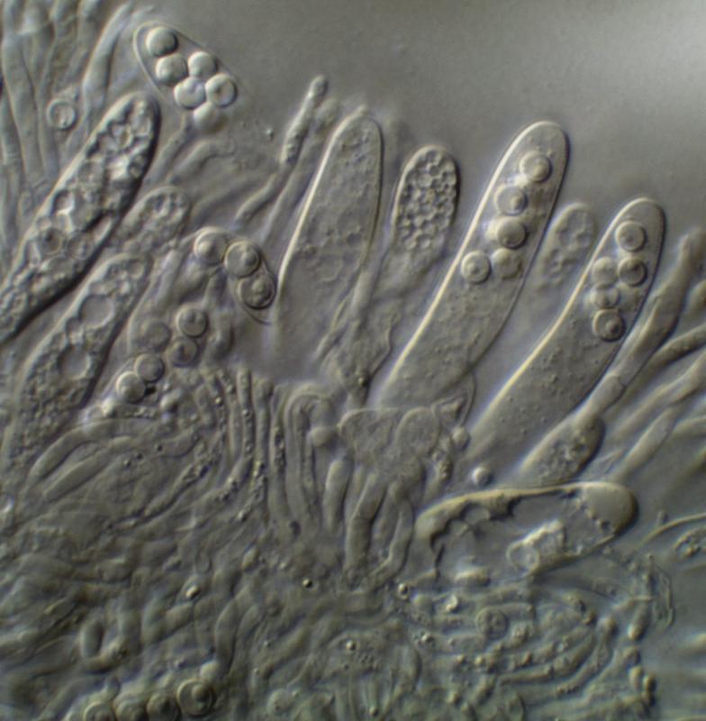
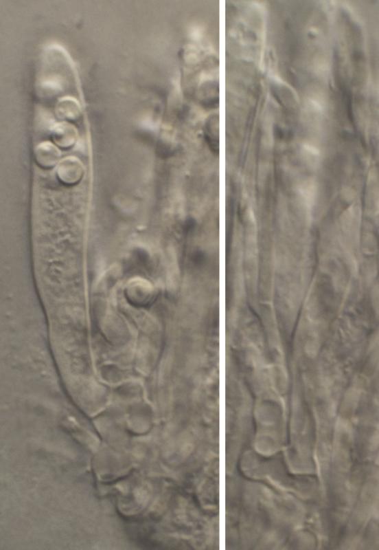
asci 8-spored J-, with croziers
ascospores spherical 2.7-3.3µm (so far too small for ilicincola ?)
paraphyses more or less parallel-sided; in MLZ (but not in IKI) they produced tiny globose iodine (?) particles, as can be seen in one of the photographs
excipulum as explained above (images will follow as a second batch)
any suggestions more than welcome
amitiés
Chris
Andreas Gminder,
01-03-2015 16:38

Re : Unguiculariopsis-like on Eutypa
Hello Chris,
may be a Hyphodiscus, like hypothejus or something similar?
best regards,
Andreas
Chris Yeates,
01-03-2015 17:14
Re : Unguiculariopsis-like on Eutypa
Hallo Andreas
that is a very interesting suggestion, for which many thanks; on checking W. Zhuang's "Notes on Lachnellula theiodea" Mycotaxon XXXI, pp. 411-416 I am pretty sure that I have Hyphodiscus theiodeus as so many of the characters fit perfectly.
It is encouraging to read in that paper a quote from Richard Korf re this taxon " . . . .the ectal excipular layer of this discomycete is unlike that known to me in any other of the 'hairy inoperculate discomycetes' " - I don't feel so bad now about being baffled initially by the structure of the excipulum. I attach the relevant paper for completeness. I shall post my final images in a while. The only 'fly in the ointment' is that I could not see an iodine reaction at the ascus apex - I shall revisit that before drying the material.
LG
Chris
 Mycotaxon-1988-31-411-0001.pdf
Chris Yeates,
01-03-2015 20:13
Re : Unguiculariopsis-like on Eutypa
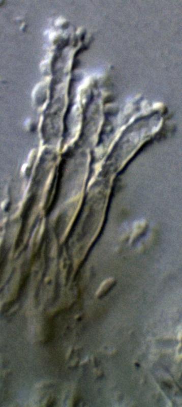
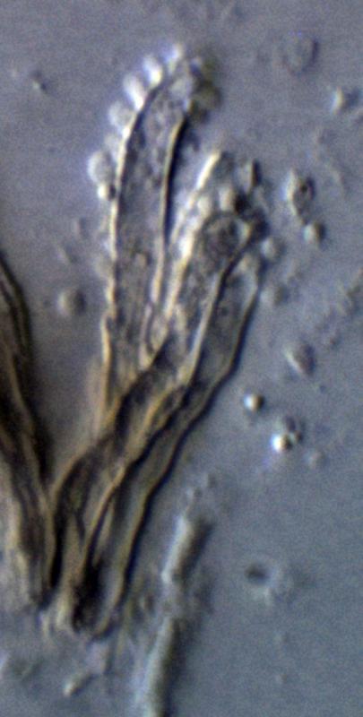
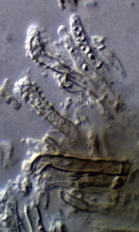
Here are images showing precipitation in paraphyses in MLZ, and the glassy gelatinised ectal excipulum of textura intricata with numerous 'hairs' warted in their upper portion.
I still cannot get a J+ reaction even with pre-treatment in KOH.
I still cannot get a J+ reaction even with pre-treatment in KOH.

Hans-Otto Baral,
02-03-2015 22:46

Re : Unguiculariopsis-like on Eutypa
Hi Chris
I did not read through your description because I thought the case is clear. Now you drew my attention by email to the inamyloid asci. Indeed, usually the species has hemiamyloid rings, while some species with ellipsoid spores exist having inamyloid asci. But I have a single collection of H. theiodeus (HB 8516b) which was also totally inamyloid. Images in Cubby, here my text to them:
Sjaelland, Allindelille Fredskov, Fagus branch c. 1.5 m above ground, on Peniophora. Ap. rehydr. 0.15-0.35 mm diam. Asci *35-45 x 5.7-6(-6.3) µm, with croziers, IKI-!, K+IKI dto. (2 ap. tested). Sp. *2.5-2.7(-3) x 2.4-2.6(-2.8) µm. Paraph. filled with 1refr. small or large guttules far down. Ectexc. near base partly covered by yellow pigment.
Zotto
Chris Yeates,
02-03-2015 23:21
Re : Unguiculariopsis-like on Eutypa
Many thanks for that information, Zotto; this is apparently only the second time this species has been recorded for the British Isles: http://www.fieldmycology.net/FRDBI/FRDBIrecord.asp?intGBNum=45183
Best wishes
Chris
Best wishes
Chris