
28-04-2026 20:07
 Lothar Krieglsteiner
... on twig in the air at standing Ceratonia siliq
04-05-2026 18:13
 Stephen Martin Mifsud
ID request for what seems to be a true aquatic fun
04-05-2026 16:39
Stephen Martin Mifsud
ID request: This specimen was collected in Malta o
04-05-2026 09:50
 Castillo Joseba
Me mandan el material seco de Galicia,(España) re
02-05-2026 12:42
Alain BRISSARDBonjour à tousJeuidi 30 avril dernier on m'a remi
02-05-2026 13:06
Pauline. PennaBonjour Please can someone help me with this id
01-05-2026 22:45
 Thierry Blondelle
Bonjour à tous, Une récolte sur bouse séchée d
14-04-2026 05:32
Ethan CrensonHi all, A few weeks back a friend pointed out som
28-04-2026 20:33
Vitus SchäfftleinHello, I found Trochila ilicina on Ilex aquifoliu
there is some uncertainty with identification of this species.
Large spores of this specimen coincide only with one species in Stomiopeltis (S. betula) though the host differs (Luttrell, 1946; Ellis, 1977). There is a species described from Ledum (S. ledi), but it has much smaller spores (Remler, 1979). It could be possible to consider S. versicolor as well, which also was collected on Rhodod. hirsutum. But it was later transferred to Microthyrium and in "British fungi keys" mentioned with spore appendages. Therefore i inclined to S. betula. Probably you will have other suggestions?
Nina.
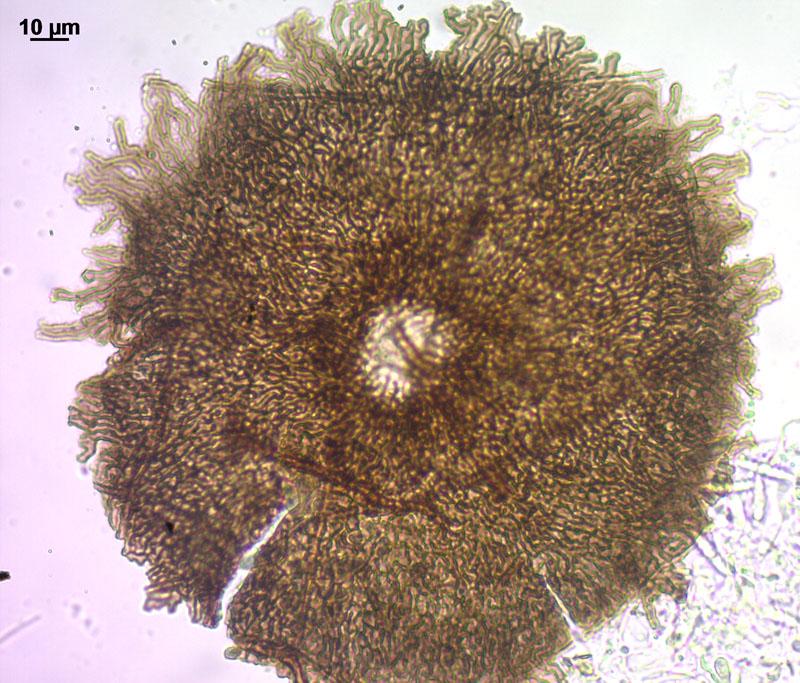
Thyriothecia scattered on upper leaf side (fallen leaves of Andromeda polifolia), dark brown, 150-200 mk in diameter, with visible pore.
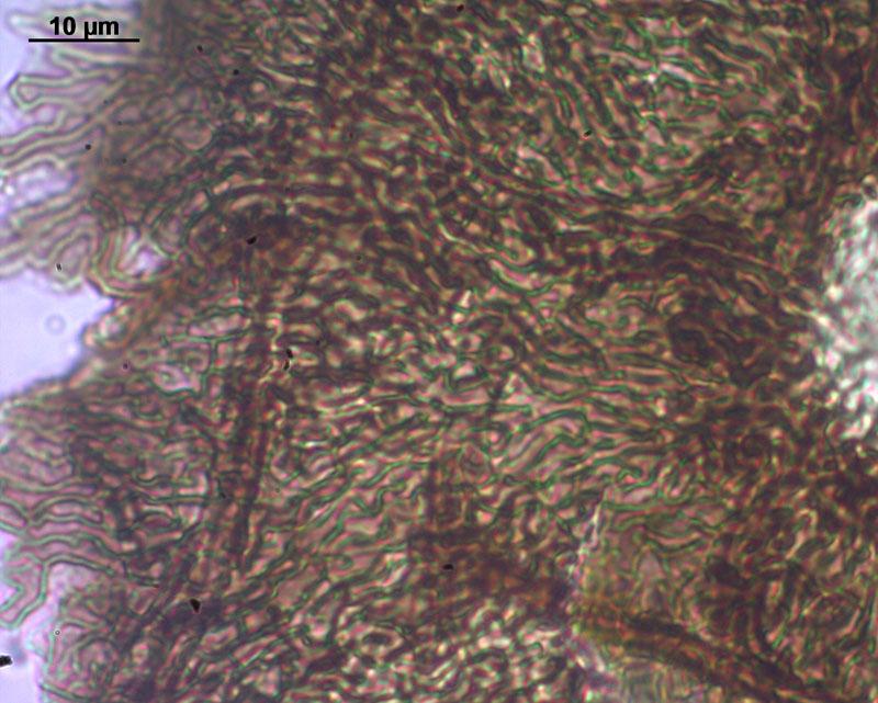
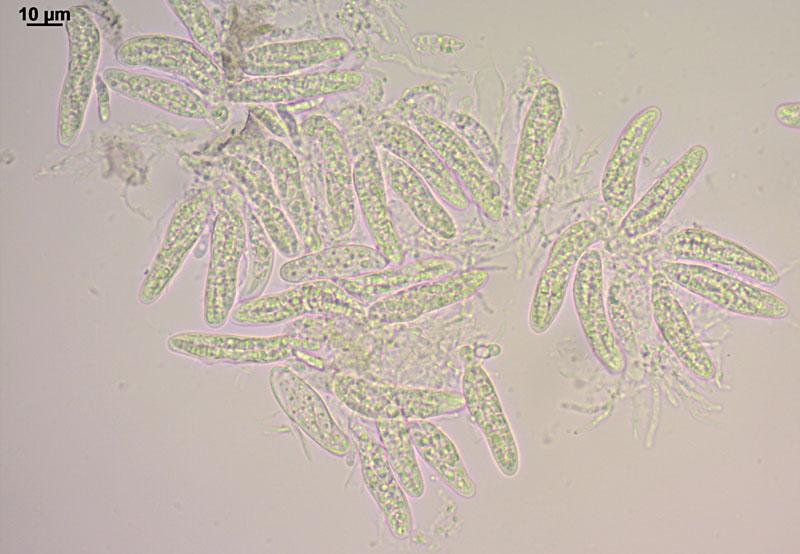
Scutellum from irregular lobed cells, in some ascocarps (probably later in development) cells to the edge become more elongate and radially arranged; asci 37–44 x 8–10 mk; pseudoparaphyses filiform, about 1.5 mk broad; spores fusoid, with many oils, slightly heteropolar, with one weak septa, some curved, 15 (13–18) x 3.2 (2.5–3.9) mk (n=23, measurements in dead state).
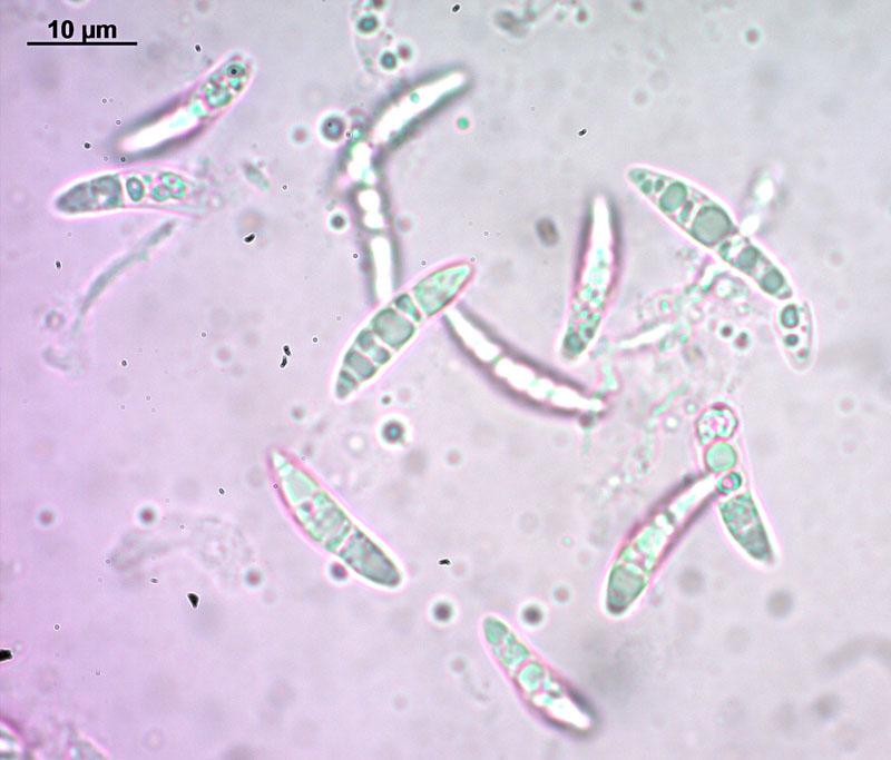

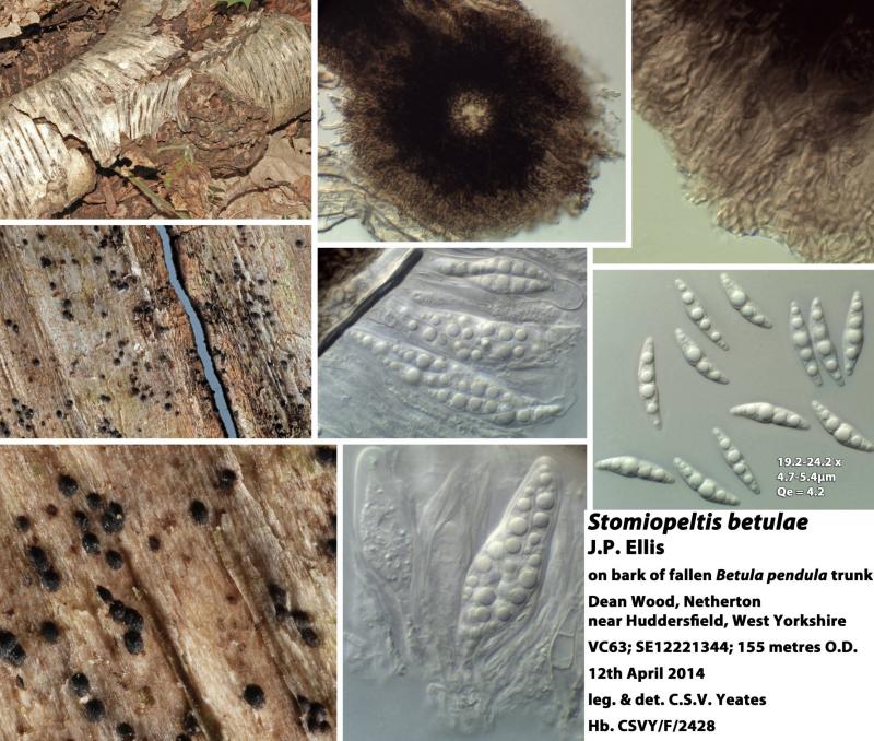
I only know S. betulae on Betula on which it is very common here. My measurements are always towards the top of the range given by Pamela Ellis - see attached image. I think your measurements are too small for S. betulae - perhaps you have an undescribed taxon. Do you have any macro-images?
Cordialement
Chris
thank you for showing me S. betulae. It looks more robust, and yes, spores in my specimen are smaller. I did not make macro-photos because it is barely seen by naked eye, very scattered dots on leaves, better to use lens for them ). I will collect more material of this group to make clear picture later, now will left it under-identified.