28-04-2026 20:07
 Lothar Krieglsteiner
... on twig in the air at standing Ceratonia siliq
11-05-2026 12:32
 Bernard CLESSE
Pourriez-vous m'aider à identifier cette héloti
29-04-2026 10:44
Lothar Krieglsteiner
growing at moist, drying-out soil at the side of a
05-04-2026 22:46
Lothar Krieglsteiner
on wood of Ceratonia, Algarve, 3.4.2026.The color
10-05-2026 16:18
brigitte vignotbonjour trouvée en Ariège sur bois une petite
10-05-2026 23:17
 Andreas Gminder
Hello,today we found in a moist steep decidous for
27-04-2026 17:16
Lothar Krieglsteiner
.. Algarve, moist lying.The conidiomata look like
10-05-2026 09:02
Buckwheat PeteHello everybody, ould this be Lachnum subvirgineu
The meaning of "trabeculate"
Björn Wergen,
28-01-2014 22:17
 Hi friends,
I have one question: what does the word "trabeculate" mean? Its mostly used to describe paraphyses/pseudoparaphyses. I have problems to decide whether the paraphyses are trabeculate or not...
In latin, trabecula means "beam". I think it could be the connections between the paraphyses/pseudoparaphyses?
Thanks in advance!
regards,
björn
Chris Yeates,
28-01-2014 23:51

Re : The meaning of "trabeculate"
From Dictionary of the Fungi:
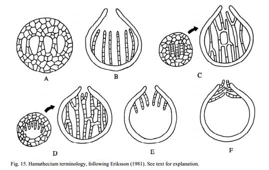
"Hamathecium (Eriksson, Opera Bot. 60: 15, 1981), a neutral term for all kinds of hyphae or other tissues between asci, or projecting into the locule or ostiole of ascomata; usually of carpocentral origin; interascal tissues. Eriksson recognized seven categories (see Fig. 14A-F - below):
(A) Interascal pseudoparenchyma, carpocentral tissues unchanged or compressed between developing asci; e.g. Wettsteinina.
(B) Paraphyses, hyphae originating from the base of the cavity, usually unbranched and not anastomosed; e.g. Pyrenula, Xylaria.
(C) Paraphysoids (trabecular pseudoparaphyses; tinophyses), interascal or pre-ascal tissue stretching and coming to resemble pseudoparaphyses; often only remotely septate, anastomosing and very narrow (see Barr, Mycol. 71: 935, 1979); e.g. Patellaria, Melanomma.
. . . . . . . . "
regards
Chris