
23-05-2026 11:44
 Charles Grapinet
Hello, I am having trouble identifying this copro
25-05-2026 16:44
François BartholomeeusenHi forum members,During an excursion organised by
25-05-2026 16:35
 Bernard CLESSE
Bonjour à toutes et tous,J'ai trouvé récemment,
22-05-2026 13:29
Gernot FriebesHi,I am curious to hear your opinion on this mater
23-05-2026 18:57
Sylvie Le GoffBonjour à tousRécolté sur une branchette de Sal
22-05-2026 14:44
 Lothar Krieglsteiner
in unripe condition citrine yellow, then soon fadi
22-05-2026 21:35
Steve ClementsBonjour, I expected this find on old wood on our
22-05-2026 18:12
Lothar Krieglsteiner
... in moist chamber from Portugal.As the fungus s
22-05-2026 20:08
Ethan CrensonHello all, Yesterday in NYC I was visiting an e
this beautifil species was collected several times at leaves of Rubus chamaemorus. Could be from Rutstroemiaceae, but i have not succeeded in finding necessary description there. Four related species which could be found at this host: Sclerotinia tetraspora, Ciboria latipes, Scleromitrula rubicola, Rutstroemia chamaemori - all have different spores (no mention of allantoid shapes).
May be somebody is familiar with this?
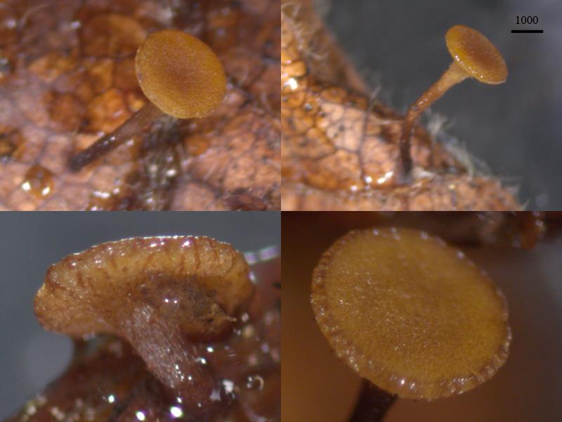
Apothecia cupulate, stipitate, 1.3–4.7 mm in diameter, stem 0.8–2 mm high, site densely at both leaf sides, without sclerotia but black stromatized lines are present at the leaf; reddish-brown, hymenial surface minutely speckled, outer surface longitudinally rugose, stem base dark to dark brown.
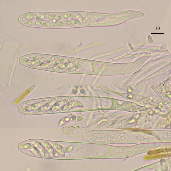
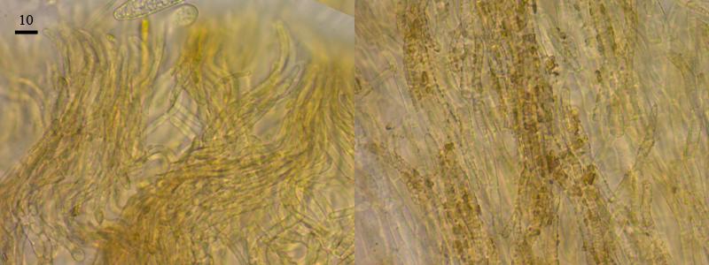
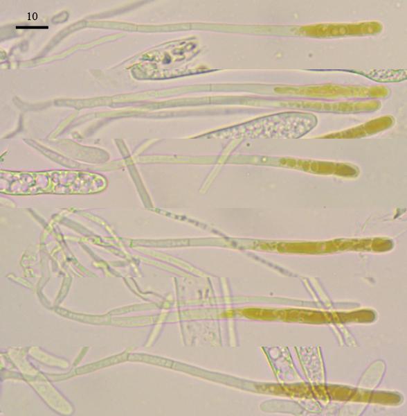
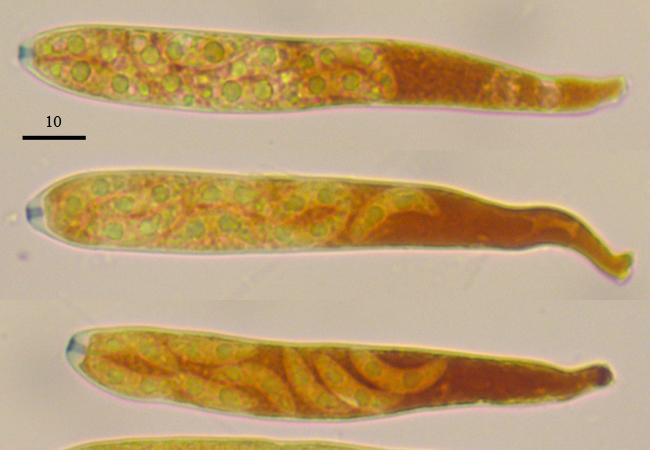
Excipulum from porrecta, outer hyphae incrusted by brown pigment, at the edge not enlarged hyphoid elements; asci with crozies, euamyloid ring, 97.6–125 x 12.5–14; paraphyses cylindrical, enlarged to upper part, rarely branched, septated, with brown content in upper part, about 113 x 4.4 (width at upper part); spores allantoid, with two medium oils and several tiny, 17 (15.4–18.9) x 4.7 (4.1–5) (n=11).
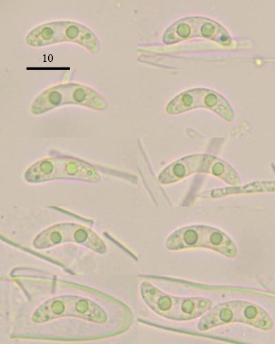

indeed very interesting! I looked up the protologue of R. chamaemori and also see that your spores are longer and much more curved. My reproduction of the photos is not very good, maybe someone has a pdf with better quality? Perhaps Chris?
It is Holm & Holm 1977, Kew Bull. 31(3):567-572
In R. firma I noticed variation in spore curvature, maybe this is the reason also here. Were the spores always such in your finds? The spores on the photo of Pl. 25C in Holm look like having only small polar guttules, but I am not sure. the description says simply "guttulatae" which does not help.
Zotto
there are raw pictures of the specimen,
https://www.cubby.com/pl/%234364/_3840835779bb44b195cf01598cd04670
I guess they all are done from one apos, but the specimen is not at hand now and i am not sure about spore shape variation (i will reply with this in three weeks when reach the collection).
In this examined apos all spores were that curved.
Nina.?