
12-06-2026 14:50
 FranûÏois Frelûˋchoux
Bonjour, Voici la brû´ve description d'une Mollis
10-06-2026 21:16
FranûÏois Frelûˋchoux
Bonsoir,Le dernier du jour, en attendant votre avi
11-06-2026 19:01
 William Slosse
Hello all,In an attempt to make a culture of a sus
11-06-2026 19:03
 Nicolas VAN VOOREN
Chers membres d'Ascofrance,Le site sera placûˋ en
10-06-2026 23:08
 ûˋric ROMERO
Bonjour tous, Je vous propose un Mollisia trouvûˋ
09-06-2026 18:32
Camille MertensSur morceau de roseau immergûˋ 0,5 - 0,7 mm de dia
10-06-2026 12:54
Steve ClementsBonjour encore, Pouvez-vous m'aider, s'il vous pl
10-06-2026 21:07
FranûÏois Frelûˋchoux
Toutes les tiges de gentianes jaunes de l'an passû
10-06-2026 13:41
FranûÏois Frelûˋchoux
Bonjour û nouveau, Voici une trouvaille d'hier.
Hymenobolus agaves anamorph
Miguel ûngel Ribes,
12-03-2013 00:39
 Good night
Perhaps someone remember this Hymenobolus agaves: http://www.ascofrance.fr/search_forum/10909
Rubûˋn has foung more collections in another Canary Island, La Gomera. In some collections, between H. agaves apothecium, are growing too a white-orange anamorph, 2-5 mm broad, relatively hard (it is posible to cut it).
Is it posible the anamorph of H. agaves? How to study this anamorph?
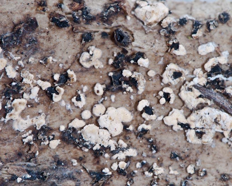
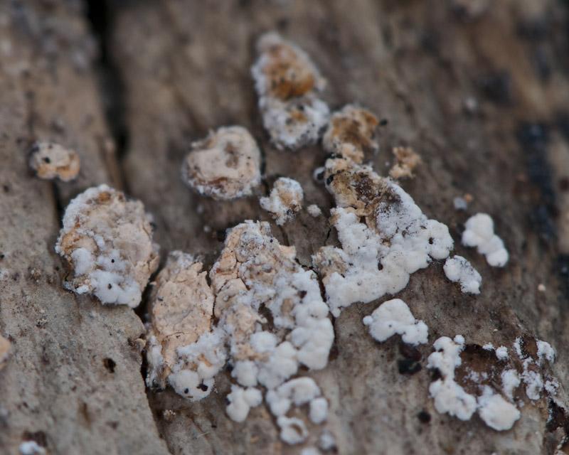
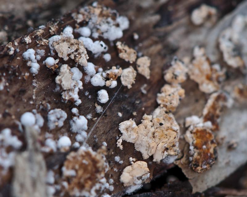
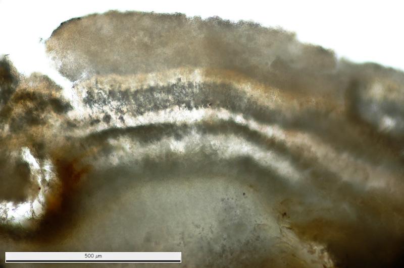
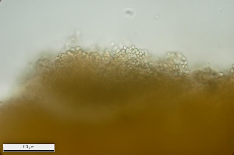
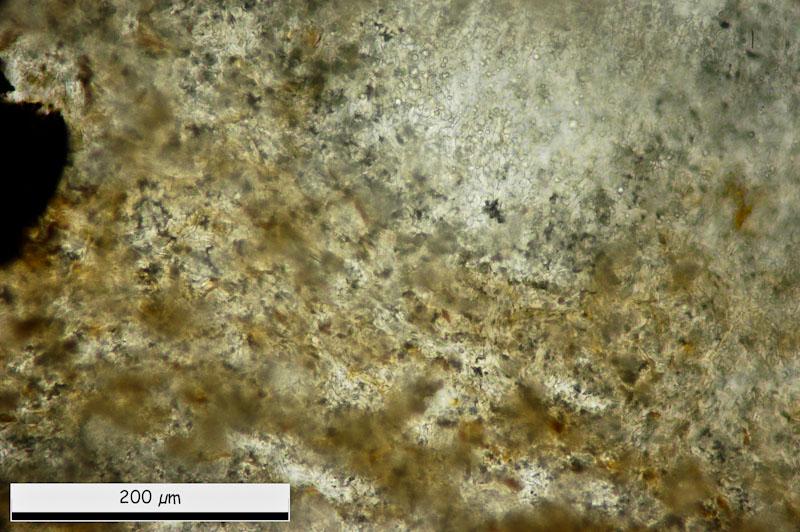
This are general views.
Thank you.
Miguel ûngel Ribes,
12-03-2013 00:44
Re : Hymenobolus agaves anamorph
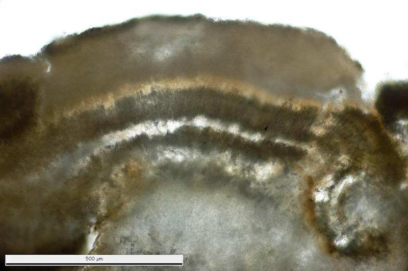
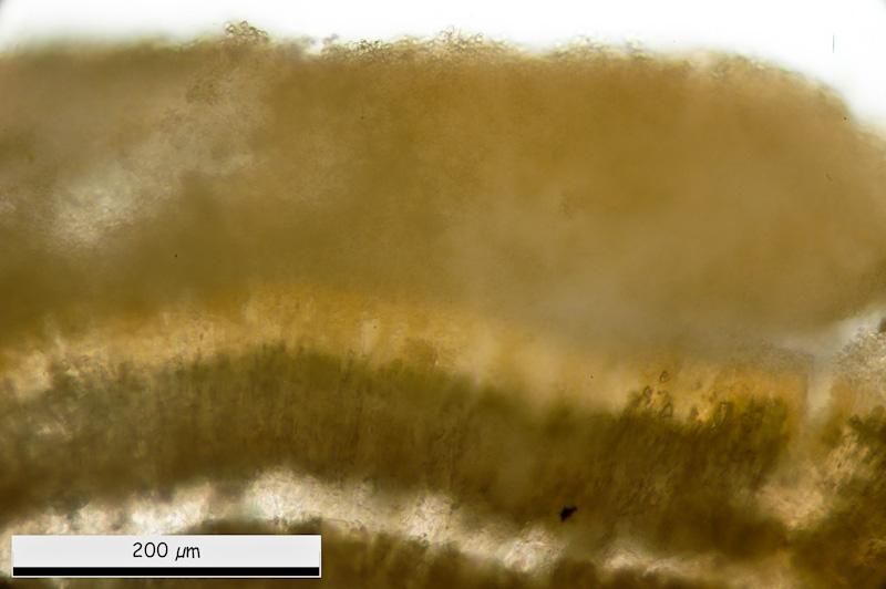
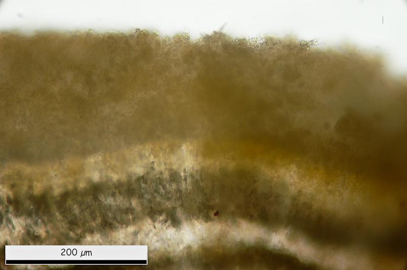
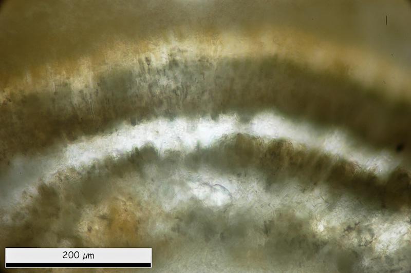
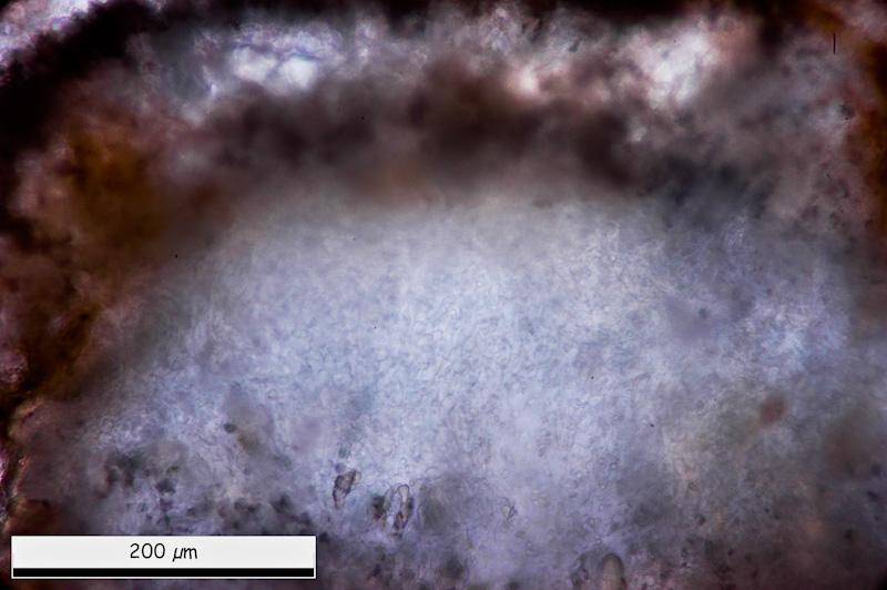
General micro views. It is posible to see some layers. External one with more-less rounded cells.
Miguel ûngel Ribes,
12-03-2013 00:50
Re : Hymenobolus agaves anamorph
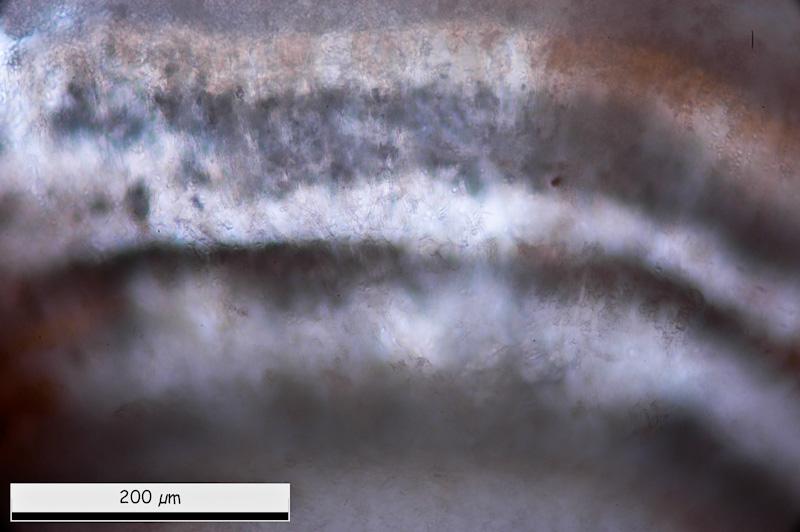
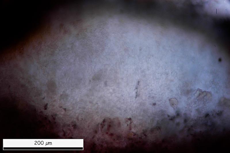
Inside, tow black lines. And in medular area a white area with globose-angular structure mixed with cilyndrical cells.
Miguel ûngel Ribes,
12-03-2013 00:53
Re : Hymenobolus agaves anamorph
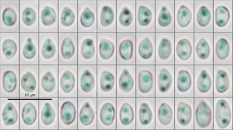
Eliptical conidiospores in water:
(4.06) 4.49 - 5.21 (6.63) x (2.87) 2.94 - 3.35 (3.57) ôçm
Q = (1.28) 1.40 - 1.70 (1.86) ; N = 52
Me = 4.88 x 3.14 ôçm ; Qe = 1.56
(4.06) 4.49 - 5.21 (6.63) x (2.87) 2.94 - 3.35 (3.57) ôçm
Q = (1.28) 1.40 - 1.70 (1.86) ; N = 52
Me = 4.88 x 3.14 ôçm ; Qe = 1.56
Miguel ûngel Ribes,
12-03-2013 00:55
Re : Hymenobolus agaves anamorph
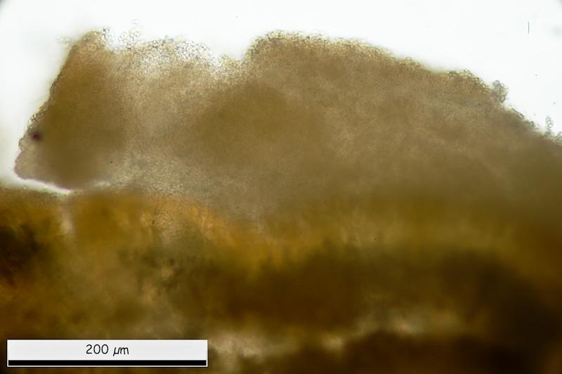
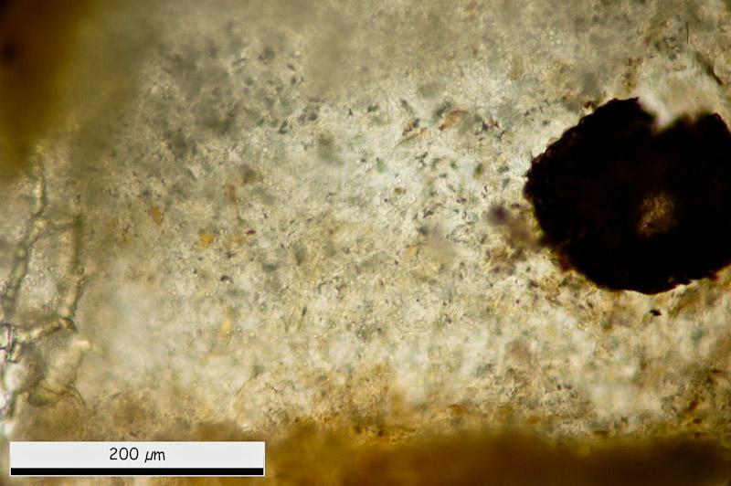
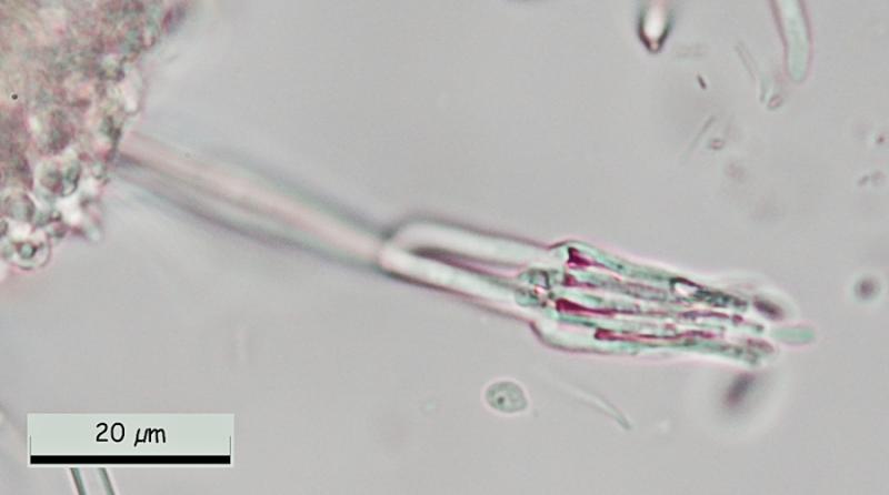
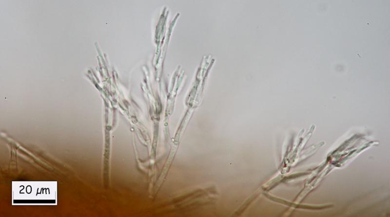
And finally, at the margin, this conidial structure.
Than you in advance.
Miguel û. Ribes
Than you in advance.
Miguel û. Ribes
Hans-Otto Baral,
12-03-2013 08:06

Re : Hymenobolus agaves anamorph
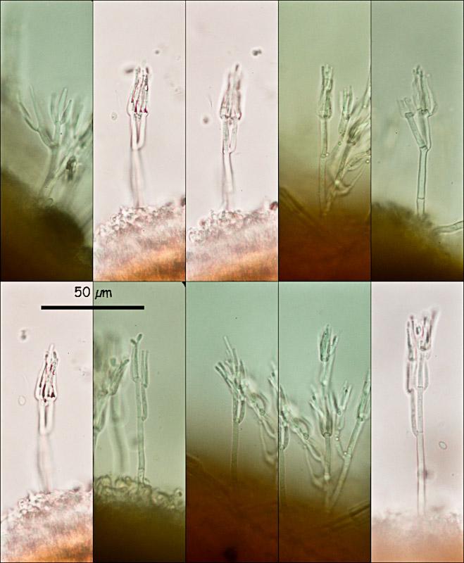
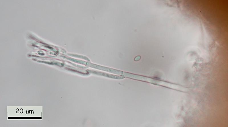
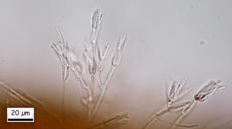
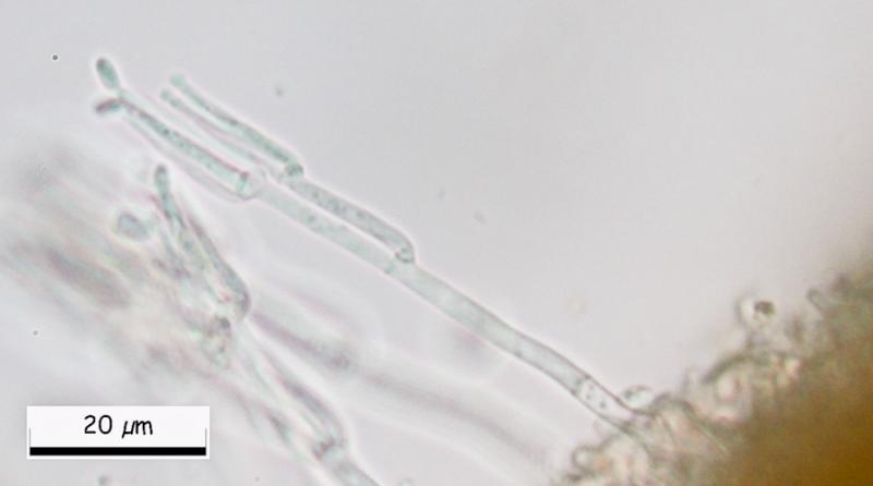
Great, Miguel! Could you please show us a closeup of the conidiogenous cells, were the conidia emerge? I assume they are phialidic. Then we can search in Genera of Hyphomycetes, or someone has an idea.
I have given the previous Hymenobolus specimen for sequencing, I am curious where it could belong.
Zotto
I have given the previous Hymenobolus specimen for sequencing, I am curious where it could belong.
Zotto
Miguel ûngel Ribes,
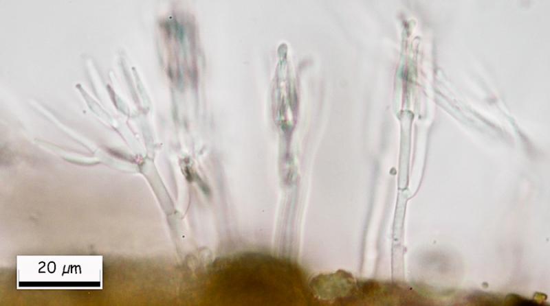
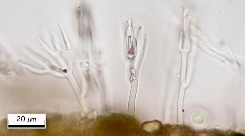
12-03-2013 11:27
Re : Hymenobolus agaves anamorph
Here there are.
Hans-Otto Baral,
12-03-2013 22:56
Re : Hymenobolus agaves anamorph
Hi Miguel
Walter Gams answered me that this is ô clearly aô Clonostachys, probably Clonostachysô solani (Harting) Schroers & W. Gams, which is quite common, often fungicolous, and the anamorph of a Bionectria. So certainly not belonging to Hymenobolus.
Zotto
Walter Gams answered me that this is ô clearly aô Clonostachys, probably Clonostachysô solani (Harting) Schroers & W. Gams, which is quite common, often fungicolous, and the anamorph of a Bionectria. So certainly not belonging to Hymenobolus.
Zotto
Miguel ûngel Ribes,
13-03-2013 00:15
Re : Hymenobolus agaves anamorph
Hi Zotto, Superb.
Thank you again to resolve this puzzle.
See you.
Thank you again to resolve this puzzle.
See you.