
05-04-2026 22:46
 Lothar Krieglsteiner
on wood of Ceratonia, Algarve, 3.4.2026.The color
05-04-2026 20:40
 Robin Isaksson
Hi!Found i Japan on bark of Abies sp. Spores 35-4
31-03-2026 21:18
 Miguel Ángel Ribes
Good evening. oes anyone have the original descrip
31-03-2026 20:57
Stefan BlaserHello everybody, I hope somebody can help me with
26-03-2026 15:31
 Åke Widgren
Hello,I found this one in October last year, on r
31-03-2026 16:20
 Mlcoch Patrik
Hello, Please about help with determination. On
31-03-2026 08:19
 Bernard CLESSE
Bonjour à toutes et tous,Pourriez-vous m'aider à
 • Macro and habitat suggest Gelatinodiscaeae.
• With spores suggests Ascotremella faginea.
• Very challenging to section.
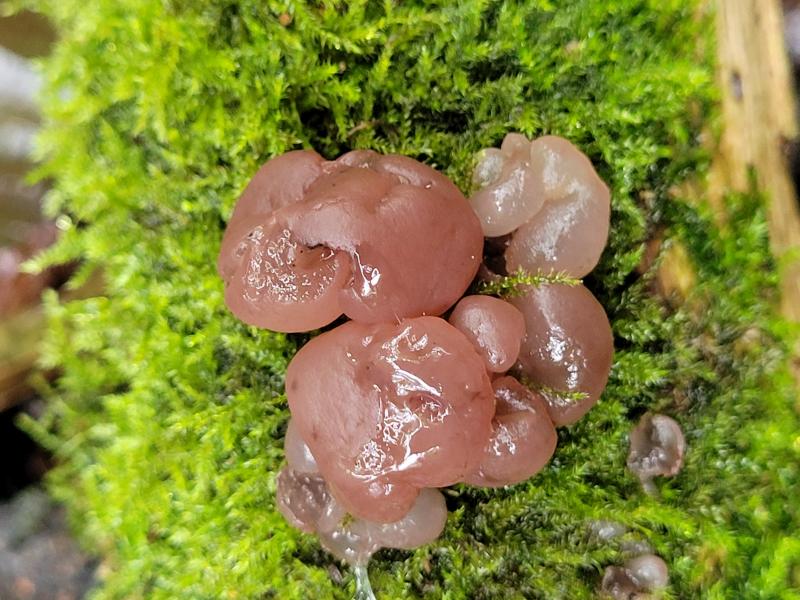
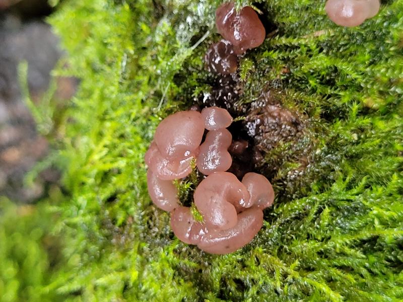
Habitat: On damp and decaying angiosperm log, medium size, with bryophytes, in a ditch, damp and shady area, near a large pond, mixed deciduous woodland, Low Weald, England, late-September, after rain.
Apothecia: Diameter < ~ 1 cm (may not be largest), pale lilac to pinkish-brown, paler when immature, broadly turbinate to more irregular-lobate, mostly caespitose, appearing more cerebriform together, superficial, substiptate (very short thick stipe), sometimes overlapping, surface with grainy appearance under low magnification, internally jelly-like, receptacle more whitish and translucent, margin indistinct, slightly out-rolled, disc more purplish, convex, opaque, with dull appearance.
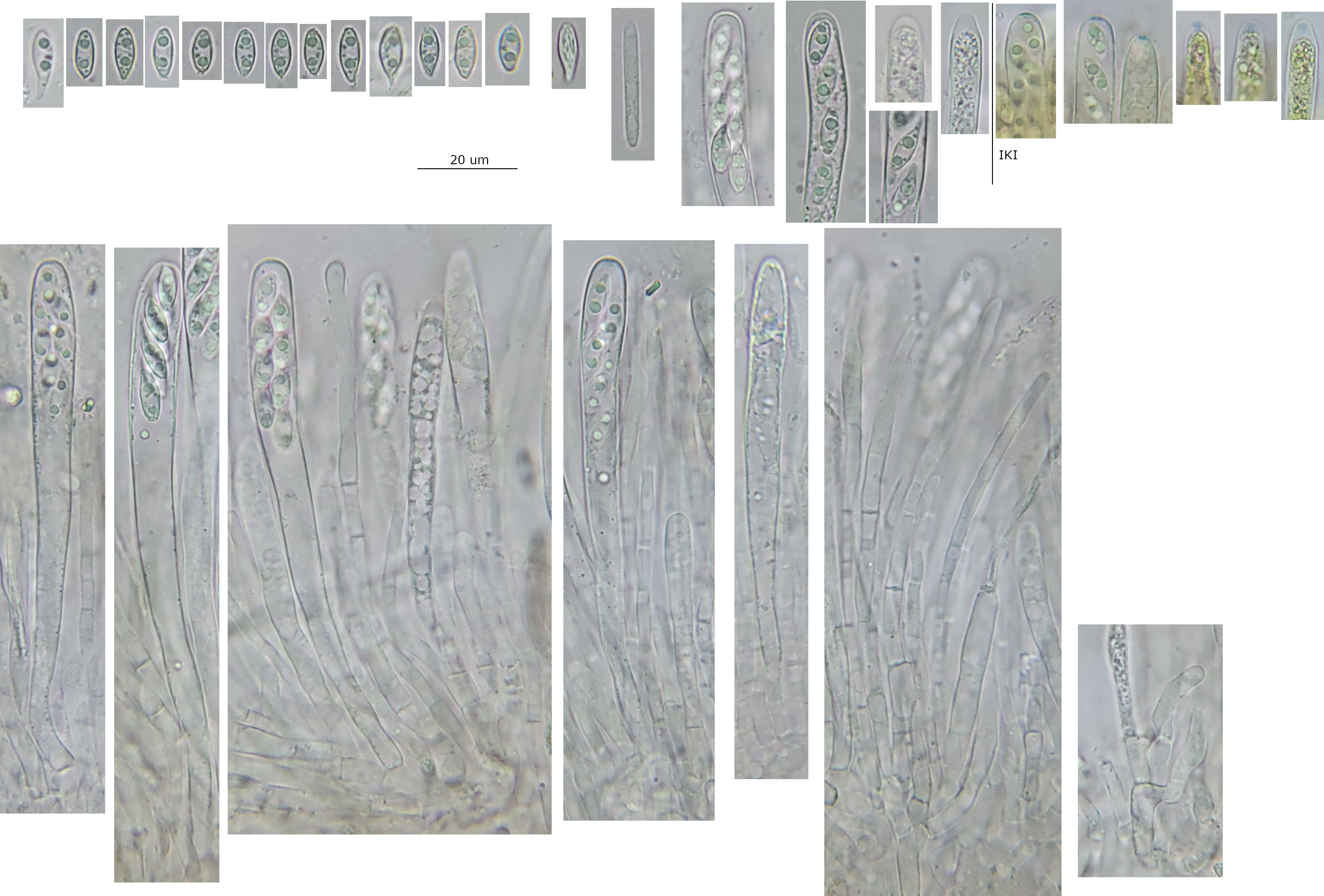
Asci: Cylindrical-clavate, simple septa, rings bb, apex rounded to subtruncate when turgid, rounded-acute when flaccid, biseriate when turgid, uncertain about spore orientation possibly lower spores inversely oriented or irregular orientations, thickening noticeable when flaccid, dome-like when immature, no discharging observed.
Spores: Ellipsoid to limoniform-lacrymiform, often heteropolar, one pole more rounded and the other more acute, acute pole sometimes slightly to distinctly mammiform (incl. within ascus), roughly symmetric in face view but slightly sub-curved or asymmetric in profile view, a medium-size LB at each pole, and many smaller ones, apparently uninucleate and aseptate, ornamentation of faint longitudinal striations, appearing whitish.
Free spores in water: (8.4) 9.2-11.0 (14.2) × 3.5-4.3 (4.7) µm, Q = (2.0) 2.2 - 2.9 (3.5), n = 30, mean = 10.0 × 3.9 µm, Q mean = 2.6.
Paraphyses: Cylindrical, multi-septate, apical cell often longer, apex sometimes irregularly inflated, no branching near the apex observed, oblong hyaline VBs, brownish en masse.
Medullary: Loose texture intricata, narrow hyphae, highly gelatinised, many small crystals and some medium and large ones, near the subhymenium denser and thicker hyphae, with patches of reddish pigment and some pigmented hyphae.
Subhymenium: Difficult to distinguish clear margin from medullary, many globose-pyriform swollen cells of varying sizes.
Ectal: Textura prismatica, marginal cells globose-pyriform.
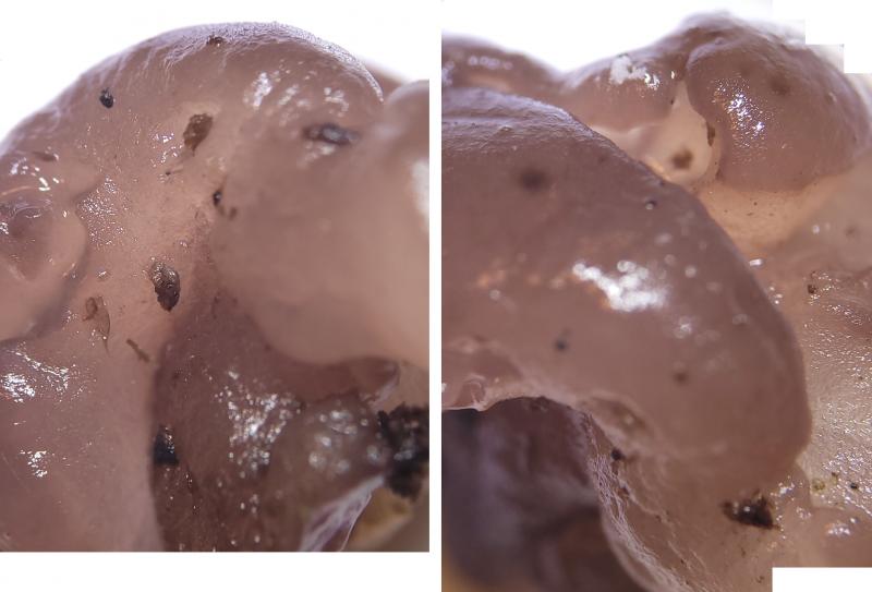
 Hymenium-0013.jpeg
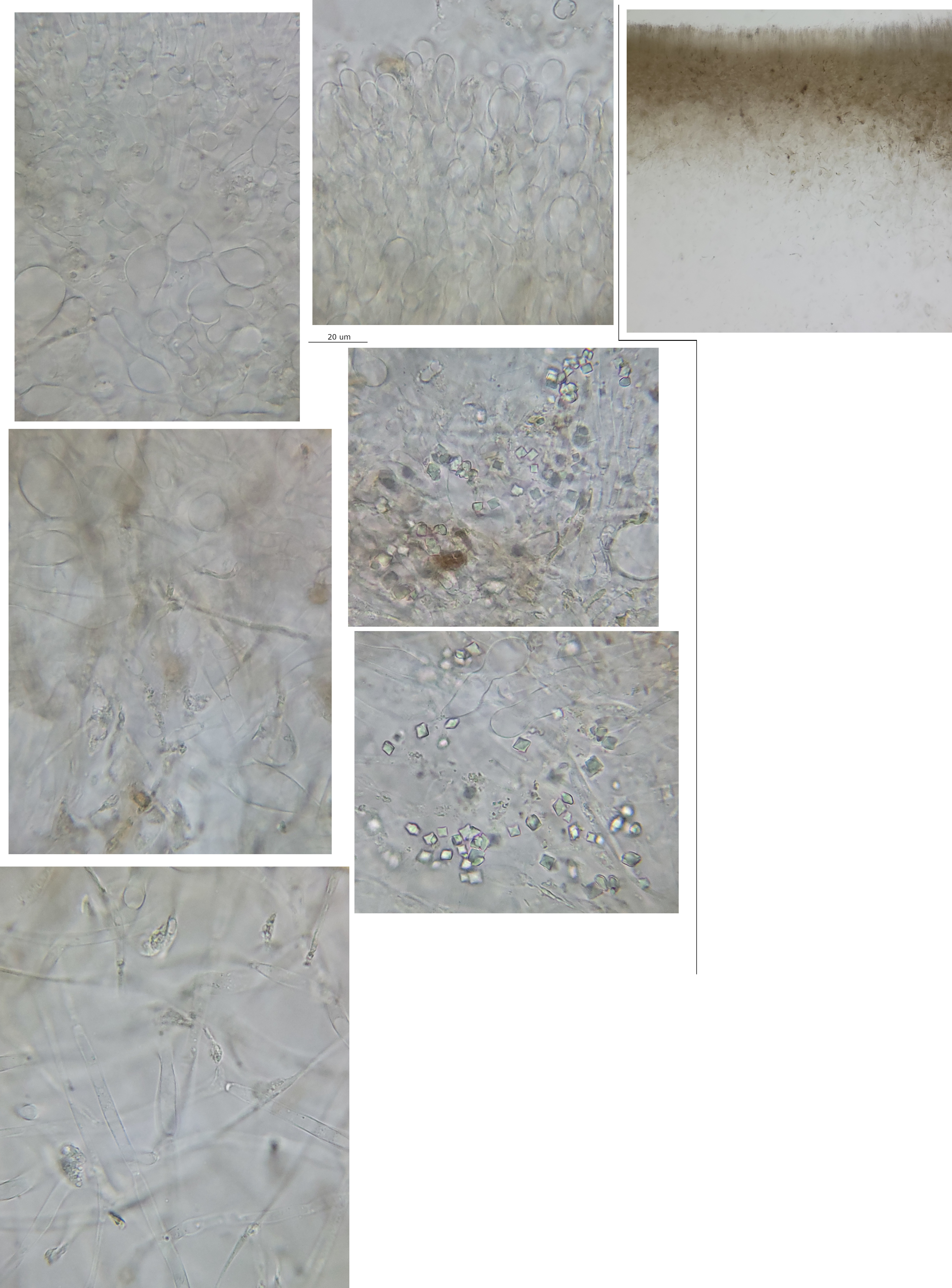 Excipulum-0006.jpeg
This was found during a local group foray on Sunday (22/09/24) in Sussex, coordinates 50.9100,-0.2684. It was mentioned then that there is some confusion between A. faginea and Neobulgaria pura var. foliacea.
It seems like it should be relatively easy to separate more irregular or overmature forms of N./Ombrophila pura from A. faginea using microscopy.
The spore ornamentation was unexpected and I was concerned that it was artifactual. So I didn't manage to get any good photos, but I've tried to collect together some where you can just make out some striations.
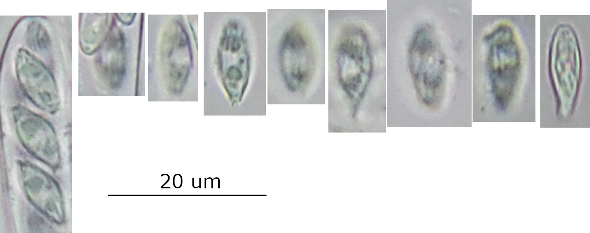 spore-ornament-0001.jpeg
You are right that some of the earlier work is a little confused, I was referring to Gamundi and Dennis (1969) and Dennis (1971). The gelatinous layer on the surface of the excipulum in Ombrophila is also mentioned that is not present in A. faginea.