
23-05-2026 18:57
Sylvie Le GoffBonjour à tousRécolté sur une branchette de Sal
23-05-2026 11:44
 Charles Grapinet
Hello, I am having trouble identifying this copro
22-05-2026 21:35
Steve ClementsBonjour, I expected this find on old wood on our
22-05-2026 18:12
 Lothar Krieglsteiner
... in moist chamber from Portugal.As the fungus s
22-05-2026 20:08
Ethan CrensonHello all, Yesterday in NYC I was visiting an e
11-01-2022 16:36
 Jason Karakehian
Hi does anyone have a digital copy of Raitviir A (
20-05-2026 17:47
Margot en Geert VullingsWe found this Mollisia on dead Juncus stems mown l
22-05-2026 14:47
Gernot FriebesHi,superficial ascomata collected on bark of a liv
22-05-2026 14:44
Lothar Krieglsteiner
in unripe condition citrine yellow, then soon fadi
22-05-2026 13:29
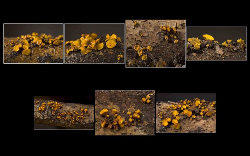
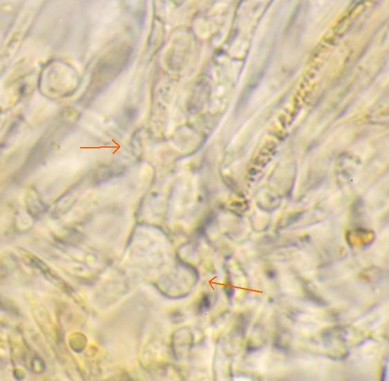
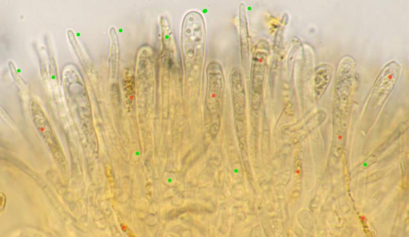
Gernot FriebesHi,I am curious to hear your opinion on this mater

Loc: Plantation Trail, Jean Lafitte National Historical Park and Preserve, Louisiana, USA
Coll: D. Newman, R. Cronce & K. Thorstad
Substrate: on dead, downed, corticate, firm/undecomposed, hardwood stick
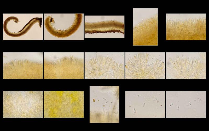
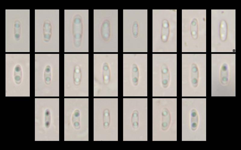
all mounts made in KOH. excipulum composed of irregularly inflated/swollen chains of cells (asexual propagules as in I. irregularis?); context composed of long, slender, even hyphae, possibly embedded in a gelatinous matrix; paraphyses filiform and short, only slightly exceeding asci; both hymenium and abhymenium appear to contain ionomidotic granules/contents; asci biseriate; spores bi-guttulate and (probably) aseptate.
Spores:
(5.1) 5.3 - 6.5 (6.8) × (2.1) 2.2 - 2.6 (2.7) µm
Q = (2) 2.2 - 2.9 (3) ; N = 20
Me = 5.9 × 2.4 µm ; Qe = 2.5
GIF of ionomidotic reaction available at https://inaturalist-open-data.s3.amazonaws.com/photos/347983283/original.gif
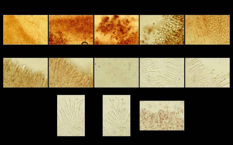

Two new things I've noticed in these recent mounts: 1: the paraphyses are multi-septate, and 2: many contain bright yellow, refractive contents. I had observed it rather vaguely in the hymenium before, but not inside the paraphyses.
Thank you for sharing these high-value images. I would like to sequence this specimen, it would help to clear the mysterious taxonomy in this group...
If you like to send a piece of specimen to our lab, please, use this address:
Kadri Pärtel
Chair of Mycology
Department of Botany
Institute of Ecology and Earth Sciences
University of Tartu
Oecologicum
J. Liivi St. 2
50409 Tartu
Estonia
kadri.partel@ut.ee
Phone +372 5226179
THANK YOU FOR COOPERATION!

https://inaturalist.org/observations?place_id=any&q=Ionomidotis%20cf.%20fulvotingens&search_on=tags&user_id=ikhom&iconic_taxa=Fungi
I kept the first two speciments.
I wonder about I. fulvotingens that grows on Pinus strobus. Has it been ever sequenced? I found a specimen in Feb 2020 and I kept it. (https://inaturalist.org/observations/38581250)
Thank you for sharing your found.
I checked my data: all specimens we have studied and sequenced are on hardwood. It would be intresting to add one from a conifer.
Best regards,
Kadri
(5.1) 5.3 - 6.5 (6.8) × (2.1) 2.2 - 2.6 (2.7) µm
Q = (2) 2.2 - 2.9 (3) ; N = 20
Me = 5.9 × 2.4 µm ; Qe = 2.5