
11-06-2026 19:01
 William Slosse
Hello all,In an attempt to make a culture of a sus
10-06-2026 21:16
 François Freléchoux
Bonsoir,Le dernier du jour, en attendant votre avi
11-06-2026 19:03
 Nicolas VAN VOOREN
Chers membres d'Ascofrance,Le site sera placé en

09-06-2026 18:32
Camille MertensSur morceau de roseau immergé 0,5 - 0,7 mm de dia
10-06-2026 12:54
Steve ClementsBonjour encore, Pouvez-vous m'aider, s'il vous pl
10-06-2026 21:07
François Freléchoux
Toutes les tiges de gentianes jaunes de l'an pass�
10-06-2026 13:41
François Freléchoux
Bonjour à nouveau, Voici une trouvaille d'hier.
10-06-2026 11:53
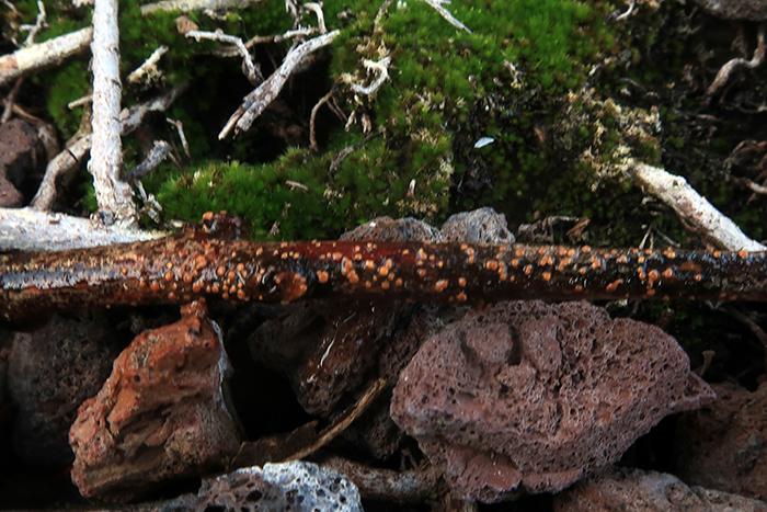
Steve ClementsBonjour, This disco is abundant on dead stems of
 Hello.
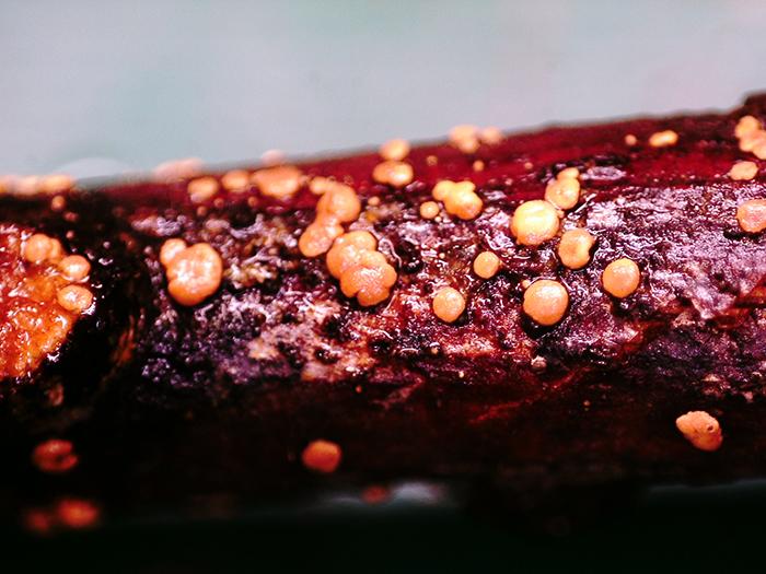
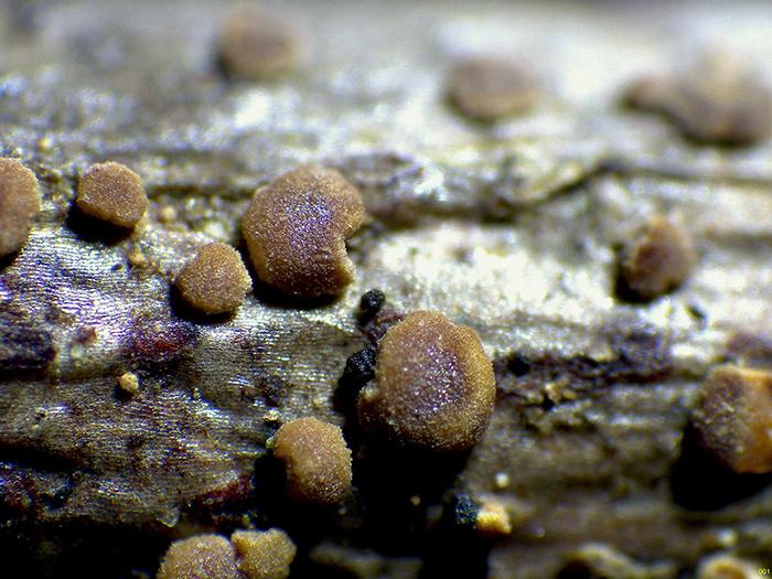
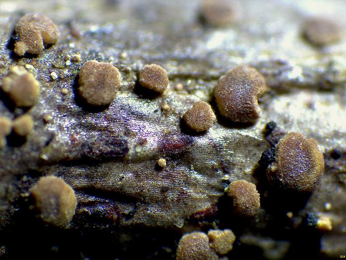
Some apothecia sprouting massively on a thin trunk of Laurus nobilis.
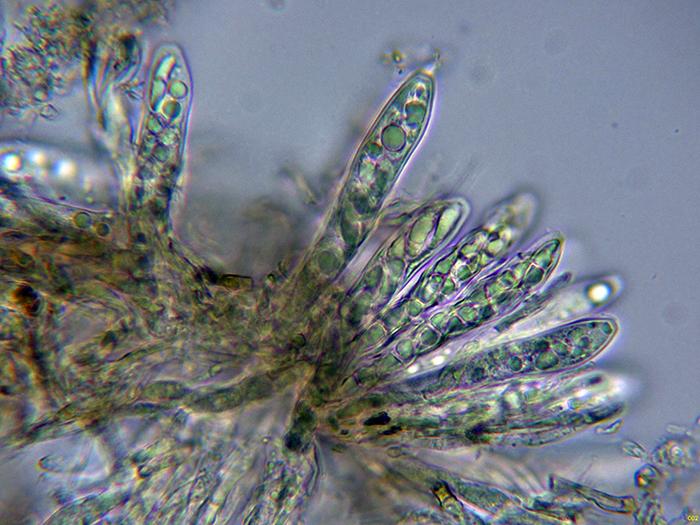
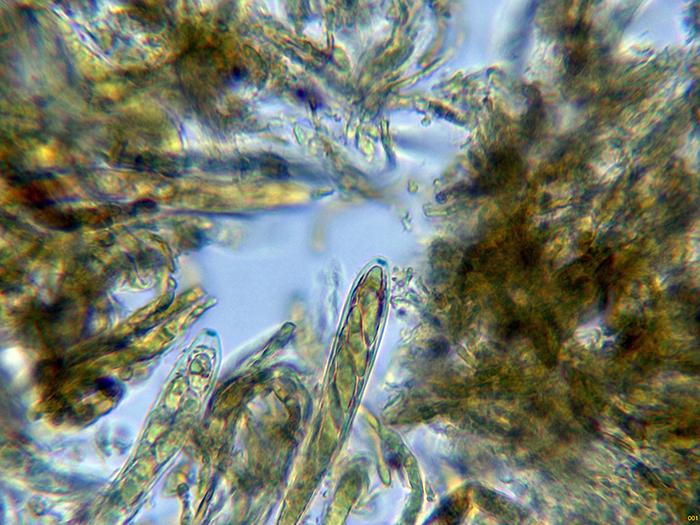
Spherical in shape, somewhat flattened, orange when wet and greenish brown when dry. with a diameter of between 0.2 to 0.7 mm.
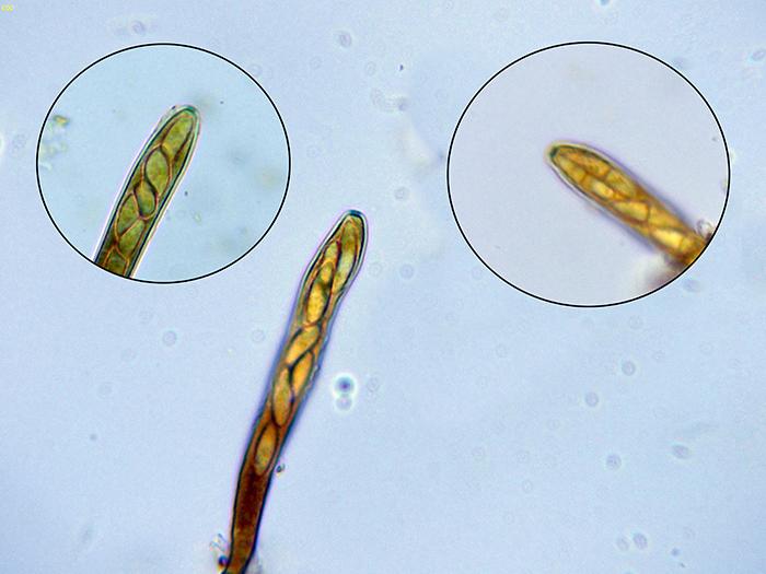
Octosporic asci, with uncinules at their base and amyloid reaction of their apical apparatus.
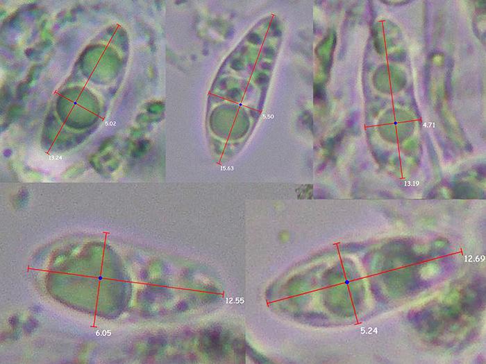
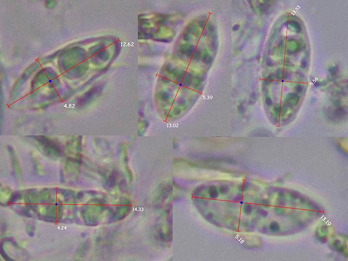
Moriform ascospores, poorly septate and with free spore measurements of (12.5) 12.6 - 14.3 (15.6) × (4.2) 4.7 - 5.6 (6) µm.
Based on the data obtained, everything seems to fit with what could be considered Claussenomyces prasinulus, but I already studied Clausennomyces prasinulus last year and the reaction of the apical apparatus to iodine was negative, and I can't find anything more similar than Claussenomyces or Vexillomyces.
Any opinion from you will be well received.
Thank you very much in advance.
Kind regards.
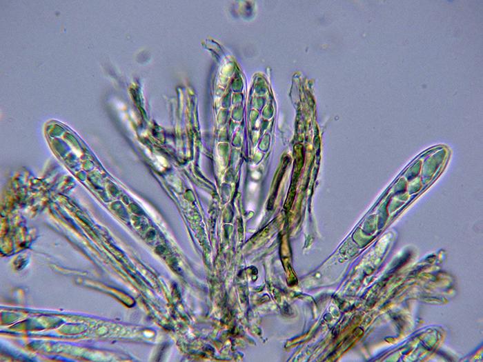

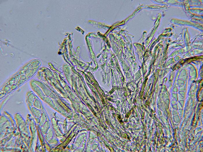
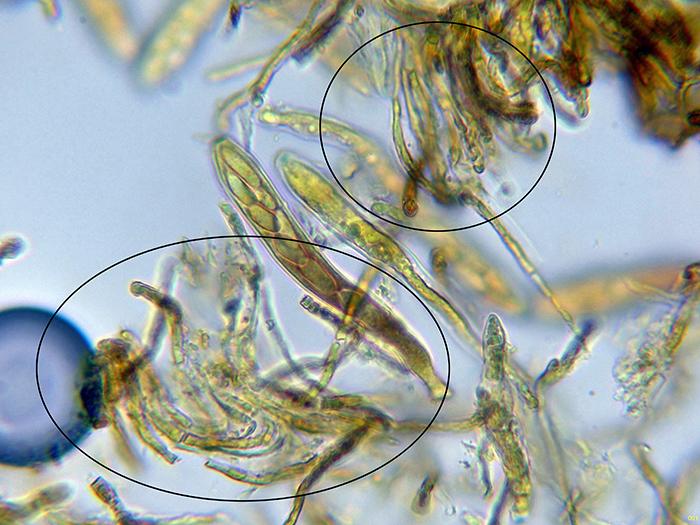
The apothecia were not at their best and unfortunately the ascos were dead.
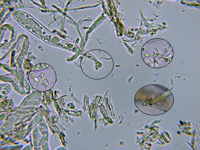
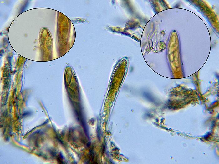
I add a couple of images of the paraphyses, one in water and the other in Lugol, these filiform, septate paraphyses, with intracellular pigment, some branched and that do not protrude above the level of the asci.
Again thank you very much for your help.
Kind regards.
It fits very well with the little information I have been able to find on Rodwayella sessilis.
Kind regards.
The information in your folder is very interesting, including a very detailed description of microscopy.
Kind regards.