
23-05-2026 11:44
 Charles Grapinet
Hello, I am having trouble identifying this copro
25-05-2026 16:44
François BartholomeeusenHi forum members,During an excursion organised by
25-05-2026 16:35
 Bernard CLESSE
Bonjour à toutes et tous,J'ai trouvé récemment,
22-05-2026 13:29
Gernot FriebesHi,I am curious to hear your opinion on this mater
23-05-2026 18:57
Sylvie Le GoffBonjour à tousRécolté sur une branchette de Sal
22-05-2026 14:44
 Lothar Krieglsteiner
in unripe condition citrine yellow, then soon fadi
22-05-2026 21:35
Steve ClementsBonjour, I expected this find on old wood on our
22-05-2026 18:12
Lothar Krieglsteiner
... in moist chamber from Portugal.As the fungus s
22-05-2026 20:08
Ethan CrensonHello all, Yesterday in NYC I was visiting an e
Discomycet on cone of Picea
Gernot Friebes,
11-04-2009 08:24
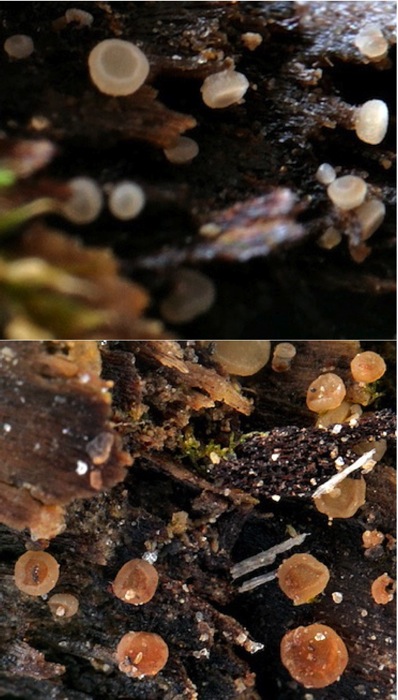
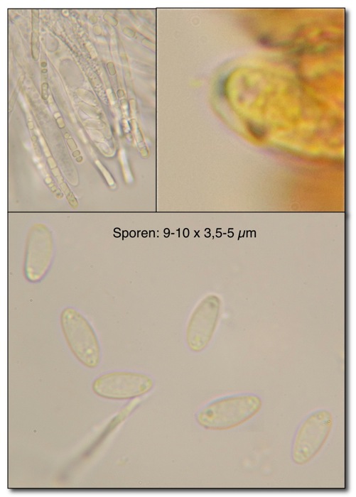
a few days ago I found this Ascomycet on a cone of Picea. When getting old the apothecia turn reddish-brownish. Asci are IKI b and the spores are 9-10 x 3.5-5 µm. The apos are sessile. I thought maybe a Calycina but I did not found a species that fits with my collection.
Best wishes,
Gernot
Gernot Friebes,
11-04-2009 08:27
Re:Discomycet on cone of Picea
here are the microscopical features. I would be grateful if someone would have an idea about my finding!
Hans-Otto Baral,
11-04-2009 17:47

Re:Discomycet on cone of Picea
The ascus apical ring looks more like Hymenoscyphus, so Calycina seems excluded. I think it is Hymenoscyphus ravus, though there the spores are a bit smaller (*6-8 x 2.8-3.5). A very similar but apparently distinct species, "Ciboria" rava, has spores which better fit to yours, but that is on catkins of Betula.
Is the width of 5 µm perhaps a bit too high? Do you have a scvale to your microphotos?
Zotto
Is the width of 5 µm perhaps a bit too high? Do you have a scvale to your microphotos?
Zotto
Gernot Friebes,
11-04-2009 21:29
Re:Discomycet on cone of Picea
Hello Zotto,
I looked at the spores again (dried, rehydrated material) and this time they were up to 11 µm long and 4,5 µm broad. I have no scale to my microphotos but tomorrow I can try to make a photo through the ocular, if necessary. What next?
Best wishes and many thanks,
Gernot
I looked at the spores again (dried, rehydrated material) and this time they were up to 11 µm long and 4,5 µm broad. I have no scale to my microphotos but tomorrow I can try to make a photo through the ocular, if necessary. What next?
Best wishes and many thanks,
Gernot
Hans-Otto Baral,
12-04-2009 10:26
Re:Discomycet on cone of Picea
This group is in bad need of a thorough study. Neither the genus is clear nor the species limits are well explored, in my opinion. Looking for croziers would be necessary, also measuring asci (now probably only in dead state), and a section of the excipulum (now difficult in the dead state). Only thorough documentation may help. It is not impossible, in my experience, that a species occurs on both Betula catkins and Picea cones.
Both Hymenoscyphus ravus and Ciboria rava were described by Svrcek 1989. Svrcek observed crystals in the excipulum of H. ravus which I never saw.
I send you the literature by email.
If you do not use the camera zoom for your microphotos, it is easy to make a scale which you can paste in every photo.
Zotto
Gernot Friebes,
12-04-2009 17:02
Re:Discomycet on cone of Picea
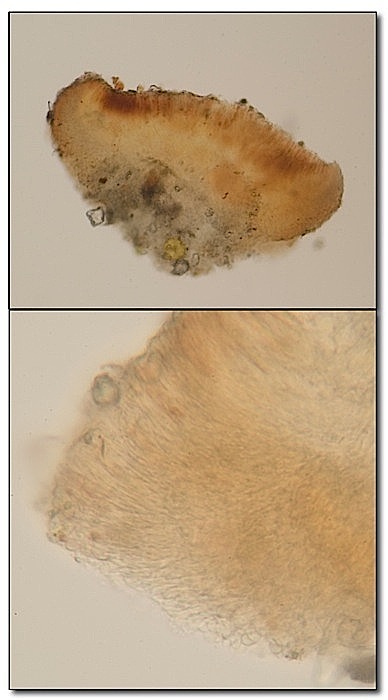
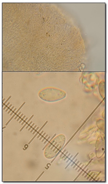
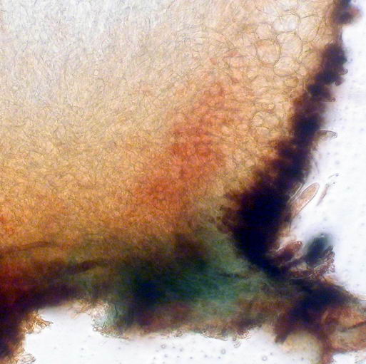
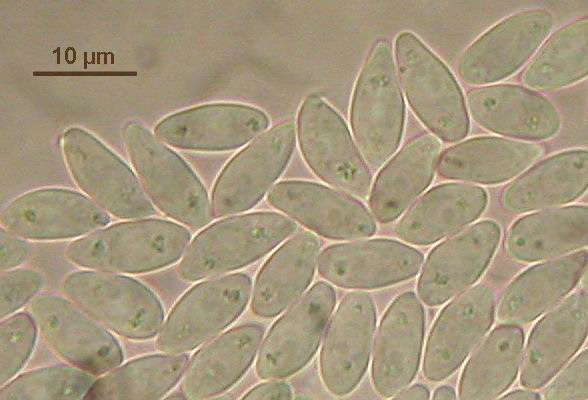
I checked my collection again: the asci have croziers, with 65-74 x 5-6 µm. The excipulum seems to be between a textura globosa and a textura angularis with elongated cells (like a textura prismatica) at the margin of the apothecia, without any crystals. I also took a photo where you can see the measuring scale and a spore with nearly 5 µm width.
The description of Ciboria rava by Svrcek (thanks for sending it to me!) seems to fit quite well except for the apothecia which he described as shortly stipitate but within mine collection the apothecia are sessile.
Best wishes,
Gernot
picture: section of an apothecium (above) and the excipulum (beneath)
Gernot Friebes,
12-04-2009 17:03
Re:Discomycet on cone of Picea
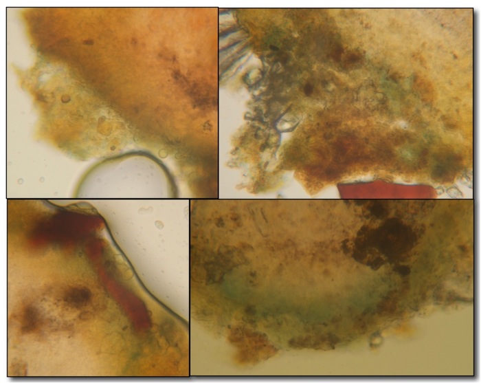
second picture:
Hans-Otto Baral,
12-04-2009 19:07
Re:Discomycet on cone of Picea
I got another, quite simple idea what it could be. Hymenoscyphus imberbis (= Phaeohelotium imberbe). This is very polyphagous and has such a spore size. What I repeatedly saw in this species is a bluing of the anhoring hyphae in IKI:
Hans-Otto Baral,
12-04-2009 19:10
Re:Discomycet on cone of Picea
The spores of this find on Fagus cupules: Sp. *11-13 x 4.5-5.3 µm, probably too long for your fungus.
I just see a drawing on the DVD: Hymenoscyphus imberbis aff., HB 6574.JPG. This is from Bulgaria, on Pinus cones. Spores 6-9 x 3.2-3.8 µm (dead state).
Zotto
I just see a drawing on the DVD: Hymenoscyphus imberbis aff., HB 6574.JPG. This is from Bulgaria, on Pinus cones. Spores 6-9 x 3.2-3.8 µm (dead state).
Zotto
Gernot Friebes,
12-04-2009 20:17
Re:Discomycet on cone of Picea
yes, there is a blueing in lugol which is even stronger in Melzer's reagent. So it really seems to be Hymenoscyphus imberbis!
Best wishes and many thanks again,
Gernot
edit: the photo in the bottom right corner was taken in Melzer's reagent.
Best wishes and many thanks again,
Gernot
edit: the photo in the bottom right corner was taken in Melzer's reagent.
Hans-Otto Baral,
12-04-2009 20:32
Re:Discomycet on cone of Picea
o.k., this is not a final proof for imberbis, but supports my idea. Such blue reactions occur also in other taxa and are perhaps not very consistent. F.ex. there is a Pezizella amyloideoexcipulata Svrcek described on Fagus cupules with spores 6.5-11 x 2.3-3.2 µm, which seems close to H. imberbis.
Zotto
Zotto
Gernot Friebes,
12-04-2009 21:00
Re:Discomycet on cone of Picea
sorry, than I was a little bit to hasty, mainly because I remember a person who told me that the blueing base in IKI would be quite a distinctive character of H. imberbis. It's interesting that this blue reaction can be seen in several taxa, I will look for it in the future more precisely.
What do you think now, is there another feature I can look for which supports H. imberbis or would it be better if I'd name the collection Hymenoscyphus imberbis cf.?
Best wishes,
Gernot
What do you think now, is there another feature I can look for which supports H. imberbis or would it be better if I'd name the collection Hymenoscyphus imberbis cf.?
Best wishes,
Gernot