
11-06-2026 19:01
 William Slosse
Hello all,In an attempt to make a culture of a sus
10-06-2026 21:16
 François Freléchoux
Bonsoir,Le dernier du jour, en attendant votre avi
11-06-2026 19:03
 Nicolas VAN VOOREN
Chers membres d'Ascofrance,Le site sera placé en

09-06-2026 18:32
Camille MertensSur morceau de roseau immergé 0,5 - 0,7 mm de dia
10-06-2026 12:54
Steve ClementsBonjour encore, Pouvez-vous m'aider, s'il vous pl
10-06-2026 21:07
François Freléchoux
Toutes les tiges de gentianes jaunes de l'an pass�
10-06-2026 13:41
François Freléchoux
Bonjour à nouveau, Voici une trouvaille d'hier.
10-06-2026 11:53
Steve ClementsBonjour, This disco is abundant on dead stems of
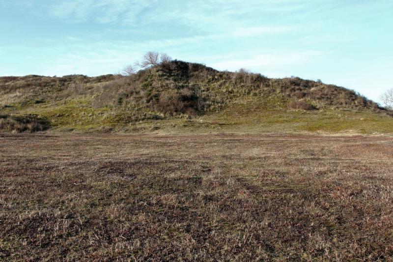
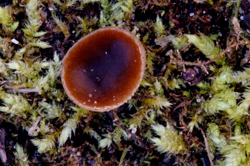
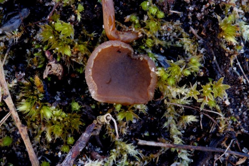
Good afternoon everyone,Yesterday 05/02 I found a large number of small Pezizas between recently mown scrub of Salix repens on the floor of a damp dune valley in the Belgian nature reserve Ter Yde, Oostduinkerke. This dune valley is extensively grazed by ponies.
I am thinking, although not immediately dung in the area, of Peziza fimeti. But I am not sure, varia is a possibility too but I never saw this species so tiny.
Anyone have any confirmation/correction?
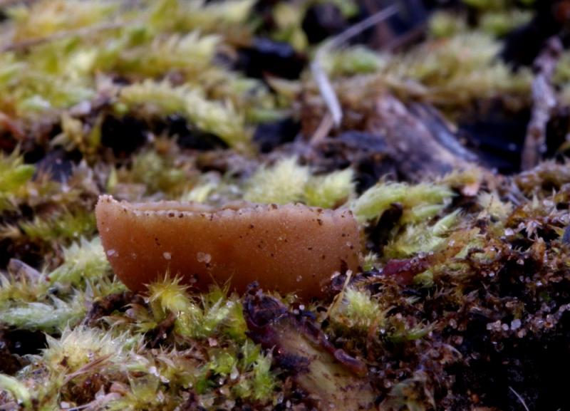
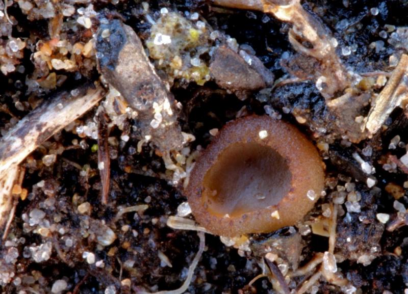
Macroscopic:
° diameter apothecium up to 12mm
° outer wall yellow-brown - faun; hymenium darker, reddish brown
° sitting, no stem
° edge apothecium flocculent
° apothecium dextrinoid (Melzer)
Microscopic:
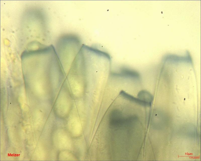
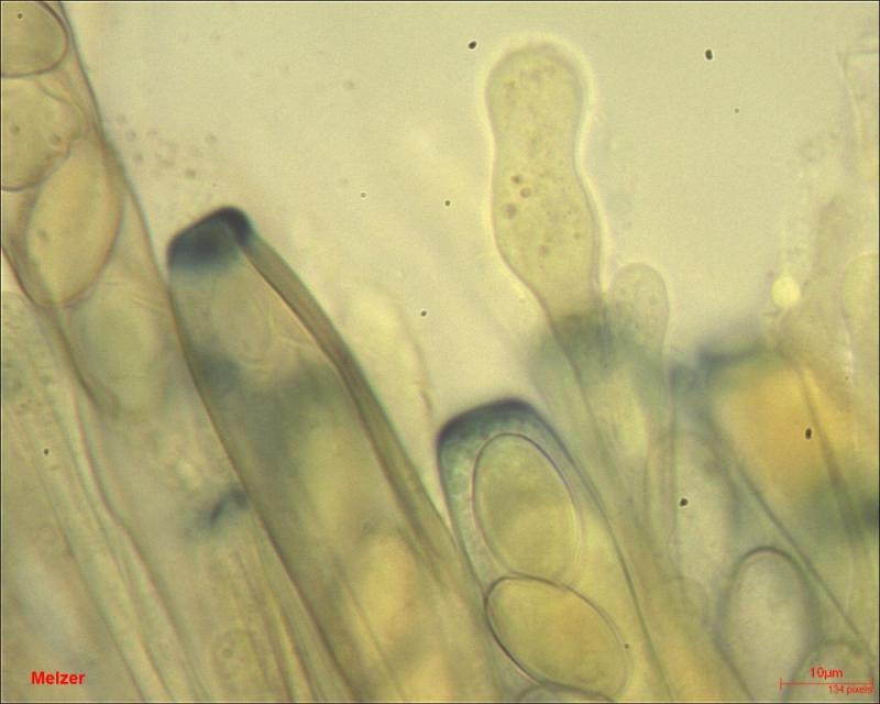
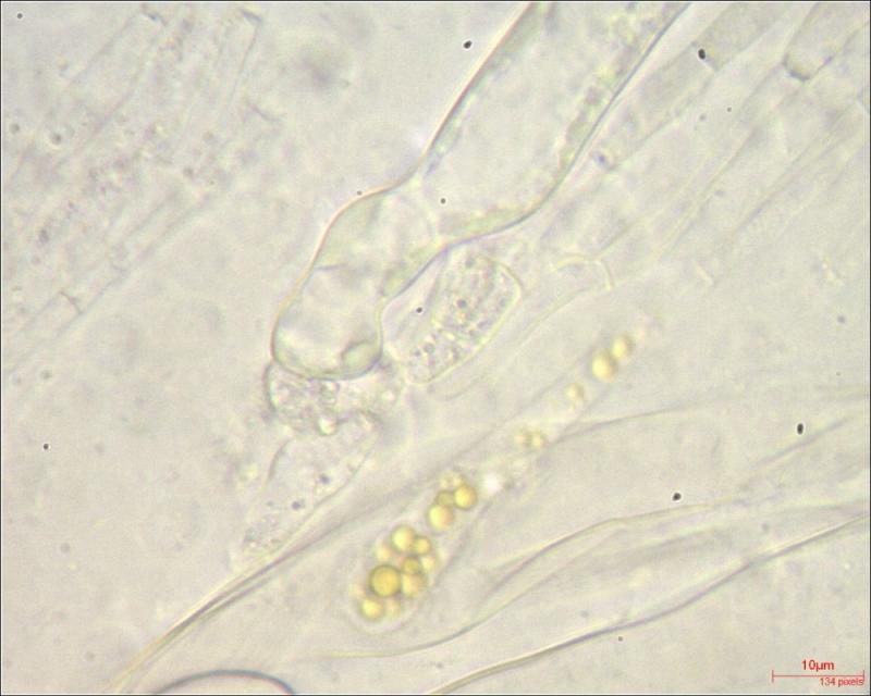
° Asci:
-amyloid type WTR
- no croziers (seems to me)
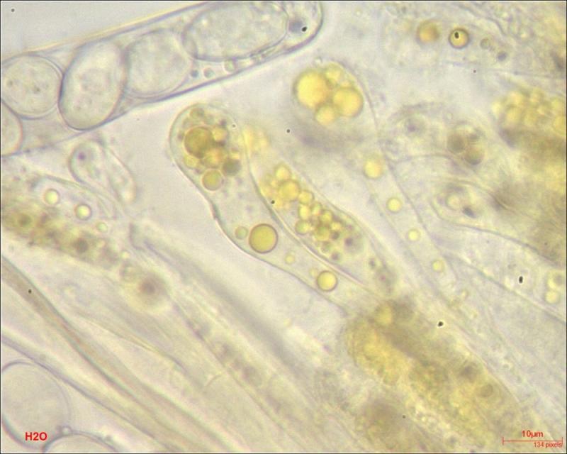
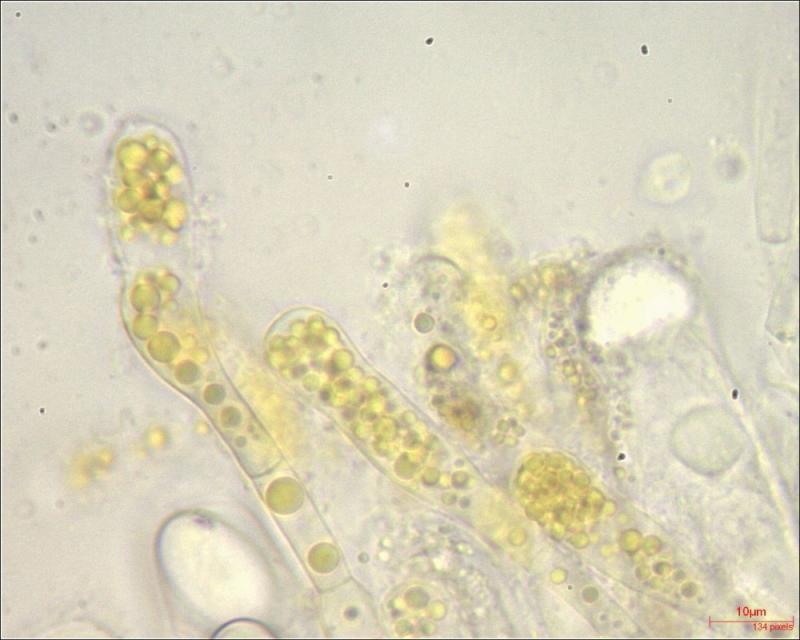
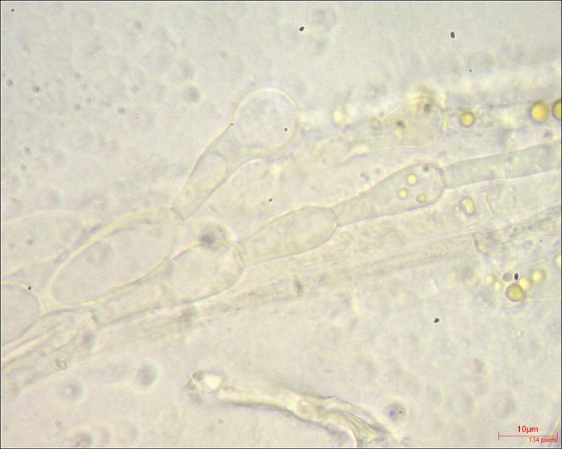
° paraphyses:
- moniliform
- VBs with yellow pigment
- ø tops 7.45 – 11.83µm
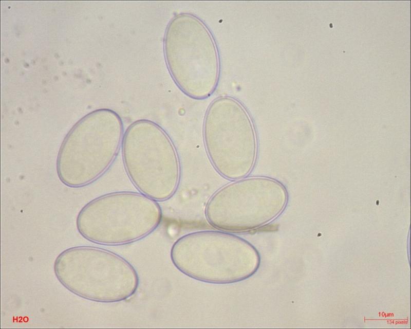
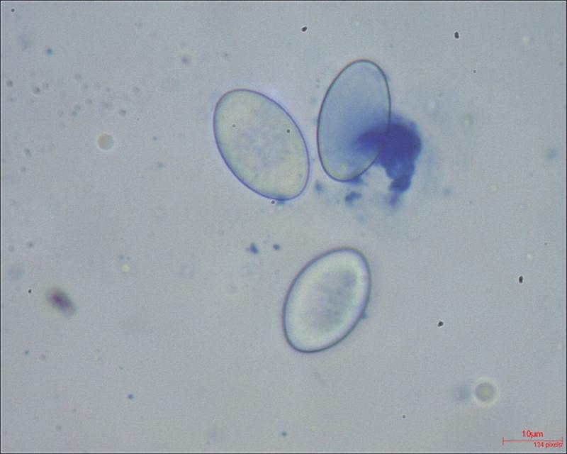
° Spores:
- smooth
- hyaline
- with nucleus & droplets at the poles
- 19.05-20.86 x 10.92-12.80µm
- Av(30) = 11.82 x 19.98µm
- Q 1.58-1.82
- Q(av30) = 1.68
Thx in advance,
William
Your description fits well P. fimeti, except the substrate. I don't have any experience of this species not growing on dung...
Two points:
- the moniliform paraphyses are rather common in Peziza s. str., this is not constant and often are due to the external moisture.
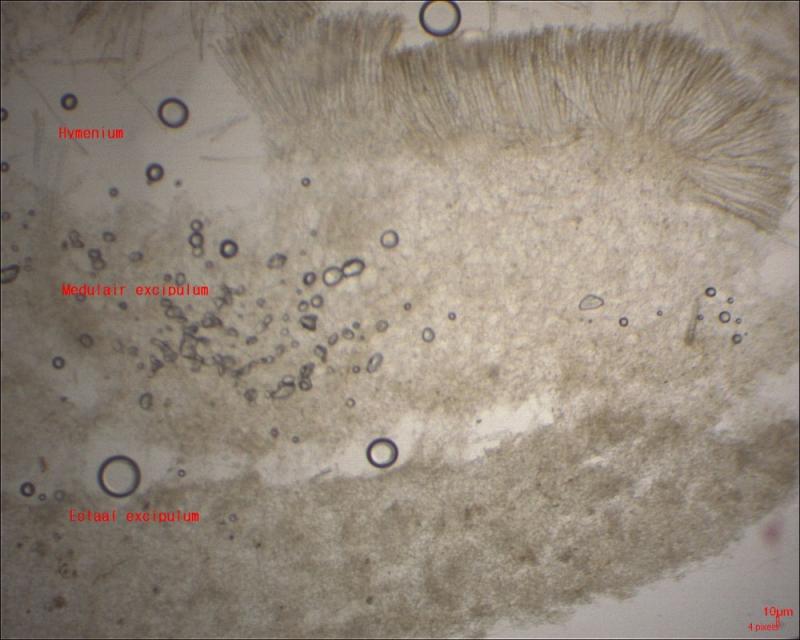
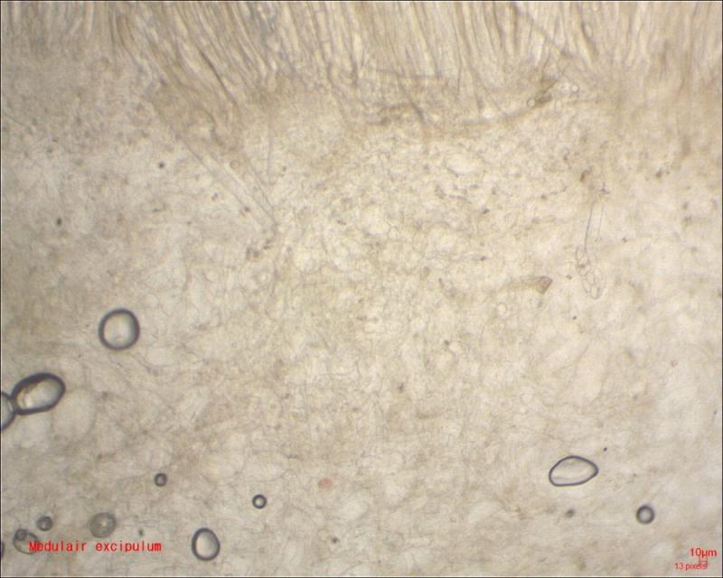
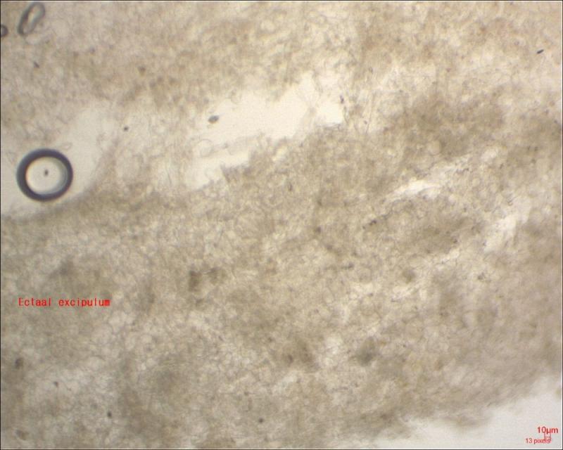
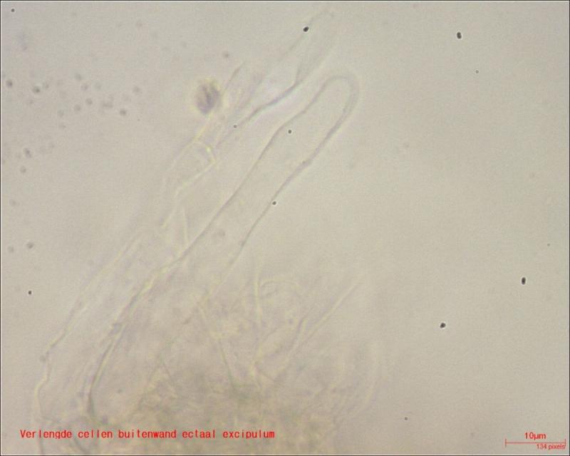
- the excipulum of P. fimeti shows 4 layers: a medullary excipulum divided in three parts because there exists a thin layer of hyphae inserted between the cells of the medullary excipulum, and an ectal excipulum of t. globulosa (some emerging hyphae can be seen in the outermost part).
Very interesting info about the molinifomr paraphyses.
I will try to make another coupe to examin the excipulum (there is still fresh material in the box).
About the substrate:
apparently the Pezizas grew on soil, not on wood or woody substrates, and at first glance even less so on dung. The vegetation on the finding place was mown with the cutter bar. As a result, there is a possibility that pony dung was grind and spread in this way and that these pezizas still developed on dung residues. But this is hard to prove, I think. The only certainty is that there is pony dung at the site of these Pezizas.