
12-06-2026 14:50
 François Freléchoux
Bonjour, Voici la brève description d'une Mollis
10-06-2026 21:16
François Freléchoux
Bonsoir,Le dernier du jour, en attendant votre avi
11-06-2026 19:01
 William Slosse
Hello all,In an attempt to make a culture of a sus
11-06-2026 19:03
 Nicolas VAN VOOREN
Chers membres d'Ascofrance,Le site sera placé en

09-06-2026 18:32
Camille MertensSur morceau de roseau immergé 0,5 - 0,7 mm de dia
10-06-2026 12:54
Steve ClementsBonjour encore, Pouvez-vous m'aider, s'il vous pl
10-06-2026 21:07
François Freléchoux
Toutes les tiges de gentianes jaunes de l'an pass�
10-06-2026 13:41
François Freléchoux
Bonjour à nouveau, Voici une trouvaille d'hier.
 Hi,
Hi,Is this just a long spore variation of Hymenoscyphus scutula (H. aff. fucatus), or is it something else?
https://inaturalist.ca/observations/57610849
On rotten leaves, white when fresh and yellowish when dried.
The stalk base was white.
Spores:
(28.6) 29.5 - 36 (36.7) × (3.5) 3.9 - 4.4 (4.5) µm
Q = (6.6) 6.9 - 8.8 (9.9) ; N = 30
Me = 32.8 × 4.2 µm ; Qe = 7.9
Asci with a simple base, IKI+b.
Best regards,
Igor

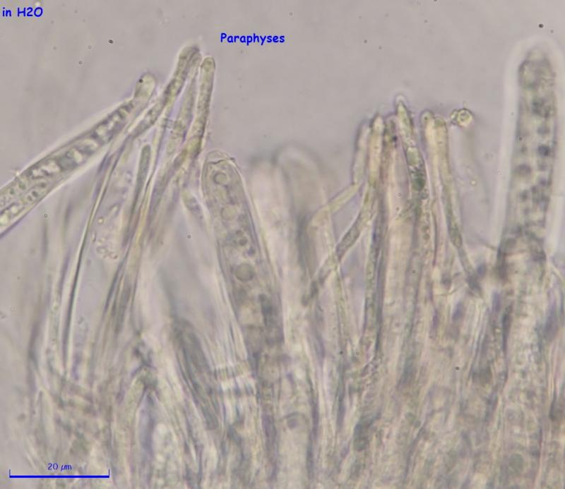
I did examine the specimen the next day, but this was more delicate than others and maybe it was too much for it. I took many pictures, but none of them shows the content of paraphyses well, but I attached the best what I have. It was my first year of hunting Hymenoscyphus like species and I'm lost in the process of recording and identification of them, so I'm not efficient, but I'm learning and maybe by the summer I will know what to do.
It is in an area where I hike a lot, so I will have a high chance of finding it again at the same spot.
Best regards,
Igor
Sorry for raising this topic again. I reexamined the specimen, looked at the textura and I think the medullary excipulum is porrecta and ectal excipulum is prismatica. I don't know if this helps. I uploaded the pictures (https://inaturalist.ca/observations/57610849).
I usually look at spores, paraphyses, and asci when they are still fresh and I take macro photos and I can do the rest later. What other steps should I do when I get fresh Hymenoscyphus-like specimen? Or what other tools should I be using?
Last year I was overwhelmed with a number of new species I was collecting. Usually, if I find something new it really slows me down and I was unable to look at all my specimens, so definitely I've missed a lot. This coming season I will revisit these places and I will get more fresh specimens. I want to be ready to handle them.
Thanks,
Igor
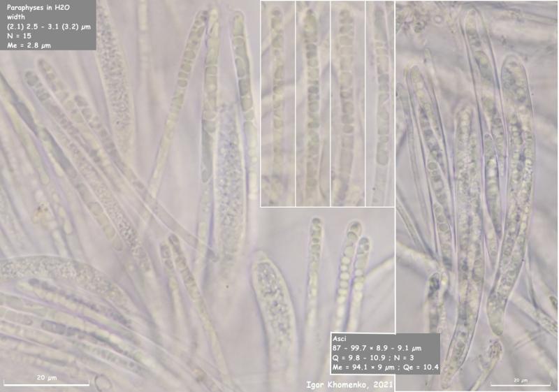
This Hymenoscyphus is back at the same spot as the last year and I collected enough to study.
This time I got a good picture of paraphyses. The spores slightly smaller ((26.7) 27 - 31.7 (35.6) × (4) 4.1 - 4.7 (4.9) µm), but the rest looks the same. I still don't know on what leaves it grows, but there is a chance that it could be Ulmus.
I looked for crystals in the stipe and couldn't find them, only saw cells with nucleus.
All new pictures are here:
https://inaturalist.org/observations/90185294
Igor
Regards,
Igor