20-04-2026 22:00
 Malcolm Greaves
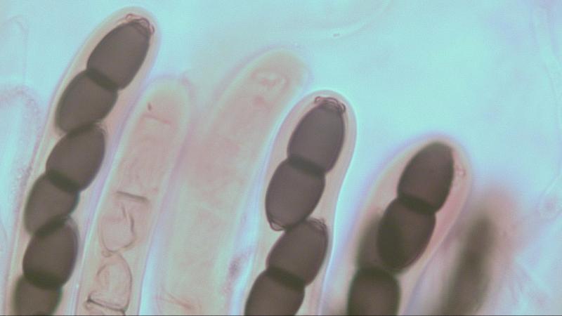
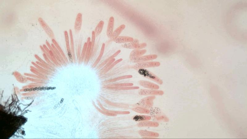
These pale yellow, hairy ascos were growing on cul
19-04-2026 21:23
Steve ClementsBonjour, I found this anamorphic fungus on old pl
19-04-2026 20:46
Steve Clements1 mm diameter approx spherical conidiophores on pl
12-04-2026 17:56
 Hardware Tony
Found on dead stems in February earlier this year
17-04-2026 19:16
 Enrique Rubio
Hi to everybodyI would appreciate any assistance r
14-04-2026 05:32
Ethan CrensonHi all, A few weeks back a friend pointed out som
17-04-2026 15:14
 Bruno Coué
Bonjour.Récoltes du 16/04/2026, sur feuilles mort
12-04-2026 15:52
Gernot FriebesHi,I'm looking for help with this anamorph collect
14-04-2026 21:52
Gernot FriebesHi,found on dead leaves of Carex elata. Conidia: 4
16-04-2026 22:09
Buckwheat PeteHello, I'd like to ask about this older specimen:
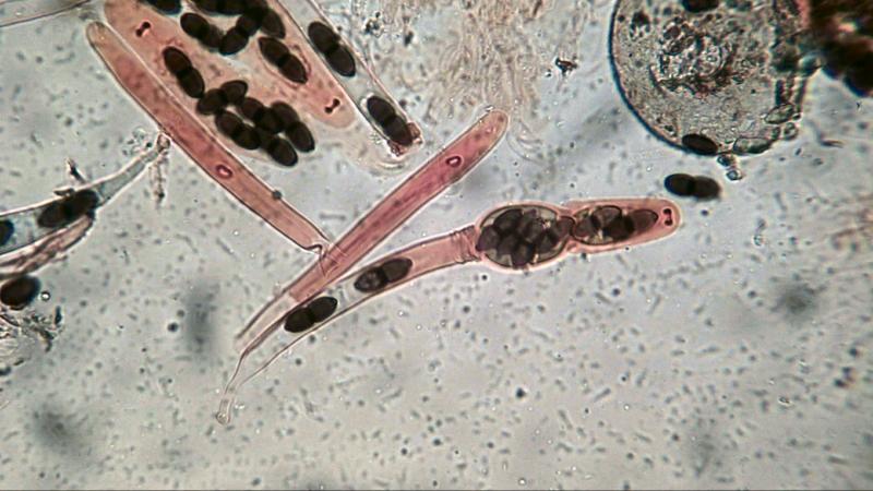
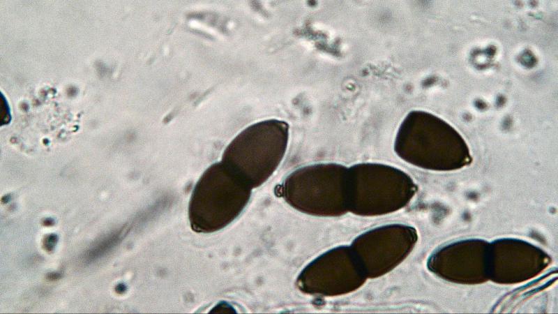
 The reason why I post all these different characteristics of Trichodelitschia bisporula is to confirm if items published in documentations are present or not by means of collected photographic material.
If anyone thinks I am telling nonsense please feel free to comment using the attached photos.
Again the photos are presented as 2D images so it takes some insight to interpret them as part of a 3D presentation.
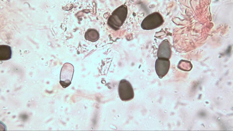
Moreau explained the expulsion of ascospores in Trichodelitschia bisporula species and in his drawing (fig. 72) the ascus swells because of water intake and lengthens; the outer wall breaches in the middle and crumbles towards the base, squeezing the lower part of the ascus making it impossible for spores left behind in the lower part to reach the upper swollen part.
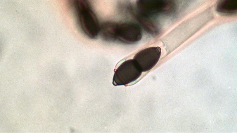
But we are missing the upper part of the outer wall still surrounding the inner wall (photo 1 thru 3).
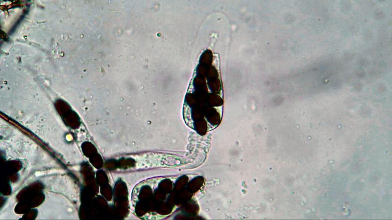
As soon as the asci comes into contact with the amount of water under the deckglass it often causes several asci to swell their upper part which takes milliseconds to complete (photo 9).
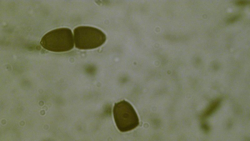
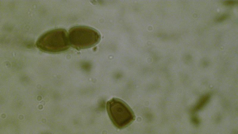
He also stated that ascospores were equiped with a curious device at each extremity that includes a clear discoid area resting on the spore and a dark cap surmounted by a small head (umbo).
It is indeed a discoid area but the top is an open ring (germ pore) (photo-4) connected to the upper part of the spore cell's outer wall which clasps the upper part of the inside cell.
Besides that it also contains a small circular ring superimposed on and clasping a gelatinous substance that is connected to the top of the inner cell. Unfortunately this is not always visible.
Umbo: 1.55-1.65 um in diameter, 1.05-1.15 um high (ring included) and a 0.30 um thick ring (photo 5).
Germ pore: 2.75-2.75 um in diameter and 3.0-3.25 um high in a 50° angle (photo 6).
The umbo is sometines seen with remnants of the gelatinous substance still attatched (photo 7) and can be seen inside the germ pore (photo 8)

By the way when you look closely at photo's 1 and 2 of my first posting you can see the difference in apical systems photo 1 is from T. bisporula and photo 2 probably from T. munkii but for the latter I need more written and/or photographic information about its apical system which in my opinion is not available.
Joop
Hello Ludovic,
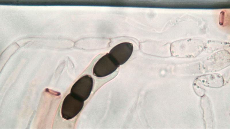
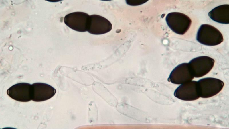
Yesterday I examined two species I preserved from my findings last year and I found a germinating spore cell (photo-1) and an indication that the umbo is connected to the inner cell (photo-2 & 3).
Photo 2 is showing the cell when in focus and photo 3 the cell is out of focus showing the inner cell and the connection of the umbo to the inner cell.
Kind regards,
Joop

As you can see on photo-4 part of the outer wall is still present at the top of the ascus.
Because the lower part of the outer wall strangles the ascus below the swelling there cannot be any pressure build up in the swelling itself.
This swelling will probably not occur within the pseudothecia for then it will block the entrance of the ostiole channel.
According to Moreau the upper part of the ascus will burst open when reaching the ostiole so spores can be discharged through the apical ring.
I do have an assumption about the function of gelatinous sheaths around the spores of Trichodelitschia species.
I already wondered why sheaths are so clearly visible inside the ascus and not outside but maybe the following will give the answer.
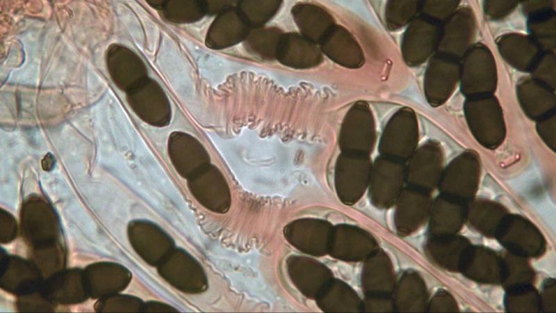
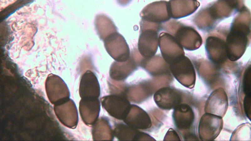
Using a mixture of water and Congo Red the following observation was possible.
When looking at photos 2 & 3 one will see the blue space in between spores, that is water.
Because pressure inside the inner ascus is still building up spores and parts of water are pushed in the direction of the apical ring.
Each spore has a certain amount of water underneath it which at at the end, when its companion spore is entering the apical ring, will make spore expulsion possible when pressure is at the required level.
The swollen sheaths surrounding the spores will block these packs of water to prevent it from streaming out, for else spore discharge will not be possible.
Once a spore has been discharged the amount of water underneath the spore will also disappear and the next spore will enter the ring and thus building up pressure for another expulsion.
Joop