19-05-2026 12:55
 Hardware Tony
After checking Gminder and Otto's library I cannot
19-05-2026 10:27
Patrice TANCHAUDBonjour, rûˋcolte rûˋcente sur terre retournûˋe i
18-05-2026 12:43
Sylvie Le GoffBonjour û tousPuis je avoir votre aide sur ce que
19-05-2026 14:56
û ge OterhalsI found this white cushion-formed ascomycete on ro

18-05-2026 10:13
 Lieve Deceuninck
Dear forum members,I identified this as the teleom

17-05-2026 19:05
Thomas FlammerI have found this tiny 200 ym cup shaped apothecia
17-05-2026 16:41
Margot en Geert VullingsWe found this Lachnum on an old Rubus stem.Fruitbo
Propolis viridis?
Miguel ûngel Ribes,
15-08-2008 01:21
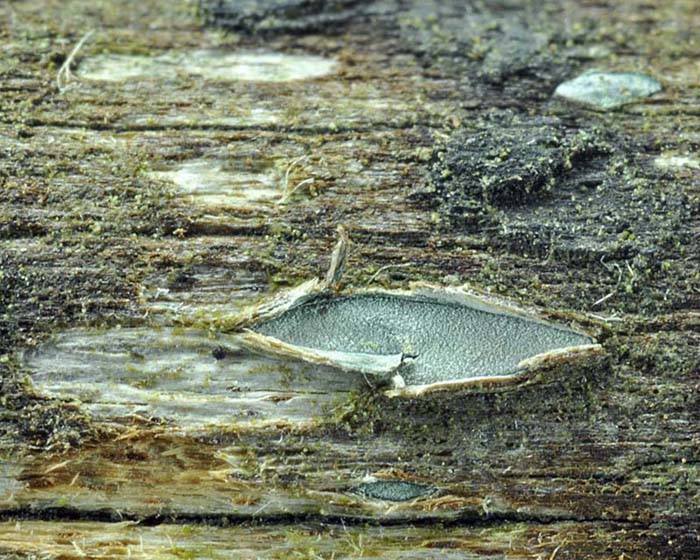
 I am going to try it again with an easier fungi, I hope.
On Eucalyptus globulus wood, 2-3 x 1 mm, green-blue color, broken the wood. Gelatinous flesh. I think it is near P. viridis, according to Baral (sporal size and shape, paraphysis with flexuous apex, inamiloid asci with a red-brown mass inside, and bluish colour). But in the CABI the actual name is P. farinosa (Pers.) Fr. with a lot of sinonyms like P. versicolor Fr. and P. viridis Dufour. Everytimes I have see P. versicolor it was white, but it is described with several colors. Is there one or two species?
Sporal measures (1000x, in water and fresh material)
21.4 [23.5 ; 24.6] 26.7 x 8.3 [9.6 ; 10.3] 11.6
Q = 1.9 [2.3 ; 2.5] 2.9 ; N = 24 ; C = 95%
Me = 24.09 x 9.98 ; Qe = 2.43
Thank you

Miguel ûngel Ribes,
15-08-2008 01:21
Re:Propolis viridis?
Another closer photo
Miguel ûngel Ribes,
15-08-2008 01:23
Re:Propolis viridis?
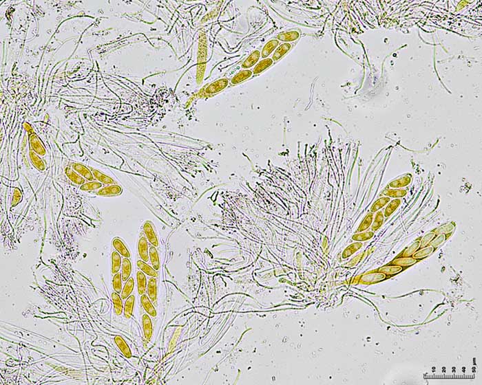
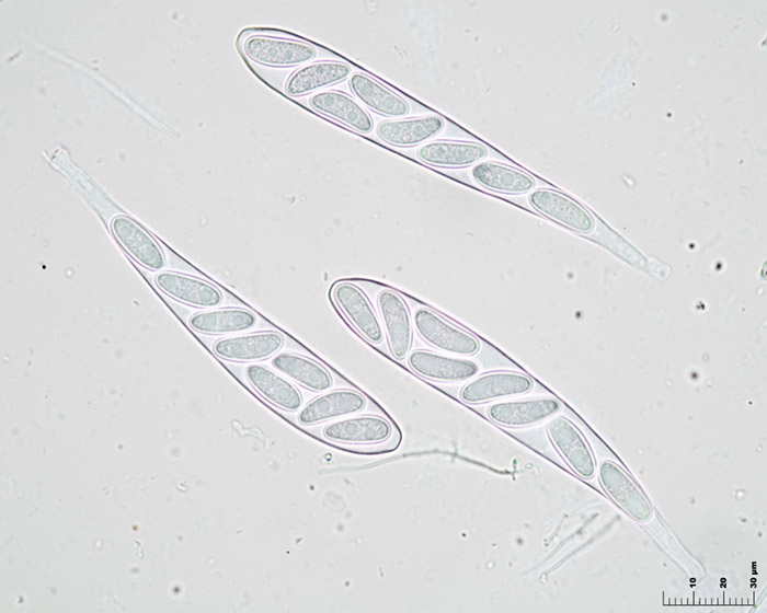
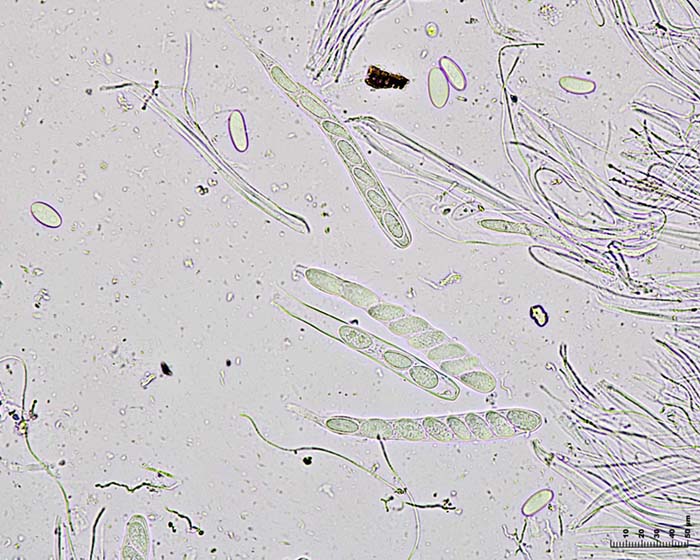
8 spores and inamiloid asci in IKI
Miguel ûngel Ribes,
15-08-2008 01:24
Re:Propolis viridis?
8 spores asci in red congo
Miguel ûngel Ribes,
15-08-2008 01:25
Re:Propolis viridis?
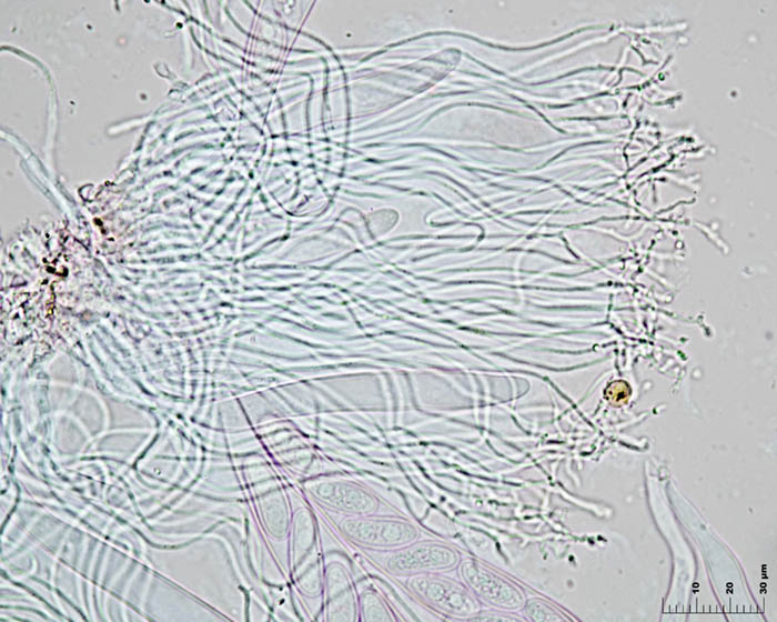
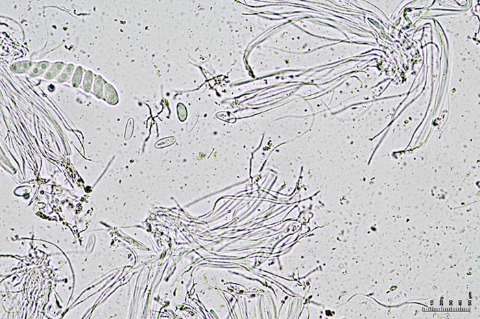
Paraphysis branched and flexuous in red congo
Miguel ûngel Ribes,
15-08-2008 01:26
Re:Propolis viridis?
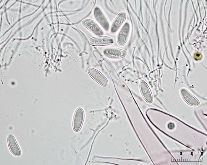
Mature spores in red congo
Miguel ûngel Ribes,
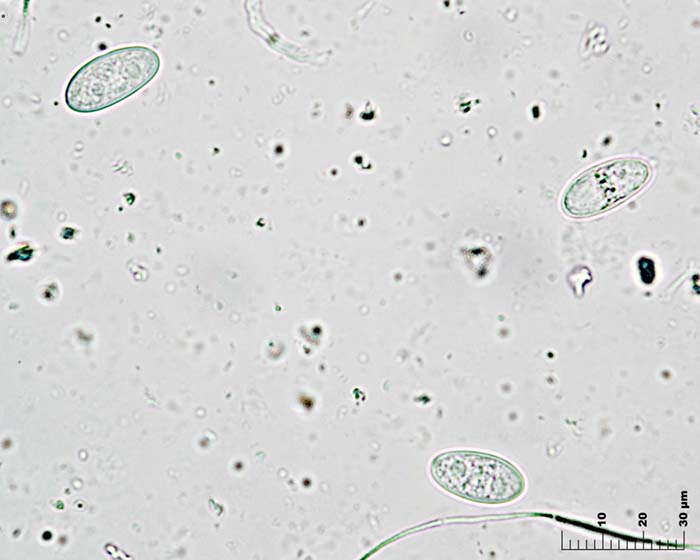
15-08-2008 01:29
Re:Propolis viridis?
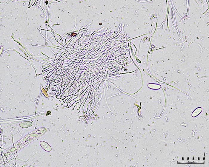
Spores in water
Hans-Otto Baral,
15-08-2008 15:28

Re:Propolis viridis?
Hi Miguel
as you can see from my DVD, there is rather strong variation among the finds I refer to P. viridis. Spore length ranges from 14-19 up to 22-31 ôçm. But those large-spored are uncertain, and mediterranean viridis ranges (14-)16-22(-26) x 5.5-8.5 ôçm. The apos may somtimes be only greenish at the margin, or even entirely white!
So what you have here is unclear to me. At least I am sure that your photos show dead spores. Why? You say the measurements are made from fresh in water? The photos not? The oil drops should be much more distinct, and the plasma not detached from the wall. Looks like in lacophenol. Propolis is xerotolerant (like Srictis), so you can rehydrate the apos many weeks later and have living spores to study, perhaps over a period of half a year.
Zotto
Miguel ûngel Ribes,
16-08-2008 12:16
Re:Propolis viridis?
Hi Zotto
I don't know why my spores are dead cause photos macro are made at 18/06/2008 and photos micro at 29/06/2008, only 11 days after, and all this time the species has been inside a fridge. The spores measuements are made with Piximetre 3.8 over photos micro in glicerine water, only free spores. I am sending more spores photos in water in order to see if are they deadô¢ô¢??
I have made a new measurement, this time with 400x photos in glicerine water, and the results are very similar:
Sporal measures (400x in water, fresh material)
20.1 [23.1 ; 24.7] 27.7 x 8.4 [10.3 ; 11.2] 13.1
Q = 1.6 [2.1 ; 2.4] 2.9 ; N = 25 ; C = 95%
Me = 23.89 x 10.74 ; Qe = 2.25
Thanks Zotto, you are very patient
Miguel ûngel Ribes,
16-08-2008 12:16
Re:Propolis viridis?
Anothe spores in glicerine water
Miguel ûngel Ribes,
16-08-2008 12:18
Re:Propolis viridis?
And another one in water with 1000x