
28-04-2026 20:07
 Lothar Krieglsteiner
... on twig in the air at standing Ceratonia siliq
04-05-2026 18:13
 Stephen Martin Mifsud
ID request for what seems to be a true aquatic fun
04-05-2026 16:39
Stephen Martin Mifsud
ID request: This specimen was collected in Malta o
04-05-2026 09:50
 Castillo Joseba
Me mandan el material seco de Galicia,(España) re
02-05-2026 12:42
Alain BRISSARDBonjour à tousJeuidi 30 avril dernier on m'a remi
02-05-2026 13:06
Pauline. PennaBonjour Please can someone help me with this id
01-05-2026 22:45
 Thierry Blondelle
Bonjour à tous, Une récolte sur bouse séchée d
14-04-2026 05:32
Ethan CrensonHi all, A few weeks back a friend pointed out som
28-04-2026 20:33
Vitus SchäfftleinHello, I found Trochila ilicina on Ilex aquifoliu
Thank you in advance,
zaca
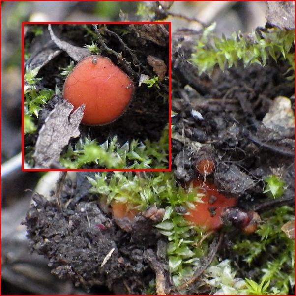
Data:
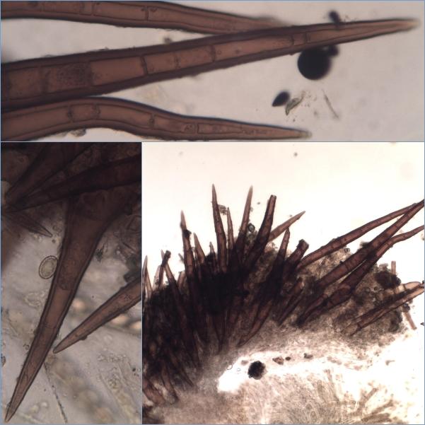
A few apothecia growing on soil under shrubs;
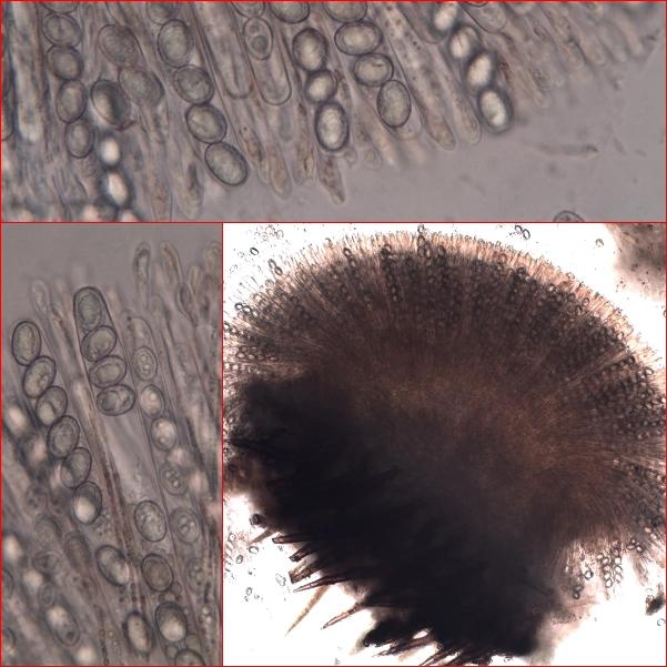
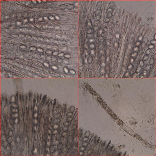
Spores:
(13.8) 16.1 - 18.8 (19.7) x (10) 11.5 - 13.8 (15.1) µm
Q = (1.3) 1.33 - 1.49 (1.5) ; N = 50
Me = 17.4 x 12.4 µm ; Qe = 1.4
Asci: 220-260 x 12-16 µm;
Paraphysis tips globose up to 10 µm in diameter;
Hairs up to 350 x 32 µm.

How are you ?
For this collection, impossible to say without spores in BC...
Probably around nigrohirtula complex, but spores seem to be not mature...
Beñat
I hope everything is right with you as well.
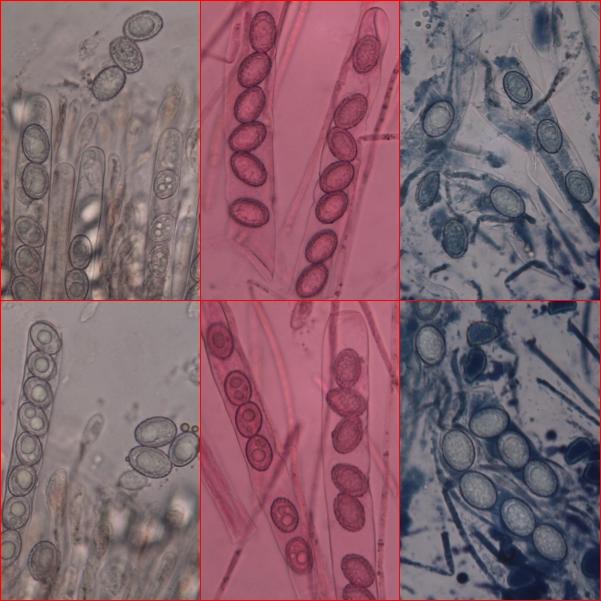
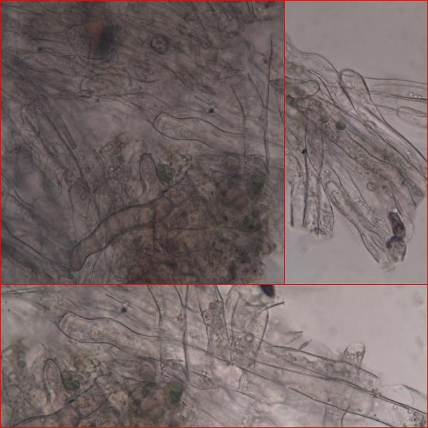
I suppose that by BC you mean "Bleu de Crésyl". I posted some photos of it in the 4th group; The stain was not very effective; Maybe I have to let the blue act for a litle longer. Anyway, I will do it again (in the last apothecia I have, the collection was really short) and post here the results.
Best regards,
zaca

The usual stain for Scutellinia spores is Lactophenol Cotton Blue.
Mal
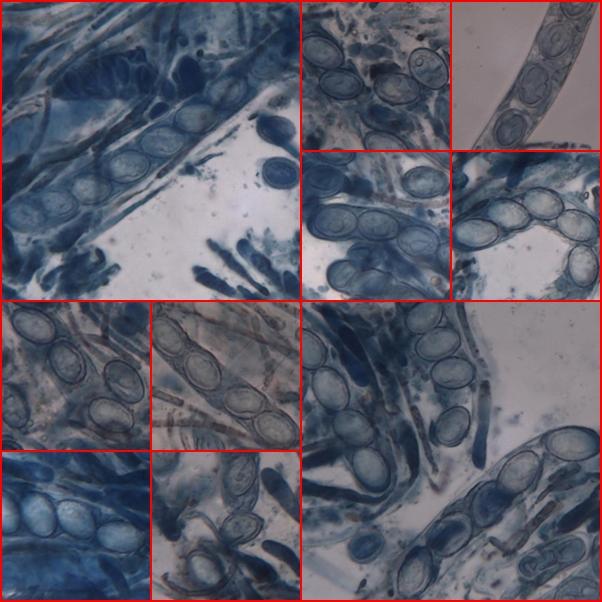
But, I experienced the same problem with Lactophenol Blue than before with Crésyl Blue, the stain is poor and is unevenly distributed. I upload the result of this try.
Regards,
zaca

but as Beñat has pointed out the spores are immature and therefore cannot be of any help. See if you can make a spore drop - with a (thin) marker pen draw a small circle on a microscope slide; turn the slide over and place the hymenial surface of the Scutellinia directly over the circle.
If you leave that for a good period of time you can check the dry microscope slide for ejected (mature) spores in the area within the circle under the compound microscope at x100 or x200; if you see spores, mount in Cotton Blue in Lactophenol, heat gently and show the results.
I hope Beñat does not disagree too much with this procedure I often use . . . .
amitiés
Chris
for your detailed explanation that certainly will be helpful in future.
Unfortunately this time it didn´t work: in fact, after an entire day I was not able to see any spore. The material was already very scarse.
Regards,
zaca