
23-05-2026 11:44
 Charles Grapinet
Hello, I am having trouble identifying this copro
25-05-2026 16:44
François BartholomeeusenHi forum members,During an excursion organised by
25-05-2026 16:35
 Bernard CLESSE
Bonjour à toutes et tous,J'ai trouvé récemment,
22-05-2026 13:29
Gernot FriebesHi,I am curious to hear your opinion on this mater
23-05-2026 18:57
Sylvie Le GoffBonjour à tousRécolté sur une branchette de Sal
22-05-2026 14:44
 Lothar Krieglsteiner
in unripe condition citrine yellow, then soon fadi
22-05-2026 21:35
Steve ClementsBonjour, I expected this find on old wood on our
22-05-2026 18:12
Lothar Krieglsteiner
... in moist chamber from Portugal.As the fungus s
22-05-2026 20:08
Ethan CrensonHello all, Yesterday in NYC I was visiting an e
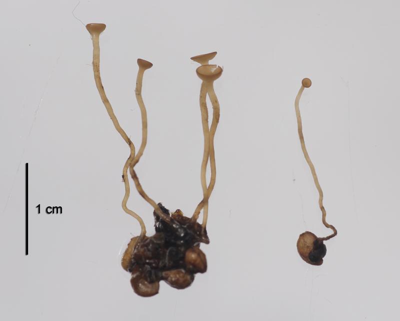
Apothecia cupulate with long stipe, up to 2.5 cm long, disc 2.5 mm across, light brown, hymenium light yellowish-brown, upper part of stipe pubsecent under lens, arise from short cylindric sclerotia, 5 apothecia collected from one old berry, sclerotia attached to seeds.
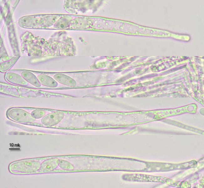
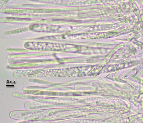
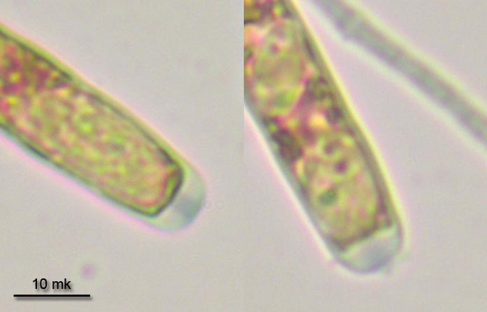
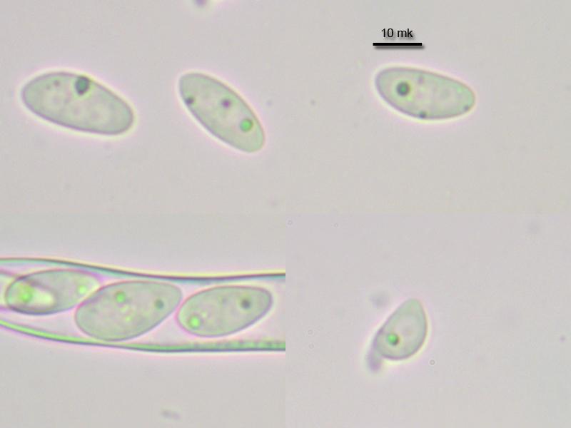
Asci cylindrical with narrowed base, with crozier, with euamyloid ring, 4-spored (but irregularly one to 6-spored asci seen, with part of spores abrupt, many of asci were also underdeveloped and empty), 108-119 x 9.5-11.3 mk; paraphyses cylindrical, moderately branched, with many small greenish guttules, 2.4-3.7 mk broad in upper part; spores ellipsoid, inequilateral, with two small guttules at both ends, 13.1-15.5 x 6-7.5 mk (n=7, only large not abrupt spores measured).
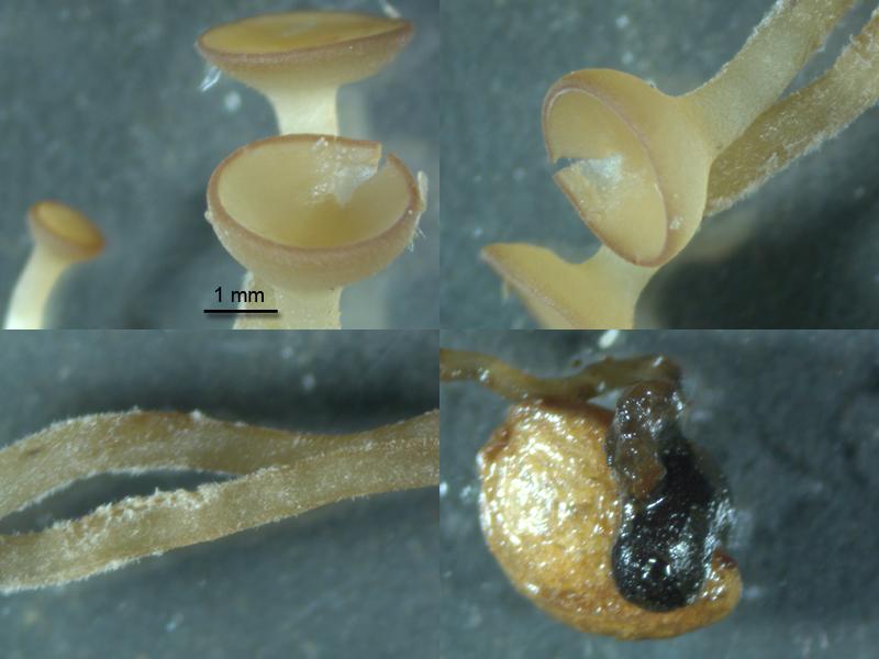

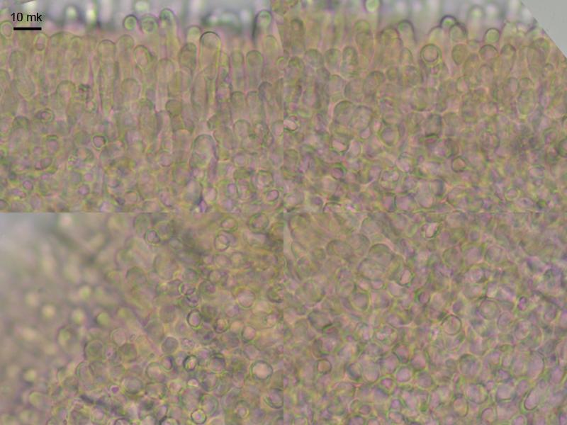
outer layer of receptacle from textura globosa, cells with thickened walles, yellowish, end cells at the edge clavate.
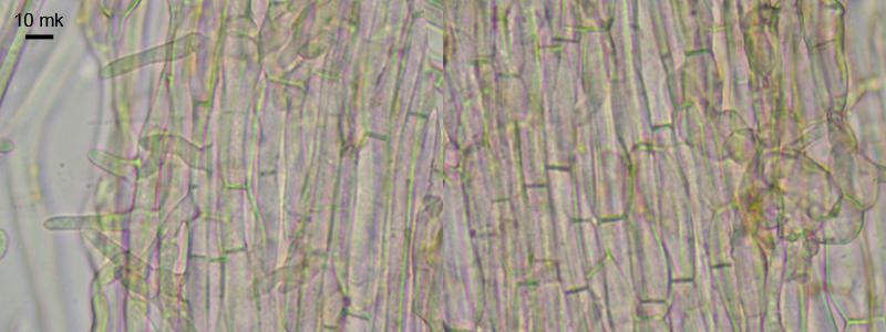
Stipe has excipulum from textura porrecta, cells brown, slightly rough from incrusting pigment, with branching hyphoid hairs.

Rutstroemia chamaemori
Ciboria latipes
Sclerotinia tetraspora
all with a similar spore size.
Did you compare all three? I do not know either of them but I suspect you are right with Sclerotinia tetraspora, because latipes does not fit and R. cham. is on leaves.
But I wonder about the excipulum. Your photo shows only the cortex of very small globose cells. Are they larger inside?
Zotto
The species has distinct scletorium (not stromatized tissues or black stroma), which points to the genus Sclerotinia.
I have seen Rutstroemia chamaemori on this plant (likely, but spores were curved which is not mentioned in descriptions) and showed it here before. http://www.ascofrance.com/search_forum/24922
Ciboria latipes - i have not met, but according to the key in Nordic Macromycetes (2000) it should have stromatized host tissues instead of a distinct sclerotium. Is it right?
I have microscopied excipulum once more, it is made from such small cells in upper part, no larger cells inside. Most inner tissue (medulla) made from interwoven hyphae. Cells become some larger at the base of receptacle. Probably the apothecia were also young, as many asci were underdeveloped.
The VBs in the paraphyses would point to a Botryotinia, by the way.